



GLP-1 diet plan
A GLP-1 diet plan is a structured eating framework for patients taking semagluti...

2
Last Updated: Oct 13, 2026

Gastrointestinal assessment follows the IAPP sequence (Inspection, Auscultation, Percussion, Palpation) – not the standard IPPA used in other body systems, because palpation alters bowel sounds.
Normal bowel sounds occur 5-34 times per minute; auscultate each quadrant for at least 3 minutes before declaring sounds absent.
Red flag symptoms including melena, hematemesis, acute abdominal rigidity, and unexplained weight loss require immediate escalation regardless of other findings.
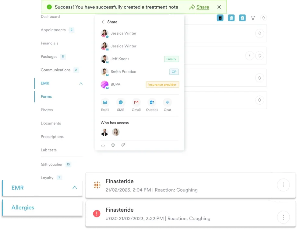
Pabau’s digital forms and AI-powered clinical documentation tools help clinicians capture structured GI findings faster, reducing the documentation burden after assessment.
Most missed GI diagnoses trace back not to clinical inexperience, but to incomplete or out-of-sequence assessment. A nurse who skips auscultation before palpation may unknowingly alter the bowel sounds they were about to document.
A clinician who rushes the subjective history misses the three-week weight loss the patient mentioned offhand. The gastrointestinal assessment is one of the few physical examinations with a mandatory sequence, and understanding why that sequence exists changes how accurately it gets performed.
This guide covers the complete gastrointestinal assessment workflow: abdominal anatomy, history-taking with OLDCARTS, the IAPP physical examination sequence, interpretation of findings, special tests, red flags, documentation, and special population considerations. It is written for RNs, nursing students, and healthcare professionals who need a reliable clinical reference.
Accurate gastrointestinal assessment depends on knowing which organs lie in each abdominal region. The four-quadrant model (RUQ, LUQ, RLQ, LLQ) is the standard anatomical reference used throughout the physical examination.
Knowing these mappings lets you correlate the patient’s pain location with likely organ involvement before you begin the physical examination.
The subjective phase establishes the clinical context for everything that follows. Use the OLDCARTS mnemonic to structure pain history, and layer in bowel habits, dietary changes, and medication use around it. Good patient consultation best practices start with letting the patient describe their chief complaint in their own words before narrowing to structured questions. Stress and mental health often drive GI symptoms, so a biopsychosocial assessment can add useful context to the history.
OLDCARTS stands for: Onset, Location, Duration, Character, Aggravating/Alleviating factors, Radiation, Timing, Severity.
Beyond OLDCARTS, ask about bowel habit changes (constipation, diarrhea, frequency), stool characteristics (color, consistency, blood, mucus), nausea, vomiting, appetite changes, and unintentional weight loss. Review all current medications, including NSAIDs, anticoagulants, and laxatives. Ask about alcohol use and family history of GI malignancy.
Sending a patient history form ahead of the appointment shortens the subjective phase. You start the consultation with the medication list, alcohol intake, and family history already on record, which leaves time for the symptom story.
Abdominal assessment uniquely follows IAPP (Inspection, Auscultation, Percussion, Palpation) rather than the IPPA sequence used in other body systems. The reason is straightforward: percussion and palpation can artificially stimulate or inhibit bowel sounds, so auscultation must happen first. According to NCBI StatPearls, this sequencing is a consistent requirement across physical examination standards.
Position the patient supine with arms at their sides and knees slightly flexed. Expose the abdomen from the xiphoid process to the pubic symphysis. Ensure adequate lighting and a warm room to minimize involuntary muscle guarding.
Stand at the foot of the bed and look across the abdomen tangentially to detect subtle contour changes. Inspect for:
Place the diaphragm of the stethoscope lightly against the skin. Bowel sounds assessment requires listening in all four quadrants, starting in the RLQ (where ileocecal activity is most reliable) and moving systematically. Listen for at least 3 minutes in each quadrant before documenting sounds as absent, per standard nursing textbook guidance cited by Lecturio’s nursing assessment guide.
Normal bowel sounds occur 5-34 times per minute. Also auscultate the aorta, renal arteries, and iliac arteries for bruits, which can indicate vascular pathology.
Percuss all four quadrants using indirect percussion (pleximeter finger on skin, strike with the middle finger of the dominant hand). The two key sounds are tympany and dullness.
Begin with light palpation (1-2 cm depth) before proceeding to deep palpation (4-6 cm depth). Always start away from any area the patient has identified as painful, and watch the patient’s face throughout for signs of discomfort.
This consolidated reference table maps each gastrointestinal assessment step to expected and unexpected findings. It improves on the fragmented tables found across competing resources by presenting all four IAPP steps in a single view, making it usable at the bedside. For a broader clinical reference, see clinical skin assessment tools for comparison to dermatological assessment workflows.
Three special tests are standard components of the gastrointestinal assessment when clinical presentation suggests specific diagnoses. These are documented alongside the main IAPP findings, not performed in place of them. For structured evaluation documentation, a structured clinical evaluation template illustrates how templated assessments capture complex multi-step findings consistently.
These symptoms, whether reported in the subjective history or discovered during physical examination, require immediate escalation regardless of overall clinical picture. Early recognition of these red flags is a core nursing competency.
Document red flag symptoms with precise timing and severity before calling the physician or escalating. A nurse who can state ‘absent bowel sounds in all four quadrants for 15 minutes, rigid abdomen, patient reports pain 9/10’ gives the receiving clinician actionable data immediately, not a vague concern.
The gastrointestinal assessment technique and expected findings vary across three key patient populations. Applying adult-standard criteria to these groups produces inaccurate interpretation.
Pregnant patients: The uterus displaces abdominal organs progressively from the first trimester onward. The appendix migrates superiorly and laterally, so RLQ pain localization for appendicitis is unreliable after 20 weeks. Bowel sounds may be diminished due to progesterone-related smooth muscle relaxation. Constipation is common and not inherently pathological. Always document gestational age and modify palpation pressure accordingly.
Pediatric patients: Children often cannot reliably localize pain with one finger, and anxiety produces voluntary guarding that mimics pathological rigidity. Approach slowly, warm hands, and palpate through clothing initially if the child is distressed. Tympany is more pronounced in infants due to air swallowing. In neonates, bilious vomiting is always abnormal and requires urgent evaluation.
Older adult patients: Classic signs of peritoneal irritation (rebound tenderness, fever, rigidity) are frequently blunted or absent in older adults even with serious pathology.
Constipation is common, but new-onset constipation in an older adult warrants investigation. Diminished pain perception means conditions like appendicitis and cholecystitis may present atypically late. Organomegaly is more difficult to palpate in obese or heavily muscled older patients.

Pabau’s digital forms and clinical documentation tools let your team capture structured assessment findings, build patient timelines, and automate documentation workflows – so clinicians spend their time assessing, not writing.

Clear, standardized documentation of gastrointestinal assessment findings protects both the patient and the clinician. Vague entries like “abdomen normal” do not convey what was actually examined. Structured clinical documentation using an objective-findings framework reduces ambiguity and supports continuity of care between shifts.
Use this SOAP-aligned structure for your GI assessment note:
For safer clinical notes practices, document the time of assessment, patient positioning used, and whether findings were unchanged from a previous assessment or represent a new development. The patient record management system in use should allow nurses to timestamp entries and link them to the relevant encounter.
Documentation is where clinical time disappears. A nurse completing a full gastrointestinal assessment across a four-patient bay during a busy shift faces a compounding problem: the more time that passes before charting, the less precise the note becomes, regardless of clinical skill.
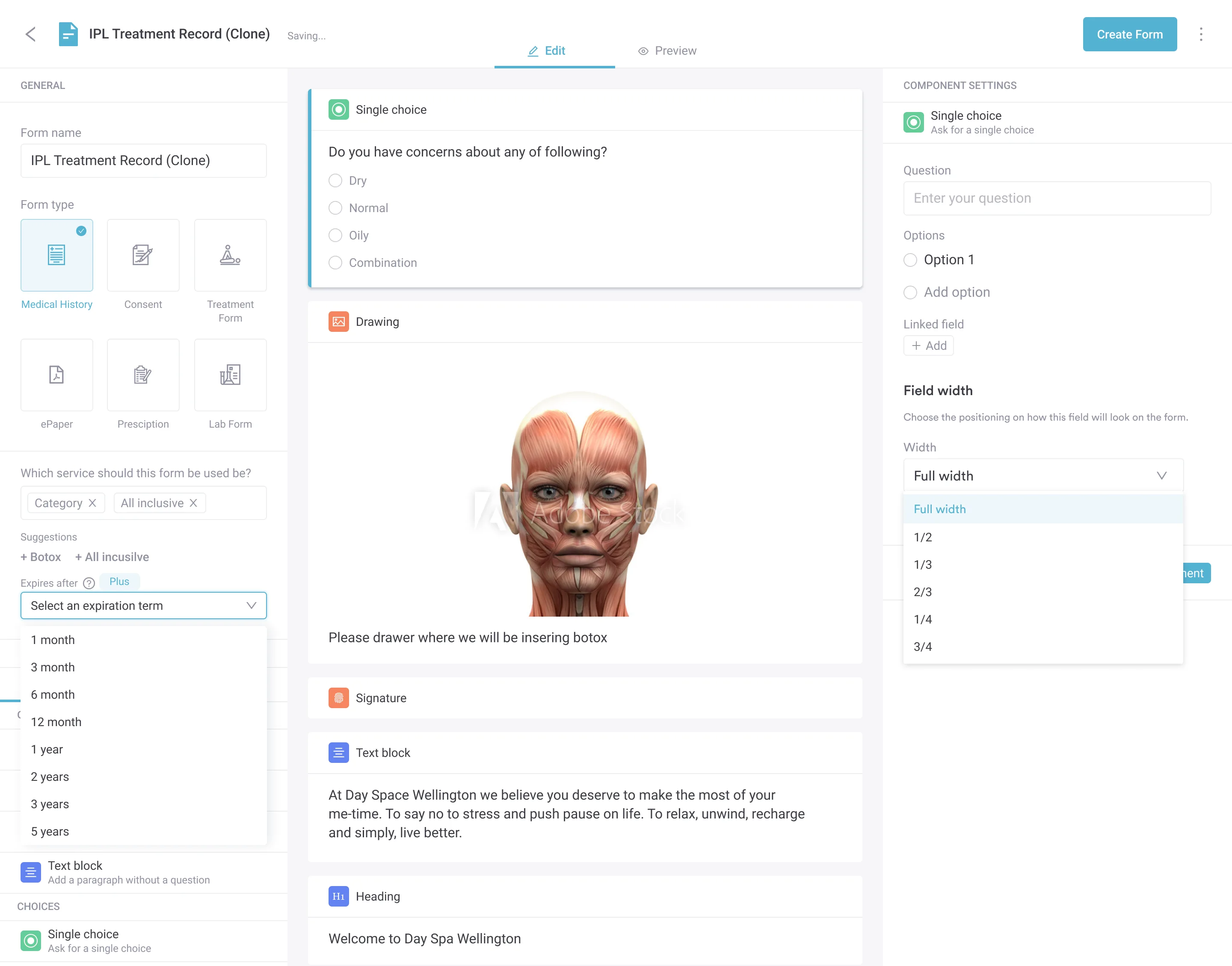
Practice management software like Pabau offers digital intake forms that let clinical teams build structured assessment templates mirroring the IAPP sequence. Instead of free-text charting, nurses complete a structured form with pre-set fields for bowel sound frequency, quadrant-by-quadrant percussion findings, and palpation results. The form enforces completeness: if a required field is blank, the clinician is prompted before submission.

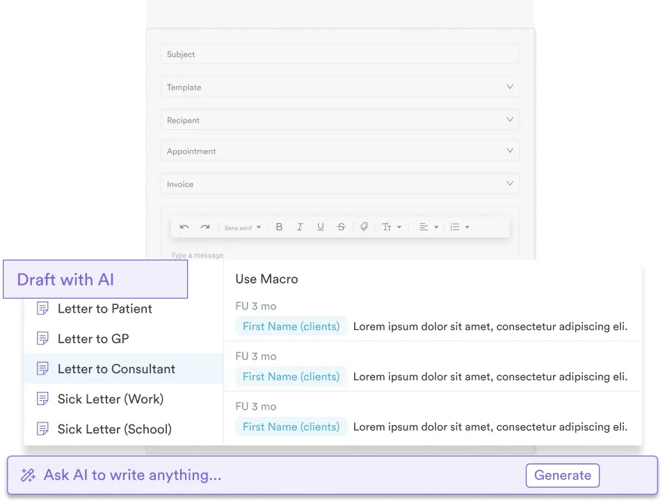
For facilities using AI-powered clinical documentation, verbal assessment findings can be transcribed directly into structured notes during or immediately after the examination, reducing the documentation lag to near zero.
Practices running high-volume GI caseloads, like GP clinics and functional medicine practices, find this particularly valuable when multiple patients require parallel assessment workflows.
The result is a timestamped, structured, auditable GI assessment record that supports handover, audit, and clinical governance without adding to clinical workload. For practices using structured capture forms, standardized medical forms across the team also reduce inter-clinician variation in what gets documented and how.
Use this checklist to confirm each component of the gastrointestinal assessment has been completed before closing the encounter. It covers subjective history, physical examination steps, special tests, and documentation requirements.
A rushed or out-of-sequence gastrointestinal assessment produces unreliable findings. The IAPP order exists for a reason, the red flag list is specific for a reason, and structured documentation matters for a reason: each element supports the one that comes after it.
For clinical teams managing high volumes of assessments, Pabau’s digital forms and structured documentation tools turn the IAPP sequence into a consistently completed, auditable workflow rather than a discretionary checklist. If you want to see how this works in practice, book a demo with the Pabau team.

Need a template to hand off patient status between shifts? Nursing shift report template covers the structure nurses use to pass on assessment findings at handover.
Want a structured way to capture the rest of the body systems review? Constitutional review of systems template extends the subjective history beyond the GI-specific questions covered here.
Caring for a patient with cirrhosis? Cirrhosis nursing care plan covers assessment data, nutritional status, fluid balance, and a day-by-day intervention grid.
A gastrointestinal assessment is a systematic clinical examination of the GI and genitourinary systems, combining a subjective history (including pain characterization, bowel habits, and medication review) with a structured physical examination following the IAPP sequence: Inspection, Auscultation, Percussion, and Palpation. It is performed by nurses and clinicians to identify normal function, detect abnormalities, and escalate red flag findings.
The correct order is IAPP: Inspection, Auscultation, Percussion, then Palpation. This differs from the IPPA sequence used in other body systems because percussion and palpation can artificially stimulate or inhibit bowel sounds. Auscultation must be completed before either technique is applied to the abdomen.
Key red flags include melena (black tarry stool), hematemesis (vomiting blood), acute abdominal rigidity, positive rebound tenderness with fever, hematochezia (bright red rectal blood), unexplained weight loss over 5% of body weight in six months, abdominal distension with absent bowel sounds, and a pulsatile midline abdominal mass. Any of these findings requires immediate escalation.
Palpation and percussion physically stimulate the bowel and can alter motility, either increasing or decreasing bowel sound frequency. Performing auscultation first captures the patient’s baseline gut activity before any mechanical interference. This is why the abdominal assessment sequence (IAPP) is the reverse of most other body system assessments.
OLDCARTS stands for Onset, Location, Duration, Character, Aggravating/Alleviating factors, Radiation, Timing, and Severity. It is the standard mnemonic used to structure pain history during the subjective phase of the gastrointestinal assessment, ensuring no key dimension of the patient’s pain experience is missed.
Normal bowel sounds occur 5-34 times per minute and are described as soft clicks or gurgles. Hyperactive sounds (high-pitched, frequent, borborygmi) suggest gastroenteritis or early obstruction. Hypoactive sounds (infrequent and quiet) may indicate post-operative ileus or peritonitis. Absent sounds across all four quadrants after 3 minutes of auscultation per quadrant signal paralytic ileus or late obstruction and require urgent evaluation.






