



GLP-1 diet plan
A GLP-1 diet plan is a structured eating framework for patients taking semagluti...

2
Last Updated: Oct 13, 2026

Caloric testing checks one ear at a time by warming or cooling the ear canal and recording the nystagmus that follows.
COWS stands for Cold Opposite, Warm Same, and it tells you which way the fast phase should beat.
Unilateral weakness above 25% and directional preponderance above 30% are usually abnormal, though lab norms vary.
The test only reads the horizontal canal at very low frequency, so vHIT still has a job to do.
Practice management software like Pabau keeps caloric bookings, pre-test screening, and VNG results in one patient record.
Dizziness rarely tells you which ear is at fault. The history sounds much the same either way, and most bedside tests fire both labyrinths at once.
Caloric testing is the way around that. It is still the only routine test that switches on one inner ear at a time. That is why it remains the reference standard for finding a weak side.
The hard part starts after the irrigation. You are left with four responses, two formulas, and cut-offs that shift from lab to lab. Get the protocol right and the numbers do most of the work for you. Get it wrong and you spend the afternoon arguing with an artifact.
Caloric testing is part of the videonystagmography (VNG) and electronystagmography (ENG) test batteries. It measures how each inner ear answers a temperature stimulus. Because you irrigate one ear at a time, you get a per-ear result that no other routine test gives you.
The mechanism is simple physics. Warm or cool water changes the temperature of the bone around the horizontal semicircular canal. That gradient sets up a convection current in the endolymph. The current deflects the cupula, the hair cells fire, and the eyes answer with nystagmus that the equipment records.
What you are reading is a very low frequency response. The endolymph movement is roughly equivalent to head rotation at 0.003 Hz, according to a review in the Brazilian Journal of Otorhinolaryngology. Everyday head movement runs between 1 and 6 Hz, so the test deliberately probes a range the canals almost never meet.
None of that works in isolation. Caloric testing sits in a battery alongside ocular motor and positional tests, and its own question is narrow. Is each ear pulling its weight?
Order caloric testing when you need to know which labyrinth is underperforming. A bedside screen such as the modified Romberg test tells you a patient is unsteady. It will not tell you which side to blame. A primary care practice sees the same presentation in many forms, so consistent recording is what makes review possible.
Screening for contraindications before you book protects the patient and the slot. A full battery runs 30 to 45 minutes, so a test abandoned halfway leaves an expensive hole in patient scheduling.
Air irrigation is the accepted fallback when the eardrum is perforated. Normative data for air and water differ. Make sure you read the result against the reference range for the modality you used.
A caloric test is four irrigations in a fixed order, with real rest between them. StatPearls and the British Society of Audiology’s recommended procedure agree on the core protocol below. Completed patient intake forms confirm medication and ear history before the patient arrives.
The patient lies supine with the head raised 30 degrees. That angle brings the horizontal canal upright, where convection works hardest. Note which ear you test first, usually the right, and record the order in the VNG software.
Standard bithermal testing uses four runs, warm (44°C) and cool (30°C) in each ear. Water irrigations last 30 seconds and air irrigations last 60. Between runs, the patient rests for 3 to 5 minutes so the nystagmus fully settles. Cut that rest short and the next response comes back artificially small.
COWS is the mnemonic that keeps you straight during live recording. Cold Opposite, Warm Same. Cool irrigation inhibits the tested canal, so the fast phase beats away from that ear. Warm irrigation excites it, so the fast phase beats toward it.
Most unusable traces are created before the water goes in. Five minutes of checking saves a repeat visit.
Monothermal screening uses one temperature, usually cool 30°C, in both ears. It suits patients who cannot tolerate a full battery, and it is quicker. If the asymmetry crosses your lab’s screening threshold, go on to full bithermal testing. Sensitivity for mild unilateral weakness is lower, so it never stands in for a diagnostic workup.
Interpretation runs off one number per irrigation, the peak slow-phase velocity (SPV) of the nystagmus. VNG software works out the rest. You still need the arithmetic, because the software will happily calculate a tidy percentage from a contaminated trace.
Unilateral weakness (UW) compares the total response from the right ear against the left. The Jongkees formula does the work.
UW% = [(RW + RC) – (LW + LC)] / (RW + RC + LW + LC) x 100
RW and RC are the right warm and right cool SPVs, and LW and LC are their left counterparts. Anything above 25% is generally treated as abnormal, subject to your lab’s own norms. A consistent result above that line points to reduced output on the weaker side.
Vestibular neuritis, labyrinthitis, acoustic neuroma, and Meniere’s disease all produce that pattern. Automated software calculates UW% for you, but reviewing the underlying traces is what catches an artifact before it becomes a diagnosis.
Directional preponderance (DP) compares the responses that beat right against those that beat left.
DP% = [(RW + LC) – (LW + RC)] / (RW + RC + LW + LC) x 100
Most labs call anything above 30% abnormal. Unlike unilateral weakness, DP does not localize to an ear. It shows a directional bias across the system, which can be peripheral or central, and it often fades as compensation takes hold.
Bilateral weakness means all four responses fall below your lab’s minimum, typically a peak SPV under 5 to 10 degrees per second. Look for ototoxic exposure first, particularly aminoglycosides and cisplatin. Autoimmune inner ear disease and bilateral Meniere’s come next. These patients usually describe oscillopsia when they move their head, which fits the finding.
The fixation suppression index (FSI) checks whether vision can damp the caloric nystagmus down. At peak response, the patient fixates on a stationary target. You then divide the SPV with fixation by the SPV without it.
A normal FSI is 0.6 or lower, which means fixation cuts the slow-phase velocity by at least 40%. An FSI above 0.6 says central suppression has failed, pointing toward the cerebellum or posterior fossa. It is one of the few caloric findings that separates central disease from peripheral disease, though it names no diagnosis on its own.
No caloric finding stands alone. Read it next to the audiogram, the history, the rest of the VNG battery, and imaging where it is indicated. The patterns below are the ones that come up most often.
Say the peak SPVs come back at right warm 22, right cool 18, left warm 9, and left cool 7 degrees per second. The right total is 40 and the left total is 16. Jongkees gives a unilateral weakness of 43% on the left, comfortably past the threshold.
Pair that with asymmetric hearing loss on the same side and the next step is imaging, not reassurance. Where the workup ends in confirmed hypofunction, most patients go on to vestibular rehabilitation, and the initial evaluation is coded with CPT code 97161.
In brain death assessment, caloric testing becomes the oculovestibular reflex check, and the protocol changes. Ice-cold water at roughly 0 to 4°C replaces the bithermal temperatures. The head stays elevated at 30 degrees, and each ear receives 20 to 50 mL.
Timing matters in two separate ways here. Watch the eyes for at least one minute after each irrigation before you record an absent response. Then leave at least five minutes before testing the second ear, so the first stimulus does not carry over.
An intact brainstem produces tonic deviation of the eyes toward the irrigated ear. In brain death, neither side moves at all. That absent response supports the diagnosis without ever confirming it alone, so follow the confirmatory steps your institution’s protocol sets out.
The video head impulse test (vHIT) is now widely available, and it often comes up as an alternative. The two tests answer different questions. vHIT reads the same horizontal canal at 2 to 6 Hz, close to everyday head movement, while caloric testing reads it far lower down.
Normal vHIT alongside 30% unilateral weakness is a common pairing after vestibular neuritis. The high-frequency response has recovered while the low-frequency one has not. Run vHIT on its own and that history disappears. Run calorics on their own and high-frequency loss slips past you.
That frequency split is why specialist practices tend to run both in the same visit rather than choosing between them.
Caloric testing has clear boundaries, and knowing them stops you leaning on one number too hard.
Those limits bite hardest when the caloric result is normal and the patient still feels awful. Orthostatic causes, cervicogenic dizziness, and central disease all sit outside what an irrigation can reach. Autonomic testing covers the orthostatic side, while cervicogenic cases often respond to targeted neck pain exercises. When neck symptoms arrive with arm paresthesia, the Halstead maneuver screens for thoracic outlet syndrome.
A caloric test takes 45 minutes and generates paperwork out of all proportion to that. There is contraindication screening beforehand, VNG numbers afterwards, a letter to the referrer, and a bill. In most practices those four things live in four different places, which is the job practice management software exists to fix.
Pabau is an all-in-one practice management system built for specialist practices. Digital intake forms collect medication and ear surgery history before the appointment, so contraindications surface before the patient is in the chair. Appointment scheduling holds the longer slot a full bithermal battery needs, with the buffer built into the service type.

After the test, unilateral weakness, directional preponderance, and FSI go into structured clinical records as fields rather than free text. Filing the report as a PDF is easy enough. Fields are what let you put last year’s numbers next to today’s at the follow-up.
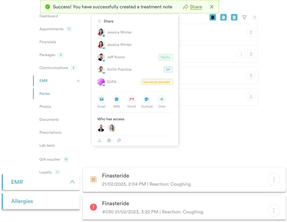
For practices billing vestibular assessments, claims and billing tools validate the insurer details on each claim before it goes out. Automated workflows then trigger the follow-up booking or the referrer letter as soon as the result is filed.
The same setup carries the rest of an ENT service line. Audiology sits next to speech therapy, and rehab referrals go out to physical therapy teams. Pulling scheduling, records, and billing together means less of the day spent moving the same information between systems.

Pabau keeps caloric bookings, pre-test screening, VNG results, and billing in one patient record, so your team stops rekeying the same information.

Caloric testing has outlasted newer technology because it still answers a question nothing else answers cleanly. Which ear is weak? The rest of the workup narrows the field, and this test names the side.
The trade-off is that it buys that answer at a frequency the patient never experiences. Treat a normal caloric result as one panel of the picture rather than an all-clear. Pair it with vHIT whenever the history and the numbers disagree.
The clinical side of this test is well documented. The admin around it usually is not, and that is where practices give back the time they saved in the test room. Book a demo to see how Pabau keeps vestibular intake, VNG results, and billing in one patient record.

Wondering what those Hz bands on an audiogram actually mean? Hearing test frequency explains what each range tells you about a patient’s hearing.
Considering independent practice as a nurse practitioner? starting a nurse practitioner practice walks through the steps to open and run an independent practice.
Need a refresher on ear exam technique? otoscope examination technique walks through technique, common findings, and documentation for a bedside ear exam.
Documenting tinnitus alongside vestibular symptoms? Tinnitus report covers the common causes and what to capture at the appointment.
Managing a patient after major trauma? primary trauma survey sets out the xABCDE framework, its steps, and adjuncts under the latest ATLS guidance.
No. The irrigation is painless, though the cool run feels cold in the ear. Most patients get 30 to 60 seconds of spinning and some mild nausea. Tell them it settles within a couple of minutes.
Usually yes, but not straight away. Ask patients to sit for 15 to 20 minutes after the last irrigation and to leave once the dizziness has cleared. Anyone still unsteady should arrange a lift home.
In the US, bithermal caloric testing with four irrigations is reported with CPT code 92537. Monothermal testing, which uses two irrigations, is CPT code 92538. Both are billed once per session rather than per ear.
An audiologist usually runs the test, with an ENT physician or neurologist interpreting the report. In hospital settings, a trained vestibular technician may carry out the irrigations. Responsibility for the diagnosis stays with the clinician.
Both record the same nystagmus during the same battery. ENG uses skin electrodes placed around the eyes, while VNG uses infrared cameras mounted in goggles. VNG is now standard, because it captures torsional eye movement that electrodes miss.






