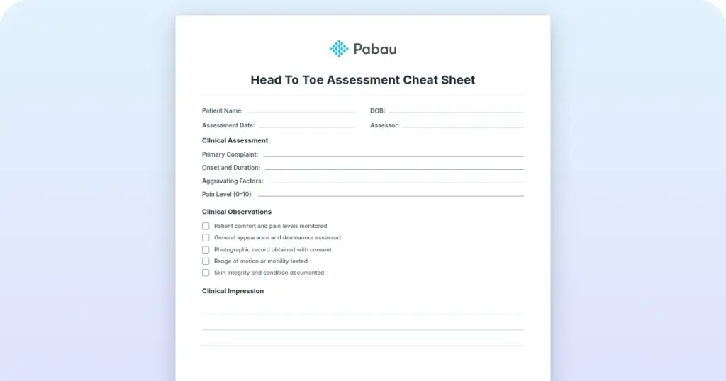
Head to toe assessment cheat sheet: Printable nursing guide
A head to toe assessment cheat sheet gives you one fixed order at the bedside, s...

2
Last Updated: Oct 23, 2026

No single rheumatoid arthritis test confirms the diagnosis, so clinicians read RF, anti-CCP, CRP, ESR, and CBC together.
Anti-CCP is the most RA-specific marker at roughly 96%, and it can turn positive years before joint symptoms start.
RF is positive in about 70% to 80% of RA patients, but also in Sjögren’s syndrome, hepatitis C, and healthy older adults.
Roughly 20% to 30% of patients have seronegative RA, so imaging and the ACR/EULAR criteria carry the diagnosis instead.
Practice management software like Pabau keeps serology, imaging reports, and clinical notes on one patient record.
Most patients expect one definitive rheumatoid arthritis test. There isn’t one. A diagnosis comes from blood markers, inflammation levels, imaging, and a careful joint exam, all read as a single picture.
Getting that picture wrong costs joints. Erosion doesn’t reverse, and the window where treatment prevents it is measured in months. The American College of Rheumatology estimates RA affects roughly 1.3 million Americans. Many wait months for a confirmed diagnosis.
The tests themselves are straightforward. Reading them together is the part that decides how fast a patient starts treatment.
A rheumatoid arthritis test is never one blood draw. Rheumatologists order a panel because RA markers overlap with lupus, Sjögren’s syndrome, and psoriatic arthritis. One positive result rarely settles anything on its own. The standard workup covers:
Imaging then fills in what serology can’t show. X-rays reveal bone erosion and narrowed joint space. MRI picks up early synovitis before any structural damage appears. Ultrasound shows soft tissue inflammation while the patient is still in the room.
Every test in the workup answers a different question. The table below sets out what each one measures, its usual reference range, and what a high result points to.
The RF test comes back positive in roughly 70% to 80% of RA patients, according to MedlinePlus. That sensitivity sounds reassuring. Specificity is the weak spot.
RF also rises in Sjögren’s syndrome, hepatitis C, chronic infections, and healthy older adults. So a positive RF on its own confirms nothing, and a negative RF rules nothing out.
Reference ranges shift from lab to lab. Most flag anything above 14 IU/mL, though context weighs more than the cutoff. Symmetric swelling, over 30 minutes of morning stiffness, and RF at 20 IU/mL point one way. An asymptomatic 75-year-old with RF at 18 IU/mL points somewhere else entirely.
Anti-CCP targets proteins that have been citrullinated, a change that happens inside inflamed joint tissue. Research in the Annals of the Rheumatic Diseases puts its specificity near 96%, against roughly 85% for RF. That difference matters when lupus, fibromyalgia, and reactive arthritis are all in play.
Anti-CCP can also show up years before the first swollen joint. A positive result in a patient with vague joint pain and a family history of autoimmune disease is worth acting on. Many rheumatologists now order anti-CCP first and treat RF as supporting evidence.
Serial results only help when they sit together. That is why an autoimmune workup belongs in structured patient records rather than a folder of lab PDFs.

Neither marker is specific to RA. Both answer a narrower question. Is inflammation active today, and is treatment moving it?
CRP rises and falls faster, so it reflects acute flares. ESR covers a longer window. Most rheumatologists track both at every visit. Borderline RF with consistently high CRP and ESR points toward active inflammatory arthritis. Isolated RF positivity with normal inflammatory markers does not.
ESR ranges also depend on age and sex. Above 20 mm/hr in men or 30 mm/hr in women may signal inflammation, though thresholds vary by lab. Anyone interpreting biomarkers in chronic disease should weigh the patient’s age, other conditions, and current medication.
Numbers only tell half the story. A simple global rating of change captures whether the patient feels better, which no CRP value shows.
Results only mean something in combination. Positive RF with negative anti-CCP and normal CRP is a different signal from strongly positive anti-CCP alongside high ESR and CRP.
Positive anti-CCP and positive RF together point strongly to RA, especially with symmetric joint swelling and long morning stiffness. Imaging and the ACR/EULAR criteria come next. A positive anti-CCP with no symptoms calls for monitoring rather than treatment.
The rheumatoid factor normal range varies by lab, with most cutoffs between 14 and 20 IU/mL. High-titer RF above 50 IU/mL tracks with more aggressive disease and a higher chance of rheumatoid nodules.
Clinicians use that titer to stratify risk at diagnosis, which shapes the first treatment decision. Across a busy patient list, lab result management keeps those thresholds visible instead of buried in attachments.
Yes, and it happens often. Seronegative RA accounts for roughly 20% to 30% of cases, according to the Hospital for Special Surgery. These patients have active synovitis, morning stiffness, and progressive joint damage while RF and anti-CCP stay negative.
Diagnosis then rests on imaging and clinical criteria. MRI can show bone marrow edema and synovitis long before an X-ray changes. Ultrasound shows synovial inflammation at the bedside.
The ACR/EULAR criteria award points for clinical features, symptom duration, and inflammatory markers. A patient can reach the diagnostic score of 6 with no positive antibody at all. Seronegative RA is not a milder disease either. It damages joints on the same timeline and needs the same treatment approach.
Blood tests find the biology. Imaging shows what that biology has done to the joint. Because the two answer different questions, one rarely replaces the other.
Which one you order depends on symptom duration, presentation, and what the practice can access. Most rheumatologists start with hand and foot X-rays. They move to MRI when early RA is suspected and those films look normal. Cardiac workups follow the same split, where thallium stress testing reveals perfusion problems that anatomy alone hides.
No blood test replaces what a rheumatologist finds by hand. Tender joint count, swollen joint count, symmetry, stiffness on waking, and features like nodules or eye inflammation all feed the diagnosis.
The squeeze test is a quick example. Compressing the second to fourth metacarpophalangeal or metatarsophalangeal joints can produce tenderness. That finding raises the question of inflammatory arthritis, according to the National Institute of Arthritis and Musculoskeletal and Skin Diseases (NIAMS).
Other findings steer the differential elsewhere. Where back or pelvic pain is part of the picture, the Gaenslen test helps point toward sacroiliac involvement, which suggests a different inflammatory arthritis.
The ACR/EULAR 2010 criteria score four domains out of 10. Six points or more classifies definite RA.
Work an example through. A patient with more than 10 involved joints, at least one of them small, scores 5 for joint involvement. Add 1 for a raised CRP and 1 for symptoms running past six weeks. The total reaches 7 with no positive antibody anywhere on the panel.
That is the point of the scoring system. It was built to catch early RA before erosion appears on film. Symptom duration is a scored domain, which is why digital intake forms that capture onset dates before the appointment earn their setup time.

The first three to six months after onset is the window that matters most. ACR guidance points one way here. Start disease-modifying antirheumatic drugs (DMARDs) inside that window and long-term joint outcomes improve. Waiting for textbook serology while symptoms progress means waiting for damage that won’t reverse.
Four presentations should trigger the panel rather than a wait-and-see appointment:
A primary care physician who suspects inflammatory arthritis should refer at that first appointment. Bilateral metacarpophalangeal and proximal interphalangeal involvement in a patient under 50 justifies the full panel straight away.
Referral speed depends on the front desk as much as the clinician. Practices running on primary care software can flag an abnormal panel the day it lands, instead of at the next review.
The same holds in functional medicine practices, where a long autoimmune workup produces results from several labs at once. Automated workflows keep the urgent ones out of a manual review queue.
Most referrals bounce back for missing detail rather than missing tests. Run through this before the letter goes out:
Two habits spoil otherwise clean referrals. A patient already on prednisone arrives with a normal CRP that hides active disease. Serology and inflammatory markers drawn weeks apart also become impossible to read side by side.
Document the symptom onset date and morning stiffness duration at every first rheumatology visit. Symptom duration is a scored domain in the ACR/EULAR criteria. Accurate notes at that first visit can move a borderline score from 5 to 6 and start treatment sooner.
Rheumatology creates a documentation load most clinical software wasn’t built for. One workup can run for months, with results arriving from several labs, an imaging center, and a specialist letter, all at different times.
Pabau pulls that into a single record. Patient record management holds the serology, the imaging reports, and the notes on one timeline. Nobody has to rebuild the history from memory at the next visit.
Forms do the groundwork before anyone sits down. Intake collects symptom onset, family history, and joint findings ahead of the appointment. Pabau Scribe, our AI scribe, then drafts the consultation note, so the clinician spends the visit examining joints instead of typing.
Reporting comes with every Pabau subscription. A multi-site group can track referral volumes, panel turnaround, and follow-up rates without exporting anything to a spreadsheet.

Pabau brings lab results, imaging reports, and clinical notes onto one patient timeline. A long autoimmune workup stays readable from the first referral through to diagnosis.

When a panel comes back muddy, more serology rarely settles it. Imaging, a careful second look at the joints, and a documented symptom timeline usually do.
That puts the pressure on documentation rather than on the lab. A practice that records onset dates, stiffness in minutes, and medication changes hands its rheumatologist a scorable case. The one that doesn’t sends the patient back for another blood draw.
Keeping serology, imaging, and notes on one record is what separates a fast diagnosis from a slow one. Book a demo to see how Pabau handles lab tracking and specialist documentation for rheumatology practices.

Comparing e-prescribing platforms for your practice? Electronic prescribing software compares seven platforms on EPCS compliance, Surescripts certification, pricing, and reviews.
Looking beyond the scale for patient results? Body composition test measures fat mass, lean mass, total body water, and bone mineral density.
Reading an abnormal complement result? C4 complement blood test explains what the test measures and how to interpret an abnormal result.
Explaining a CO2 result to a patient? Carbon dioxide blood test explains what CO2 levels on a lab panel mean and why they turn up abnormal.
Investigating a patient’s unexplained diarrhea? Ova and parasite test explains what the stool test finds under a microscope, and what it misses.
No. RF, anti-CCP, CRP, ESR, and CBC all run on a normal blood sample, so you can eat and drink as usual. Fasting only comes up when the same draw includes cholesterol or glucose tests, so check the lab slip for those.
Yes. RF, anti-CCP, CRP, ESR, and CBC can all be ordered in primary care, and doing it early saves weeks. A rheumatologist is still the one who interprets the panel alongside imaging and starts disease-modifying treatment.
Often, yes. Antibodies can turn positive later, so many rheumatologists repeat RF and anti-CCP weeks after a negative panel when symptoms persist. Imaging is usually the more useful next step, since it can show synovitis while serology stays clear.
It can change the inflammation markers. Steroids and NSAIDs lower CRP and ESR, which can make active disease look quiet. A recent infection pushes both the other way. List current medication on the request form so the results are read in context.