Pabau GO app

The new Pabau GO is heredownload on the App Store

Download on the App Store
Book a demo Book a demo
Uncategorized

ineffective airway clearance nursing care plan

Key Takeaways

Key Takeaways

Ineffective airway clearance is a NANDA nursing diagnosis describing the inability to clear secretions or obstructions from the respiratory tract, maintaining airway patency.

Related factors include excessive mucus production, artificial airways, neuromuscular dysfunction, infection, and conscious-level changes affecting cough reflex.

Assessment requires systematic breath sound auscultation, SpO2 monitoring, respiratory rate evaluation, and observation of cough effectiveness and sputum characteristics.

Pabau’s digital forms and clinical documentation features help nurses standardise airway clearance care plans, track patient outcomes in real time, and automate follow-up assessments.

Download Your Free Ineffective Airway Clearance Nursing Care Plan Template

Ineffective Airway Clearance Nursing Care Plan

A comprehensive, ready-to-use care plan template covering patient assessment, NANDA diagnosis statement, related factors, defining characteristics, expected outcomes, evidence-based interventions with clinical rationale, and structured evaluation criteria.

Download template

Ineffective airway clearance compromises respiratory function and patient safety in acute, critical, and chronic care settings. Nurses managing this diagnosis must document assessment findings systematically, establish measurable goals, and implement evidence-based interventions-often under time pressure and complex clinical conditions. This guide provides a complete structured template and clinical framework to help you deliver consistent, high-quality airway clearance care plans.

Customizable consent and intake forms

What Is Ineffective Airway Clearance? NANDA Definition

Ineffective airway clearance is a NANDA-I nursing diagnosis defined as the inability to clear secretions or obstructions from the respiratory tract to maintain airway patency. This diagnosis applies when patients lack the strength, coordination, or mechanisms to expel pulmonary secretions effectively-risking hypoxemia, infection, and airway obstruction.

The diagnosis reflects a gap between the patient’s respiratory physiology and their capacity to self-manage airway clearance. It differs from ineffective breathing pattern (which describes irregular ventilation mechanics) and is critical to identify early, particularly in post-operative, ICU, and neuromuscular disease populations where retained secretions quickly compromise gas exchange. Physical rehabilitation settings often manage this diagnosis as patients regain mobility after surgery or stroke.

NANDA-I recognises multiple related factors that contribute to ineffective airway clearance. Identifying the specific etiology guides intervention selection and helps tailor the care plan to each patient’s clinical presentation.

  • Excessive mucus or secretion production – from infection (pneumonia, bronchitis), inflammation (asthma, COPD), or environmental irritants
  • Presence of artificial airway – endotracheal or tracheostomy tubes that bypass natural defences and impair cough reflex
  • Foreign body in airway – aspirated material, food, or object obstructing airflow
  • Neuromuscular dysfunction – weakness, paralysis, or loss of consciousness impairing cough mechanics and airway protection
  • Musculoskeletal impairment – rib fractures, spinal injury, or post-operative pain limiting deep breathing and cough effort
  • Allergic airway inflammation – bronchospasm or mucous membrane swelling narrowing the airway lumen
  • Reduced level of consciousness – sedation, stroke, or altered mental status preventing active airway clearance
  • Smoking history or environmental exposure – chronic irritation increasing baseline secretion production

Understanding these etiology factors prevents generic care plans and ensures interventions target the root cause. A patient with excessive secretions needs hydration and nebulisation; a patient with neuromuscular weakness needs positioning and manual support.

Defining Characteristics: How to Identify Ineffective Airway Clearance

Defining characteristics (signs and symptoms) are the observable cues that indicate the nursing diagnosis is present. Assess for:

  • Abnormal breath sounds – crackles (fine or coarse), wheezes, rhonchi, or silent areas on auscultation indicating secretion accumulation or airway narrowing
  • Ineffective or absent cough – weak effort, no productive output, or inability to cough voluntarily
  • Dyspnoea or shortness of breath – increased work of breathing, use of accessory muscles, or complaint of breathing difficulty
  • Cyanosis – blue discolouration of lips, tongue, or nail beds signalling hypoxaemia
  • Altered respiratory rate or rhythm – tachypnoea, bradypnoea, or irregular pattern reflecting respiratory distress or fatigue
  • Productive cough with sputum – presence of secretions that the patient cannot adequately expectorate
  • Gurgling or rattling sounds – audible secretions in the airway without stethoscope
  • Elevated oxygen therapy requirements – increasing FiO2 or flow rates needed to maintain SpO2
  • Restlessness or anxiety – patient distress from sensation of airway obstruction

Nursing Assessment: Systematic Approach to Airway Clearance Evaluation

Effective assessment requires a systematic approach that captures both objective and subjective data. Use these steps as your foundation:

  1. Auscultate breath sounds bilaterally at the lung apices, mid-axillary line, and bases. Document location, character (clear, crackles, wheezes, rhonchi), and whether sounds are symmetrical or diminished in specific areas.
  2. Evaluate cough effectiveness by asking the patient to cough and observing effort, sound, and sputum production. Is the cough weak, absent, or strong? Is sputum productive or dry?
  3. Measure oxygen saturation (SpO2) via pulse oximetry at rest and with activity. Note supplemental oxygen in use and target saturation parameters.
  4. Assess respiratory rate and pattern – count breaths per minute, note depth (shallow or deep), and observe for use of accessory muscles (intercostal, abdominal, neck muscles).
  5. Inspect sputum characteristics – colour (clear, white, yellow, green, blood-tinged), consistency (thick, thin, frothy), and volume produced. Document amount (scant, moderate, copious).
  6. Check for signs of hypoxaemia – cyanosis, restlessness, confusion, tachycardia, or anxiety. These indicate inadequate oxygenation.
  7. Review level of consciousness and ability to cooperate – patients who are sedated or confused cannot actively participate in airway clearance and require more frequent suctioning or positioning.
  8. Palpate chest wall for tenderness, rib fractures, or subcutaneous emphysema that might limit cough effort or deep breathing.

Document all findings objectively in the patient record. This assessment becomes the baseline against which you measure intervention effectiveness during clinical documentation and determines your expected outcomes.

Comprehensive EMR & patient record management

Expected Outcomes and Nursing Goals for Airway Clearance

Goals must be SMART (specific, measurable, achievable, relevant, time-bound) and derived from the patient’s assessment findings. Frame them as patient outcomes, not nursing actions.

  • Short-term (within 24-48 hours): Patient maintains clear airway as evidenced by bilateral breath sounds, effective cough, and ability to expectorate secretions without assistance.
  • Short-term: SpO2 remains ≥95% on current oxygen therapy; patient does not require escalation of supplemental oxygen.
  • Short-term: Patient demonstrates effective cough technique and understands when to request suctioning support.
  • Long-term (before discharge): Patient independently maintains airway clearance using cough, deep breathing, or position changes without nursing intervention.
  • Long-term: Patient demonstrates understanding of risk factors and prevention strategies for future airway clearance problems (e.g. smoking cessation, hydration, activity tolerance).

Nursing Interventions and Rationale for Ineffective Airway Clearance

Evidence-based interventions address the patient’s specific related factors and deficits. Each intervention must have a documented rationale-the “why” that connects action to outcome.

Intervention Clinical Rationale
Elevate head of bed 30-45° Facilitates lung expansion, promotes gravitational drainage of secretions, and reduces aspiration risk. Semi-Fowler or high Fowler position is optimal for patients unable to sit upright independently.
Encourage deep breathing and coughing q2h and after position changes Voluntary deep inspiration followed by forceful expiration mobilises secretions and increases intrathoracic pressure, facilitating expectoration. Frequency ensures ongoing clearance, especially after prolonged bed rest.
Provide adequate hydration (1.5-2 L/day, unless contraindicated) Increases fluid intake hydrates respiratory mucosa and thins pulmonary secretions, making them easier to expectorate. Prevents inspissated (thick, sticky) secretions that occlude the airway.
Administer aerosol therapy/nebulised medications as ordered Delivers bronchodilators, mucolytics, or corticosteroids directly to the airway, reducing bronchoconstriction, thinning secretions, and facilitating clearance.
Perform or assist with chest physiotherapy and percussion (if appropriate) Mechanical vibration and percussion loosen secretions adhered to airway walls. Postural drainage uses gravity to mobilise secretions toward larger airways for expectoration. Contraindicated in rib fractures, pneumothorax, or haemoptysis.
Suction airway (oropharyngeal, nasopharyngeal, or endotracheal) as needed Removes secretions that the patient cannot expectorate voluntarily. Indicated when abnormal breath sounds persist despite coughing, or SpO2 drops below target. Use sterile technique for endotracheal suctioning; apply negative pressure only during withdrawal to prevent mucosal damage.
Rotate patient position q2h; encourage mobilisation as tolerated Position changes prevent secretion pooling in dependent lung areas and promote gravitational drainage. Early mobilisation (sitting, standing, walking) enhances respiratory muscle function and spontaneous coughing.
Instruct on and encourage use of incentive spirometry (post-op, respiratory patients) Incentive spirometry provides visual feedback for deep inspiratory effort, expanding alveoli, recruiting collapsed lung segments, and mobilising secretions. Reduces post-operative atelectasis.
Manage pain effectively (analgesia before breathing exercises) Pain limits deep breathing and cough effort, particularly post-operative. Adequate analgesia enables patients to participate in airway clearance interventions without splinting (voluntary shallow breathing to avoid pain).
Monitor and report response: breath sounds, SpO2, respiratory rate, sputum character Continuous evaluation determines intervention effectiveness and guides escalation (e.g. suction frequency, oxygen adjustment, medical team notification) if airway clearance deteriorates.

Evaluation Criteria: Determining Goal Achievement

Evaluation answers the question: “Are the patient’s expected outcomes being met?” Use objective, measurable criteria tied to your initial goals.

  • Goal met: Clear bilateral breath sounds on auscultation; SpO2 ≥95% on current oxygen; effective, productive cough; patient reports breathing easier.
  • Goal partially met: Some adventitious sounds persist but are reduced from baseline; SpO2 stable but requires oxygen support; patient tires with coughing effort but does produce sputum.
  • Goal not met: Abnormal breath sounds unchanged or worsening; SpO2 continues to drop despite interventions; patient unable or unwilling to cough; airway requires frequent suctioning to maintain patency.

If goals are not met within the expected timeframe, reassess the patient, review related factors, and revise interventions. Escalate to the medical team if airway clearance is deteriorating-retained secretions can rapidly progress to respiratory compromise. Structured clinical notes help track these reassessments and ensure care plan continuity across shifts.

Ineffective Airway Clearance vs Ineffective Breathing Pattern: Key Differences

These two NANDA diagnoses often co-occur but describe distinct problems. Understanding the difference ensures accurate diagnosis and appropriate intervention.

Aspect Ineffective Airway Clearance Ineffective Breathing Pattern
Definition Inability to clear secretions or obstructions from the respiratory tract Inspiration or expiration that does not provide adequate ventilation
Problem Focus Clearance of existing secretions or obstruction Mechanics of breathing (rate, depth, rhythm)
Common Defining Characteristics Crackles, wheezes, productive/ineffective cough, abnormal breath sounds, gurgling Tachypnoea, bradypnoea, dyspnoea, use of accessory muscles, irregular breathing pattern
Related Factors Example Pneumonia with excess mucus production; post-operative patients with weak cough Anxiety-induced hyperventilation; pain limiting deep breathing; neuromuscular weakness
Primary Intervention Suctioning, positioning, hydration, chest physio to mobilise and remove secretions Breathing retraining, anxiety management, pain control, positioning to support ventilation
Evaluation Signal Clear breath sounds, effective cough, patent airway maintained Respiratory rate within normal range, regular breathing pattern, adequate ventilation (normal pH, PaCO2)

A patient may have BOTH diagnoses simultaneously (e.g. pneumonia with anxiety-driven hyperventilation). The key is to treat each problem specifically: address secretion clearance with suctioning and hydration; address breathing pattern with anxiety reduction and breathing exercises. Mental health integration in clinical workflows helps identify and manage anxiety components of breathing disorders.

Documentation Guidelines for Ineffective Airway Clearance Nursing Care Plans

Accurate, timely documentation is both a legal requirement and a clinical tool. Document the following in the patient record:

  • Assessment findings: Auscultated breath sounds with location and character; respiratory rate and pattern; SpO2; cough effectiveness; sputum colour, consistency, amount; presence of adventitious sounds; patient tolerance and cooperation level.
  • Nursing diagnosis statement: “Ineffective airway clearance related to [specific etiology] as evidenced by [specific defining characteristics].” Example: “related to excessive mucus production and weak cough as evidenced by crackles bilaterally and inability to expectorate sputum.”
  • Expected outcomes/goals: Specific, measurable, time-bound outcomes (e.g. “maintain SpO2 ≥95% on room air within 48 hours”).
  • Nursing interventions implemented: All actions taken (positioning, suctioning, nebuliser therapy, cough assistance). Record time, frequency, and patient response.
  • Intervention responses: Patient tolerance, outcome of suctioning (sputum colour/amount), change in breath sounds post-intervention, SpO2 response, patient report (e.g. “breathing easier after positioning”).
  • Evaluation of goals: At end of shift or regularly, state whether goals are met, partially met, or not met. If not met, note barriers and plan for next shift (e.g. “crackles persist; patient weak and tires with cough; will increase suctioning frequency and use positioning for drainage”).
  • Communication to medical team: Notify MD if SpO2 drops below goal, if sputum character changes (blood-tinged), or if respiratory distress increases. Document the report and response.

Use clinical documentation tools that allow real-time charting during care delivery, not retrospective recording. This prevents omission of critical findings and reduces charting burden.

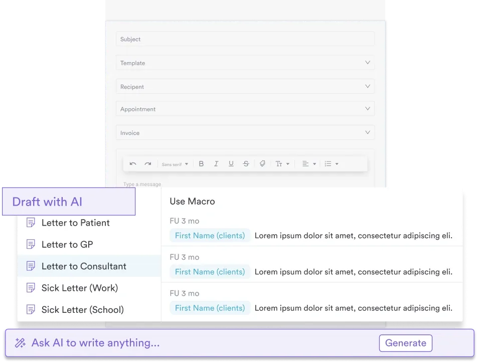
AI powered patient letters

How to Use This Ineffective Airway Clearance Care Plan Template

The downloadable template at the top of this guide follows the five-step nursing process framework that clinical teams use daily. Here is how to apply it to your patient:

  1. Assessment section: Complete the assessment questions (breath sounds location and type, SpO2, cough quality, sputum description, level of consciousness). Record baseline vital signs and any risk factors specific to your patient (post-op, ICU, neurological condition).
  2. Diagnosis statement: Select or write the NANDA diagnosis, filling in the etiology (the “related to” clause) based on your assessment. Use the related factors list provided in this guide to identify the primary driver of ineffective airway clearance for this patient.
  3. Planning section: Enter 2-3 measurable short-term goals (24-48 hour timeframe) and 1 long-term goal (discharge or outcome focus). Make goals SMART: “Patient maintains clear airway as evidenced by bilateral clear breath sounds and effective cough within 48 hours.”
  4. Interventions and rationale: Choose the 5-8 most relevant interventions from this guide (positioning, hydration, suctioning, respiratory exercises, medication administration). Write a 1-2 sentence rationale for each, explaining the scientific basis and how it addresses the patient’s specific etiology.
  5. Evaluation: At the end of each shift or per facility policy, assess whether goals are being met. Document changes in breath sounds, SpO2, cough quality, and patient report. If goals are not met, identify barriers and plan adjustments for the next shift or revision of the care plan.

Print the template or complete it electronically in your EHR. The template is designed to fit a single page or screen for quick reference during shift handoff and interprofessional rounds.

Standardise Your Airway Clearance Documentation

Pabau's clinical documentation system helps nursing teams create, track, and evaluate care plans collaboratively-reducing charting burden and improving patient outcomes. See how automated workflows and structured templates support better respiratory care.

Pabau clinical documentation dashboard

Conclusion

Ineffective airway clearance is a high-acuity nursing diagnosis that demands systematic assessment, evidence-based intervention, and continuous evaluation. This downloadable care plan template provides the structure-define the problem accurately, set measurable goals, implement targeted interventions with clear rationale, and evaluate progress objectively. The template above delivers all five components nurses need to deliver safe, consistent, quality care.

Your clinical documentation directly supports patient safety. Whether you use this template in paper or digital format, ensure every assessment finding, intervention, and evaluation outcome is recorded promptly and accurately. Early recognition of airway clearance problems and timely escalation to the medical team can prevent respiratory compromise. See how digital care planning systems streamline documentation, reduce charting time, and improve team communication around airway clearance goals.

Continue your research

Continue your research

Need structured respiratory assessment tools? Clinical assessment frameworks help standardise physical examination findings and improve diagnostic accuracy.

Looking for SOAP note guidance for respiratory patients? Writing effective clinical notes ensures your findings are documented clearly and are legally defensible.

Want to improve team communication during respiratory crises? Automated clinical workflows alert team members to critical changes in patient status and ensure timely escalation.

“` #

Frequently Asked Questions

What is the NANDA definition of ineffective airway clearance?

The NANDA-I definition is the inability to clear secretions or obstructions from the respiratory tract to maintain airway patency. This diagnosis applies when a patient lacks the strength, coordination, or cough mechanism to expel pulmonary secretions effectively, risking hypoxaemia and respiratory infection.

What are the most common related factors for ineffective airway clearance?

The most common related factors are excessive mucus production (from infection like pneumonia or inflammation like COPD), presence of an artificial airway (endotracheal or tracheostomy tube), neuromuscular dysfunction (stroke, spinal injury, sedation), and reduced level of consciousness. Pain and musculoskeletal impairment (rib fractures, post-operative states) also frequently contribute by limiting cough effort.

How do you document defining characteristics in a care plan?

Document specific, objective findings from your assessment: auscultated breath sounds (e.g. “bilateral crackles at lung bases”), respiratory rate (e.g. “tachypnoea at 24 breaths/min”), SpO2 (e.g. “SpO2 91% on room air”), cough quality (e.g. “weak, non-productive cough”), and sputum character (e.g. “thick, yellow sputum, copious amounts”). Include subjective data if available (patient reports difficulty breathing). Never write vague statements like “poor cough” without context.

When should you suction versus encourage deep breathing and coughing?

Suction when the patient is unable to expectorate secretions voluntarily-signs include abnormal breath sounds that persist despite coughing, SpO2 dropping below target, or visible/audible secretions in the airway. Encourage deep breathing and coughing first for patients with intact cough reflex and consciousness, as it is less invasive. Suction every 2-4 hours as needed or when assessment findings indicate secretion accumulation, but avoid routine suctioning that traumatises the airway mucosa.

What is the difference between ineffective airway clearance and ineffective breathing pattern?

Ineffective airway clearance focuses on the inability to remove secretions or obstructions; ineffective breathing pattern focuses on irregular breathing mechanics (rate, depth, rhythm) that do not provide adequate ventilation. A patient with ineffective airway clearance has crackles and a weak cough. A patient with ineffective breathing pattern has tachypnoea or irregular breathing. A patient may have both diagnoses simultaneously-treat each with specific interventions.

How often should you reposition a patient with ineffective airway clearance?

Reposition every 2 hours at minimum, or more frequently if the patient is immobile or high-risk. Frequent position changes prevent secretion pooling in dependent lung areas, promote gravitational drainage, and support spontaneous coughing. Alternate between semi-Fowler (HOB 30-45°), side-lying, and upright positions if tolerated. Coordinate repositioning with nursing care to maximise patient engagement and minimise disruption.

×