



GLP-1 diet plan
A GLP-1 diet plan is a structured eating framework for patients taking semagluti...

2
Last Updated: Oct 13, 2026

Hoffman’s sign test screens for upper motor neuron lesions by flicking the middle finger and watching the thumb and index finger.
A positive result suggests corticospinal tract dysfunction above C8, but it is a sign rather than a standalone diagnosis.
False positives occur in anxious patients and in people with generally brisk reflexes, so laterality and symptoms decide the urgency.
Pooled accuracy is modest, at roughly 58% sensitivity and 69% specificity for cervical myelopathy across published studies.
Recording the result, the side tested, and its reproducibility is what makes the finding useful six months later.
Hoffman’s sign test is a quick upper motor neuron screen performed during a neurological examination.
The clinician flicks the middle finger’s distal phalanx downward and watches for involuntary flexion of the thumb and index finger. According to StatPearls (NCBI Bookshelf), a positive finding points to corticospinal tract dysfunction.
Interpretation is where the test goes wrong. A positive result is a sign and not a diagnosis, and false positives turn up in anxious or hyperthyroid patients. This guide covers the technique, what the result means, the conditions behind it, and how to document the finding.
Hoffman’s sign test is a neurological screening tool that detects upper motor neuron (UMN) lesions in the upper limb. The eponym honors the German neurologist Johann Hoffmann. The description reached print in the early 20th century, when his assistant Hans Curschmann published it in 1911.
The test works by exploiting the stretch-reflex arc at the C8 level. When the corticospinal tract is damaged above the anterior horn cell, inhibitory control over spinal reflexes is lost. Flicking the middle finger’s distal phalanx stretches the flexor tendons suddenly. Without that inhibition, the thumb and index finger flex involuntarily.
The sign carries most weight when it is paired with other UMN findings: hyperreflexia, spasticity, clonus, and the Babinski response. No single sign should drive a diagnostic decision on its own.
Reliable elicitation needs consistent positioning and a clean flicking technique. Sloppy technique is the leading cause of both false positives and false negatives.
Seat the patient with the arm relaxed and the wrist neutral or slightly extended. Ask the patient to let the hand hang loose, because active muscle contraction interferes with the reflex response. Some examiners prefer a pronated forearm and others use supination. Either works, as long as you stay consistent across repeat assessments.
Grip the patient’s middle finger gently between your index finger and thumb, holding the middle phalanx. With your other thumb or fingernail, briskly flick the patient’s fingernail downward and release quickly. The motion is snappy, much like snapping your fingers. Avoid pressing on the flexor surface, which can produce a spurious response.
Repeat the flick two or three times per hand before you record a result. A single equivocal response does not count as positive. PhysioPedia notes that reproducibility across repetitions raises the clinical significance of a positive finding. Write down what you observed straight away, rather than trusting recall at the end of a busy day.
Watch specifically for flexion of the thumb at the IP or MCP joint, plus flexion or adduction of the index finger. A subtle twitch counts as a positive response. Flexion of the other fingers without thumb involvement is less specific and deserves caution. Test both hands in every assessment.
A positive Hoffman sign means involuntary thumb and index finger flexion was reproducibly observed. A negative Hoffman sign means no such response occurred. Both carry meaning, though neither settles the question on its own.
A negative result does not exclude UMN pathology, particularly in early or mild disease. Combine the Hoffman sign with deep tendon reflexes, plantar responses, and motor strength before drawing conclusions.
Always record which hand was tested and whether the result held across repetitions. That detail is what makes progression visible a year later, and structured digital examination forms make it easy to capture.

A positive Hoffman sign on one side raises the possibility of a lateralized lesion affecting the contralateral corticospinal tract. A bilateral positive sign generally carries more weight, suggesting a diffuse process in the cervical cord or higher.
In some neurologically healthy people, particularly those who are constitutionally hyperreflexic, a bilateral Hoffman sign is a normal variant. Context decides. An isolated bilateral positive sign in an asymptomatic 25-year-old athlete carries little urgency. The same finding in a 60-year-old with progressive hand clumsiness and gait disturbance is a different matter.
Whichever pattern you observe, record it precisely. “Hoffman positive left, negative right” is a clinically useful entry, while “Hoffman positive” without a side is not. Examination templates in software for physical therapists prompt for a side, which removes the omission at source.
A positive Hoffman sign implicates the corticospinal tract, so any pathology along that pathway can produce the finding. The most common associations are below.
Cervical myelopathy is the most frequent cause of a positive Hoffman sign in adults over 50. Compression of the cervical cord, typically at C3-C5 or C5-C6, disrupts the corticospinal tract above the C8 reflex arc.
Patients often present with the sign alongside progressive hand clumsiness, trouble with fine motor tasks, and a wide-based gait. Early recognition matters, because untreated progressive myelopathy can cause irreversible cord damage. A new positive sign in a patient reporting those symptoms warrants urgent cervical MRI.
MS lesions in the cervical cord or brain are a recognized cause of UMN signs, including the Hoffman response. The sign may appear during a relapse and partly resolve in remission. In an MS patient already under neurology follow-up, a newly positive or newly asymmetric sign is worth flagging.
ALS involves both upper and lower motor neuron degeneration. A positive Hoffman sign alongside fasciculations, wasting, and asymmetric weakness raises concern. That combination should prompt urgent neurological referral. The sign alone is not diagnostic of ALS.
If your practice sees these presentations, the follow-up imaging and referral pathway matters as much as the examination finding itself.
No clinical sign has perfect diagnostic accuracy, and Hoffman’s is no exception. Published figures vary widely with the population studied and the comparator diagnosis.
The pooled figures come from a meta-analysis of nine studies. The across-study ranges come from a 2025 scoping review in Spinal Cord. A 94% sensitivity figure circulates widely online, but it belongs to the Trömner sign rather than Hoffmann’s.
The practical reading is simple. A test that misses four in ten cases cannot rule myelopathy out. One that is wrong on three in ten positives cannot rule it in either. That is why the sign is read alongside the rest of the examination.
False positives are well documented in specific situations. Anxiety alone can produce generalized hyperreflexia that mimics a positive response. Hyperthyroidism does the same, as does a heavy caffeine load before the examination. Poor technique also produces spurious flexion, such as pressing the flexor surface instead of flicking the nail.
One rule covers most cases. A positive sign in a completely asymptomatic patient with brisk reflexes throughout needs monitoring rather than referral. The same sign in a patient with neck pain, hand clumsiness, and a wide-based gait needs imaging.
Both signs indicate UMN dysfunction, but they test different limbs and different levels of the corticospinal tract.
The two signs complement each other rather than substitute for each other. A patient with cervical myelopathy may show a positive Hoffman sign and a negative Babinski sign. The compression sits above the level needed to produce a Babinski response. Finding both points to more extensive corticospinal involvement.
The Trömner sign is closely related but technically distinct. Instead of flicking the dorsal nail surface, the examiner flicks upward against the volar surface of the middle finger. The positive response, thumb and index flexion, is identical.
Some clinicians treat the Trömner as a more sensitive variant, and others treat the two as interchangeable. The distinction matters when you read accuracy figures, because secondary sources often pool the two signs together.
Homan’s sign and Hoffman’s sign are different tests that get confused in search queries and in clinical records. Homan’s sign, also written Homans’ sign, is a largely abandoned screen for deep vein thrombosis.
The examiner dorsiflexes the ankle and compresses the calf, and calf pain counts as a positive result. It tests the lower limb and has no neurological function.
Hoffman’s sign tests the upper motor neuron pathway in the upper limb. Different eponym, different test, different anatomy, different question. If your software prompts for a “Homan’s test” inside a neurological examination, the field is mislabeled.
Accurate terminology matters for medicolegal records and for the next clinician who reads the notes. Structured patient records built on standardized clinical terms remove this category of error.

A positive Hoffman sign should prompt referral when the clinical picture adds up to more than an isolated reflex finding. Use the thresholds below.
Read across those thresholds and a pattern appears: the same positive sign maps to three different actions.
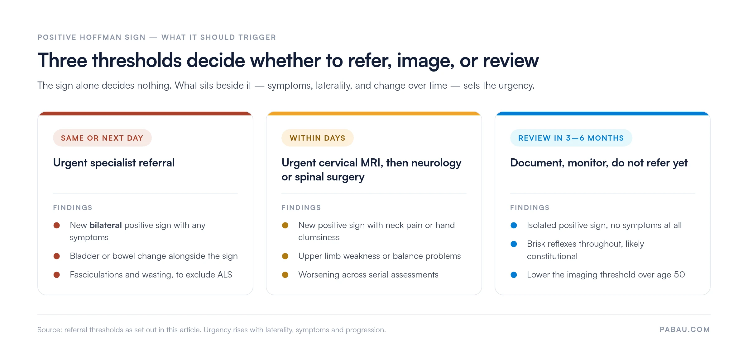
No published guideline recommends referral on a Hoffman sign alone. The decision rests on the whole picture, meaning symptoms, other UMN signs, functional status, and imaging.
Test both hands on every neurological assessment and record the result for each separately. Documenting ‘bilateral Hoffman negative’ is meaningfully different from ‘Hoffman negative’ with no laterality stated. The distinction matters if the patient returns six months later with new unilateral symptoms.
A neurological examination finding is only as useful as the record that captures it. A note reading “Hoffman’s checked, fine” cannot support progression tracking or defend a clinical decision later.
Structured documentation should capture the result, which hand or both, whether the finding repeated across attempts, and any associated UMN signs from the same assessment.
Practice management software like Pabau lets you build the neurological examination template once, with toggle fields for each sign. The examiner picks “Positive”, “Negative” or “Equivocal”, then “Left”, “Right” or “Bilateral”, instead of typing free text that the next reader has to interpret.
The entry then lives in the patient record in a searchable, auditable format. For practices running neurology, chiropractic, osteopathy, or musculoskeletal services, that structure cuts documentation time and keeps continuity across appointments.
If your team dictates notes rather than typing them, Pabau Scribe is our AI medical scribe. It captures spoken examination findings and files them into the right fields. That helps most in high-volume assessment sessions, where typing during the examination interrupts rapport with the patient.
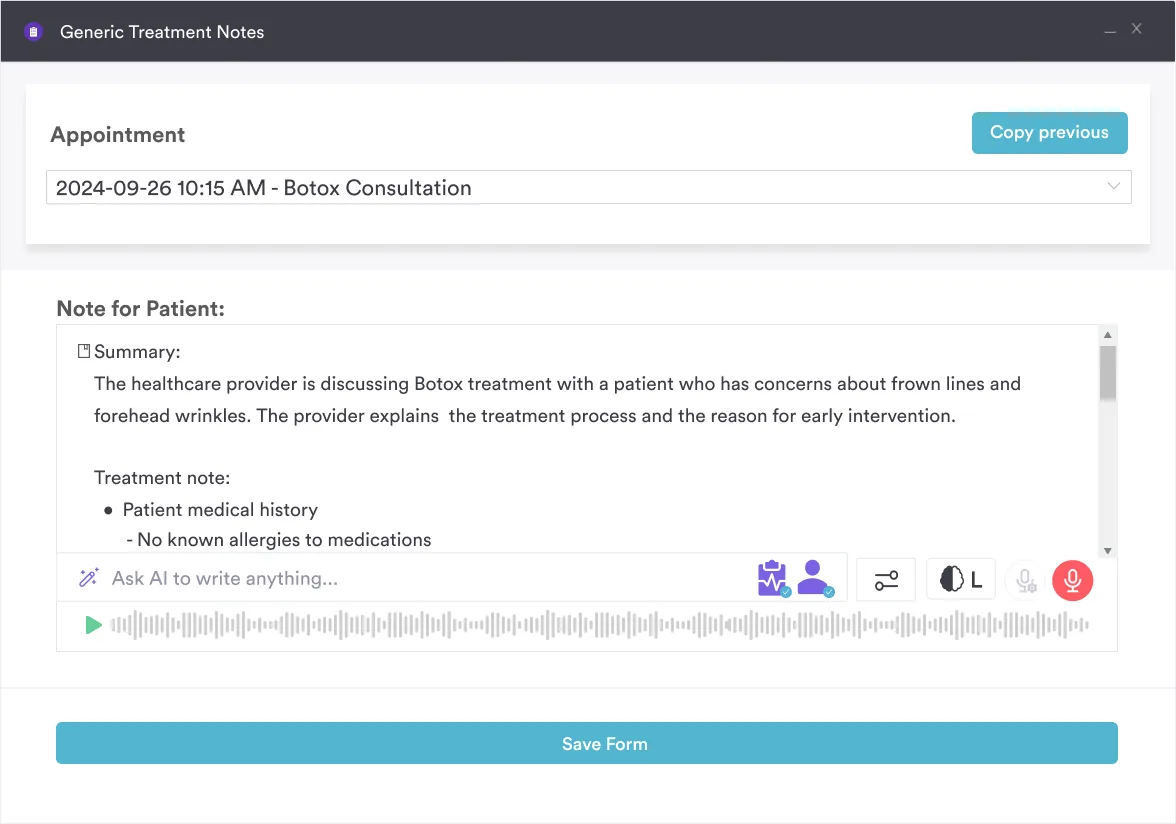
Practices already evaluating physiotherapy clinic software should check that the examination template library includes Hoffman sign fields alongside the other standard UMN markers. A template that prompts for the side removes the most common documentation miss in this examination.

Pabau’s clinical note templates and digital forms capture Hoffman sign results, laterality, and associated findings in a consistent, auditable format. That gives neurology and musculoskeletal practices a record that holds up at review.

Hoffman’s sign test is a fast, equipment-free screen for upper motor neuron dysfunction, and any neurological examiner should be able to elicit it reliably. Its accuracy is modest, so it earns its place as one input among several rather than as a decision-maker.
The judgment worth keeping is this. Symptoms, laterality, and change over time decide what a positive sign means, and the record is what makes change visible. A finding you cannot compare against last year’s note tells you very little.
Set your examination template up so the result, the side, and the reproducibility are captured by default. Book a demo to see how Pabau keeps neurological findings comparable across appointments.

List Item #1
List Item #2
Hoffman’s sign test is a bedside neurological maneuver that screens for upper motor neuron lesions. The examiner flicks the distal phalanx of the middle finger and watches for involuntary thumb and index finger flexion. A positive finding suggests corticospinal tract dysfunction above C8, most often from cervical myelopathy, multiple sclerosis, ALS, or stroke. It is a sign, not a diagnosis.
A positive Hoffman sign indicates probable upper motor neuron dysfunction affecting the corticospinal tract above C8. Urgency depends on whether the finding is new, bilateral, progressive, or accompanied by symptoms such as hand clumsiness, neck pain, or gait disturbance. An isolated positive sign in an asymptomatic patient may represent constitutional hyperreflexia rather than pathology.
No, a positive Hoffman sign is not always serious. In an asymptomatic patient with generalized hyperreflexia, it can be a normal variant. The finding becomes significant when it is new, bilateral, progressive, or associated with neck pain, upper limb weakness, gait problems, or bladder changes. Those combinations warrant urgent imaging and specialist referral.
Pooled figures put sensitivity at about 58% and specificity at about 69% for cervical myelopathy. Individual studies report sensitivity between 58% and 89%, and specificity between 41% and 100%. A 94% sensitivity figure often quoted online belongs to the Trömner sign instead. The sign cannot rule myelopathy in or out on its own.
Hoffman’s sign tests the upper limb by flicking the middle finger’s distal phalanx, and a positive response is thumb and index finger flexion. Babinski’s sign tests the lower limb by stroking the lateral plantar surface, and a positive response is great toe extension with toe fanning. Both indicate UMN dysfunction, but they test different cord levels and complement each other.
Yes, a unilateral positive Hoffman sign can occur. It typically suggests a lateralized lesion affecting the contralateral hemisphere or the ipsilateral cervical cord above C8. Asymmetry should be documented carefully with the side specified. A new unilateral positive finding alongside ipsilateral motor or sensory deficits warrants prompt neurological evaluation.
Hoffman’s sign is a neurological upper limb reflex test for UMN lesions. Homan’s sign is a largely abandoned lower limb test for deep vein thrombosis, involving ankle dorsiflexion with calf compression. The names look similar, but the two tests examine entirely different systems and should never be confused in clinical documentation.






