



GLP-1 diet plan
A GLP-1 diet plan is a structured eating framework for patients taking semagluti...

2
Last Updated: Oct 13, 2026

The Lhermitte sign test is performed by flexing the patient’s neck forward, either passively or with the patient leading.
A positive result is a brief electric shock traveling from the neck down the spine and into the limbs.
The finding points to posterior column irritation or demyelination, most commonly from multiple sclerosis or cervical myelopathy.
Cervical spondylosis, B12 deficiency, radiation myelopathy, and cord compression all produce the same sensation, so imaging decides the cause.
Treatment targets the underlying condition, and the neurological deficit sets how urgently the patient needs referral.
The Lhermitte sign test is a clinical assessment that provokes a brief, electric shock-like sensation down the spine when the patient flexes their neck forward.
A positive result points to irritation or demyelination of the posterior columns of the cervical spinal cord.
According to StatPearls (NCBI), it is one of the most recognizable signs in neurology. It is associated most often with multiple sclerosis (MS) and cervical myelopathy.
This guide covers how to perform and interpret the test, what causes a positive result, how to document it, and when to refer. It is written for clinicians and clinical learners.
Lhermitte’s sign is a transient, electric shock-like paresthesia that travels from the neck down the spine, often reaching the arms or legs. Forward neck flexion provokes it.
It is also called Lhermitte’s phenomenon or the barber chair phenomenon. The second name comes from the sensation patients describe when they recline into a barber’s chair.
The finding works as both a sign and a symptom. Clinicians elicit it during examination, and patients report it spontaneously when they look down. Some sources reserve the word sign for the provoked version, though the literature uses both terms interchangeably.
A positive result is a neurological red flag that calls for further investigation. On its own it names no diagnosis. Clinicians in neurology, primary care, and physical therapy practices need to separate it from other cervical spine signs and act on it.
Neck flexion mechanically stretches the posterior columns, also called the dorsal columns, of the cervical spinal cord. Where those columns are demyelinated or structurally compromised, the stretch triggers ectopic electrical impulses in the affected axons. Neurologists call this ephaptic transmission.
Myelin normally insulates axons and prevents cross-talk between nerve fibers. Once the myelin sheath is damaged, adjacent axons fire spontaneously and propagate signals in unusual directions. That produces the characteristic electric sensation.
The finding reflects posterior column dysfunction directly. That is why the sensation usually radiates caudally, from the neck toward the limbs, rather than staying local at the cervical level.
The test has two accepted variants. In passive elicitation the clinician guides the movement, and in active elicitation the patient moves their own neck. Both are valid, and passive is preferred where cervical instability is not suspected.
Contraindication: do not perform passive cervical flexion where cervical instability is known or suspected. That includes atlantoaxial subluxation in rheumatoid arthritis, a trauma history, and known ligamentous laxity. Image first, then examine.
A positive result does not confirm a diagnosis. It confirms posterior column involvement at or above the cervical cord, which narrows the differential considerably. Further investigation still decides the cause.
Three interpretation points matter at the bedside:
Diagnostic accuracy varies by population. In multiple sclerosis cohorts, sensitivity for cervical cord demyelination is reported between 9% and 41%. A negative test therefore does not rule the condition out.
Specificity is higher, which makes a positive result more informative. Interpret it in clinical context rather than in isolation, and compare it against the patient’s earlier examinations where you have them.

Several conditions disrupt the posterior columns and produce the same sensation. Multiple sclerosis and cervical myelopathy account for most cases, and the wider differential runs from vitamin deficiency to radiation injury.
The association with multiple sclerosis carries the most clinical weight. The sign can be an early presenting symptom, sometimes appearing before a formal MS diagnosis. In this population it reflects active cervical cord demyelination and often tracks relapse activity.
Heat can worsen the sensation in patients with MS. That overlaps with Uhthoff’s phenomenon, where a raised body temperature temporarily worsens neurological symptoms through impaired conduction in demyelinated fibers. The distinction matters when you counsel patients about exercise and bathing.
Cervical spondylosis and disc herniation compress the posterior columns mechanically rather than demyelinating them. The clinical picture can look identical to MS-related Lhermitte’s, which makes imaging essential.
MRI with and without contrast is the investigation of choice for separating structural compression from inflammatory demyelination. Older patients with degenerative cervical spine changes are the group to watch.
Lhermitte’s sign shares clinical territory with several other examination findings. Knowing the differences keeps the workup targeted and stops any single test from carrying too much weight.
The direction of travel is the clearest separator. Lhermitte’s radiates caudally from the neck into the limbs, while Spurling’s test produces ipsilateral radicular arm pain with no caudal radiation. The modified Romberg test adds a proprioceptive read on the same posterior columns.
Running all three in one session gives a far richer picture of the level and nature of cord involvement. That matters most after neck trauma, where a single positive finding is easy to over-read.
A positive result starts the workup rather than ending it. What follows decides whether the cause is demyelinating, structural, metabolic, or vascular, and whether the patient needs urgent intervention.
The path from cervical flexion to a referral decision runs through four stages.
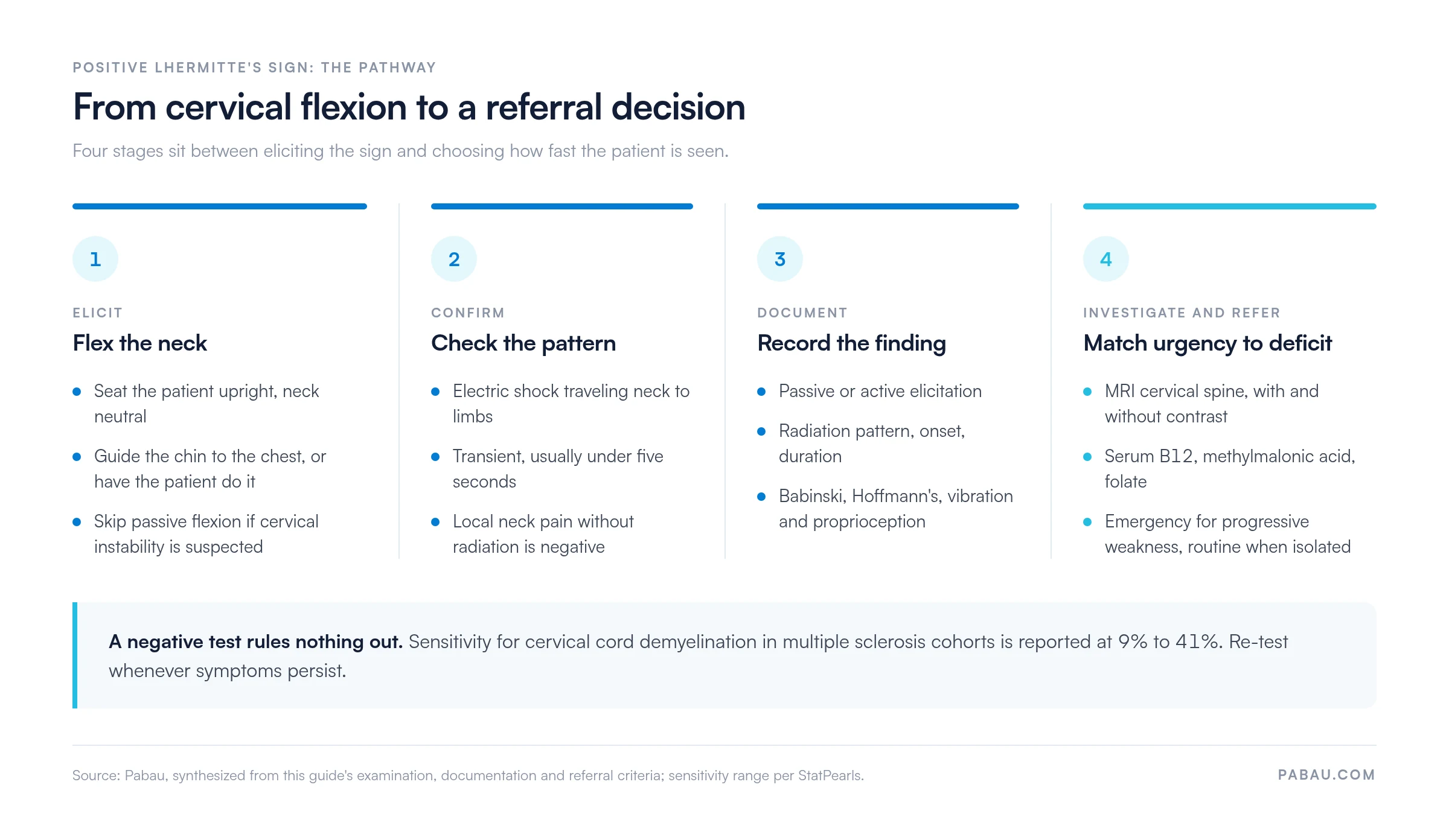
Record the reflex findings from step four in the same place as the sign itself. A structured deep tendon reflex exam keeps the biceps, triceps, and lower limb responses beside the Lhermitte’s entry, which is what the neurologist reads first.

Treatment is always directed at the underlying condition. No intervention removes the sign on its own, though targeted management often reduces how often it fires and how hard it hits.
A patient waiting for a neurology appointment can deteriorate with nobody watching. Build a follow-up touchpoint between the formal appointments, so symptom progression is recorded as it happens rather than reconstructed from memory months later.
Record the clinical reasoning behind each management pathway in the patient’s notes, not only the decision you reached. A referral letter to neurology should name the elicitation method and the radiation pattern. Add the onset and duration, plus any upper motor neuron signs found at the same examination.
The wording of the entry carries the referral, and later it carries the medico-legal record. A reviewer reading the note a year afterward needs the detail the neurologist needed on the day. The framework below sets that out.
In a SOAP note or clinical record, a positive finding should carry:
Name the dermatomal level in that entry wherever the examination supports it. “Into the right arm” and “C6 distribution” are read very differently by a neurologist.
Negative findings deserve the same care. Write “Lhermitte’s sign not elicited on passive cervical flexion, no electric or radiating sensation reported”. A documented negative carries as much weight as a positive when you track progression over time.
An isolated positive sign rarely needs an ambulance, and the same sign with progressive weakness does. The table below separates the emergency from the routine, so the referral matches the deficit in front of you.
Making the referral decision is the straightforward part. Chasing it is where patients get lost. Someone can wait weeks for imaging while the referral sits unacknowledged, so track the acknowledgment rather than assuming it.

A positive Lhermitte’s sign usually lands in a free-text note, and the detail is retyped into the referral letter later. The elicitation method and the radiation pattern drop out somewhere between the two. The neurologist then reads “positive Lhermitte’s” with no supporting detail.
Pabau stores the examination as a structured note against the client record. The finding you documented in March sits beside the one you documented in September. A change in the radiation pattern is then visible rather than remembered.
Pabau Scribe, our AI scribe, drafts the note from the consultation itself. The elicitation method and the associated findings land in the record while the examination is fresh, and referral letters pull from that same note.
Follow-up tasks and recalls run off the same timeline, so a patient waiting on an MRI stays in view. The practice finds out about a deterioration from its own diary rather than from a complaint.

Pabau stores neurological examination findings against the client record and carries them into referral letters, so the detail you documented reaches the neurologist intact.

The Lhermitte sign test costs ten seconds and a hand on the occiput, and it changes what you do next. A positive result narrows the field to posterior column pathology and sets the investigation in motion.
The judgment worth keeping is that the sign does not grade severity on its own. Sensitivity is low enough that a negative test changes little, and the urgency comes from the deficit sitting beside the sign.
Write the finding down the way you would want to read it in someone else’s referral letter. Book a demo to see how Pabau keeps neurological examination findings and referral follow-up in one client record.

Need the radiculopathy side of the differential? Spurling’s test covers the technique and interpretation for cervical nerve root compression.
Assessing proprioception alongside the cord? Modified Romberg test explains how to score postural sway and what a positive result localizes.
Checking cervical endurance after a neck complaint? Cervical extensor endurance test gives the hold times and the normative values to compare against.
Running a physical therapy or musculoskeletal practice? Mandatory compliance for physiotherapy clinics outlines the documentation and regulatory requirements for neurological and MSK assessments.
Testing the deep neck flexors after a cervical complaint? Cranio-cervical flexion test sets out the pressure stages and a scoring sheet to record them on.
Lhermitte’s sign is a transient, electric shock-like sensation running from the neck down the spine. It appears when the neck is flexed forward and can reach the arms or legs. The finding indicates irritation or demyelination of the posterior columns of the cervical spinal cord. Treat it as a neurological red flag that warrants further investigation.
Seat the patient upright and guide their chin toward the chest, or ask them to do it themselves. A positive result is any report of an electric, radiating sensation down the spine or into the limbs. It appears during or immediately after flexion. Avoid the passive version where cervical instability is suspected.
No. Multiple sclerosis is the most common cause, but it is far from the only one. Cervical spondylosis, subacute combined degeneration from B12 deficiency, radiation myelopathy, disc herniation, spinal cord tumors, and vascular conditions all produce it. A positive result always needs imaging and further workup to find the cause.
Lhermitte’s sign is the finding a clinician elicits through passive or active cervical flexion during a formal examination. Lhermitte’s symptom is the same sensation reported spontaneously by the patient, for example while looking down. The clinical interpretation is similar either way. The literature notes the distinction but uses the terms interchangeably.
No treatment eliminates the sign directly. Management targets the underlying condition. That means disease-modifying therapies and heat avoidance for multiple sclerosis, surgical decompression for structural myelopathy, and B12 replacement for subacute combined degeneration. Radiation myelopathy is managed supportively. Carbamazepine or gabapentin may be considered for symptom relief in MS, under specialist guidance.
Refer to emergency services or acute neurosurgery where there is rapidly progressive weakness or paralysis. Neurosurgery within days is indicated when MRI confirms cord compression with neurological deficit. A suspected new MS relapse warrants neurology review within one to two weeks. An isolated positive sign with no motor deficit and pending imaging can go to routine neurology outpatients.






