



GLP-1 diet plan
A GLP-1 diet plan is a structured eating framework for patients taking semagluti...

2
Last Updated: Oct 13, 2026

Hand nerve tests check the median, ulnar, and radial nerves through sensory mapping, motor testing, and provocative maneuvers.
No single provocative test confirms carpal tunnel syndrome, so combine two or three and record what each one reproduced.
Durkan’s compression test scored 87% sensitivity in its original 1991 study, while pooled data from later studies sits near 64%.
Static two-point discrimination under 6mm at the fingertip is normal, and higher values suggest reduced innervation density.
Practice management software like Pabau keeps exam findings in the client record, so the six-week comparison takes seconds.
Nerve compression rarely announces itself. A patient mentions pins and needles at 3am, blames the pillow, and books in months later. By then the thenar eminence has started to flatten, and the window for a clean recovery has narrowed.
A structured hand nerve exam catches it much earlier. It takes about five minutes and needs nothing more than a pin, some cotton wool, and a discriminator. It tells you which nerve is involved and roughly where.
A useful exam comes down to technique and an honest reading of the published accuracy. It also depends on a note someone else can still read six weeks later.
The hand nerve exam tells you which of the three nerves is affected, and at roughly what level. That is usually enough to choose between splinting, referral, and an electrodiagnostic study.
However, the exam does not grade severity objectively, and it does not rule out a second compression higher up.
The median, ulnar, and radial nerves each follow their own course, supply their own muscles, and cover their own patch of skin. Compression anywhere along that route produces a pattern you can recognize on examination.
Early compression is quiet. A patient with tingling in the thumb and index finger may show no wasting at all. A positive Phalen’s test with reduced two-point discrimination flags carpal tunnel syndrome before axons are lost.
Recording those first findings matters as much as producing them, because the follow-up exam only means something next to a baseline. A physical therapy EMR keeps both readings in the same client record.
Work through three domains in a fixed order: sensory, then motor, then provocative. Least provocative first, so you are not reading symptoms you just caused.
The table below pairs each nerve with its sensory patch, its key muscles, and the deficit you see when it is compressed.
Innervation is rarely this tidy in the room. Anatomical variants, dual innervation zones, and a second compression further up all blur the boundaries. Treat the table as a starting map and expect exceptions.
Median testing starts at the carpal tunnel, because that is where most compression sits. Lesions at the elbow, such as pronator syndrome, also produce median deficits, and forearm findings are what tell the two apart.
Three provocative tests cover most presentations.
Phalen’s test reproduces carpal tunnel symptoms through sustained wrist flexion, which raises pressure inside the tunnel and loads the median nerve.
A systematic review of 60 studies puts Phalen’s at 68% sensitivity and 73% specificity. Individual studies scatter widely either side of that, mostly because they recruited different patients.
Percuss over the carpal tunnel at the wrist crease with a fingertip or a reflex hammer. A positive sign is tingling or an electric shock running into the median territory.
On the same pooled data, Tinel’s is the weakest of the three at roughly 50% sensitivity and 77% specificity. Percussion force varies a lot between examiners, and that accounts for much of the spread.
Apply direct thumb pressure over the carpal tunnel and hold it for 30 seconds. A positive result reproduces the patient’s own symptoms in the median distribution.
The figures here need care. Durkan’s original 1991 study reported 87% sensitivity and 90% specificity in 46 affected hands. Pooled results from later studies land lower, near 64% and 83%. Which provocative test is most sensitive remains unsettled, so no single one deserves your whole diagnosis.
Pre-built assessment templates in occupational therapy software fix which tests get recorded, and in what order. That is what stops the third test being skipped when the appointment overruns.
Ulnar testing has to place the lesion at either the cubital tunnel at the elbow or Guyon’s canal at the wrist. Management differs between the two, so careful motor and sensory mapping earns its time.
Froment’s sign tests adductor pollicis, the muscle behind a strong key pinch, supplied by the deep branch of the ulnar nerve.
That IP flexion is the diagnostic feature. The patient is recruiting flexor pollicis longus, a median-supplied muscle, to cover for weak adductor pollicis. A formal pinch grip test puts a number on the same weakness for the record.
Wartenberg’s sign is a little finger that rests in abduction, caused by weakness of the third palmar interosseous. On its own it points to ulnar involvement of the intrinsic muscles.
Confirm it directly. Test finger abduction and adduction against resistance. Weak spreading, or an inability to grip paper between the ring and little fingers, points at the interossei.
Test the little finger and the ulnar half of the ring finger, on both the palmar and dorsal surfaces. The dorsal reading is the one that decides the level.
Spared dorsal sensation with a palmar deficit suggests Guyon’s canal, below the point where the dorsal cutaneous branch leaves. Loss across both surfaces points higher, at or above the cubital tunnel.
Write the two surfaces down separately. “Reduced ulnar sensation” throws away the only finding that localized the lesion. A predictable SOAP note structure keeps that detail in the same place every visit.
Radial compression happens most often at the spiral groove of the humerus, the Saturday night palsy pattern. The other common site is the proximal forearm, where the posterior interosseous nerve (PIN) passes under the supinator.
Those two patterns look different on examination, and mixing them up changes the referral.
Test wrist extension against resistance first. A complete radial lesion at the spiral groove gives a wrist drop, with the fingers unable to extend as well.
Then test brachioradialis power as elbow flexion in mid-pronation. Intact brachioradialis alongside a true wrist drop places the lesion below that branch, in the radial nerve trunk or the proximal forearm.
PIN syndrome looks different. It spares wrist extension, because extensor carpi radialis longus keeps working, so the wrist extends with radial deviation rather than dropping. The visible failure is finger drop at the MCP joints. A genuine wrist drop therefore argues against isolated PIN involvement.
The radial nerve supplies the dorsal thumb web space. It also covers the dorsal surface of the radial three and a half digits, which varies between patients. Test it with a wisp of cotton wool.
The tension test loads the nerve mechanically. Position the shoulder in internal rotation, the elbow extended, the forearm pronated, and the wrist and fingers flexed. Cervical side-flexion away from the test side adds tension.
Radial-territory symptoms in that position suggest a mechanosensitive nerve rather than a fixed block. Read it next to the motor findings, never on its own.
Sensory testing gives the most precise localization of the three domains, and it is the one most often cut short. Ten seconds per territory is not a sensory exam.
Use cotton wool or a Semmes-Weinstein monofilament for light touch. Map each nerve territory and compare it with the other hand. Asymmetry is the finding, not absolute sensitivity. Shading the result on a hand pain diagram beats describing it in a sentence.
For pin-prick, apply a fresh disposable pin or Neurotip lightly to the skin. Blunted sharpness inside a defined territory confirms sensory loss. Discard the pin straight afterwards.
Apply two points of a discriminator to the palmar fingertip at the same time, narrowing the distance step by step. Record the smallest separation the patient calls correctly and reliably.
Normal static two-point discrimination at the fingertip is under 6mm. Some sources use 5mm, depending on the instrument and the patient’s age. Readings above 6mm suggest reduced innervation density at the fingertip.
Moving two-point discrimination usually runs 1-2mm better than static, and it picks up early compression sooner.
Use the same instrument and hand position every visit, since a threshold only earns its keep against a comparable earlier reading. Validated cut-offs carry the same weight in other exams, including the Ottawa ankle rules.
Not accurate enough to stand alone. Every provocative test for carpal tunnel syndrome misses cases. The published figures also move around, depending on who was recruited and how the diagnosis was confirmed.
Pooled estimates from one systematic review sit in the table below, so the three tests are comparable. Where an original validation study reported something different, both numbers appear.
Two positives are worth more than one. The AAOS clinical practice guideline for carpal tunnel syndrome recommends combining clinical findings rather than resting a diagnosis on any single maneuver.
Run Durkan’s compression test before Phalen’s in the same appointment, and note which came first. A full minute of wrist flexion sensitizes the tunnel, so a Phalen’s-first sequence can inflate whatever you test next. Order changes the finding, which is exactly why the record should carry it.
Refer when the exam cannot answer the question the next decision rests on. Splinting an obvious mild case does not need a study. A surgical opinion almost always does.
Nerve conduction studies (NCS) measure how fast and how strongly signals travel along a peripheral nerve. That is how severity gets graded objectively rather than described.
Electrodiagnostic testing is the usual reference standard for confirming carpal tunnel syndrome and grading it. It still misses some early cases, so a normal study does not close the door on a convincing history.
Order NCS and EMG when any of these apply:
Needle EMG usually runs alongside NCS. It reads the electrical activity of individual muscles, and it detects axonal degeneration that conduction studies alone can miss in early or moderate compression.
The performing provider bills by how many nerves were studied, under codes such as 95911.
A referral that gets triaged quickly carries the exam with it. Check the note holds all five of these:
Conservative care carries on while the referral sits in a queue. Splinting, activity changes, and structured hand exercise programs give you a documented trial to point at if surgery is discussed later.
A nerve exam note earns its place when a colleague can repeat the exam from it and compare the results. That means numbers, sides, and timings. Adjectives do not survive the six weeks.
Grade motor strength on the Medical Research Council (MRC) scale, 0 to 5. Anything at 4 or below in thenar abduction deserves a formal entry and a repeat at follow-up.
For sensation, the Semmes-Weinstein monofilament scale gives an ordinal grade from normal, through diminished light touch, to absent. Standard scoring sheets do the same job in other neurological exams, the NIHSS score sheet being a familiar example.
“Some weakness” cannot be compared with anything at the next visit. A grade of 4 can.
Capture these five components in the same order every time, so serial comparison is quick:
Add symptom duration, occupation, and handedness to the same note. Insurance, employer, and legal reports all ask for those three, and chasing them months later is slow. A reference on safer clinical notes is worth reading before you set up a template.
Five recurring ones, in rough order of how often they turn up in notes:
Most practices already know what to record. The friction is where it lands. Findings get typed into free text, monofilament values end up in one clinician’s shorthand, and the follow-up exam is written partly from memory.
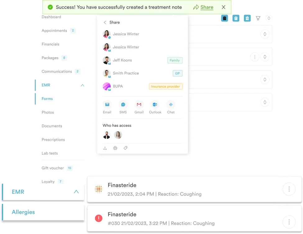
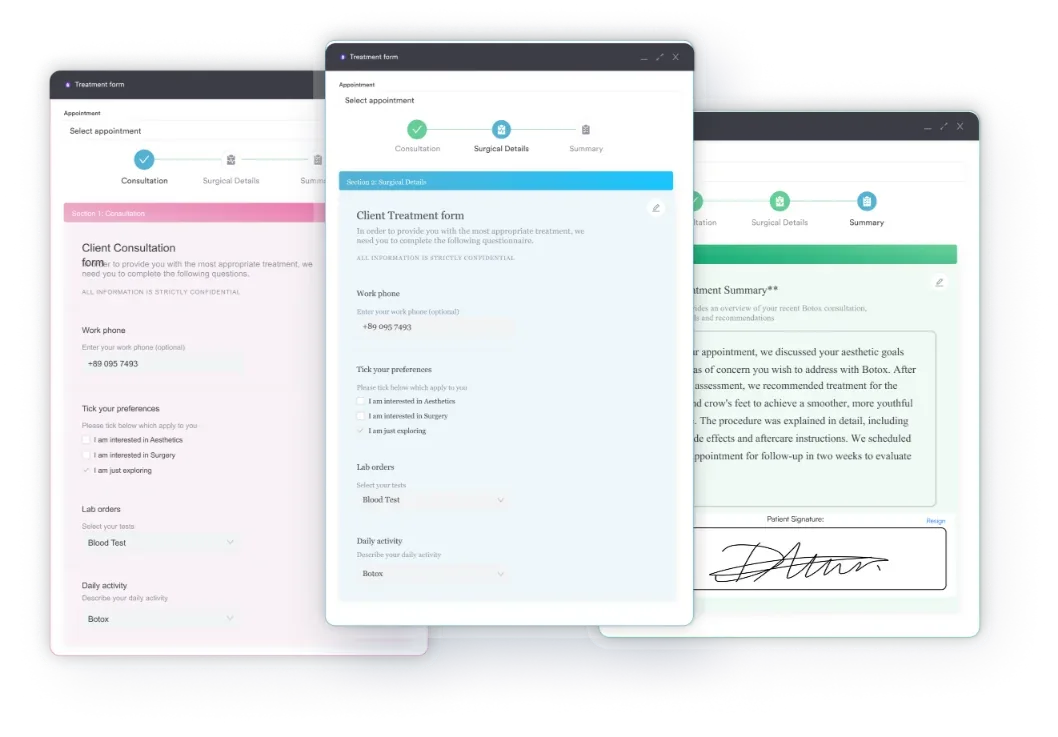
Practice management software like Pabau moves the exam into a structured form instead. Digital intake forms collect symptom duration, occupation, and handedness before the appointment starts. The exam fields then sit in the same client record as the photos, the appointment history, and the referral letter.
Dictation is handled too. Pabau Scribe, our AI scribe, turns spoken exam findings into a structured note through AI-assisted documentation, which you review and approve before anything saves.
So at the six-week review, last visit’s MRC grades and 2PD values sit on screen beside today’s. Outcome tracking charts the change between them. The decision to refer or discharge then rests on measurements rather than recall.
Forms do the same work at the front of the visit. Symptom duration, occupation, and handedness arrive before the patient sits down, which leaves the appointment for the exam itself.

Pabau stores structured exam fields, digital forms, and clinical photos in the same client record. Follow-up comparisons and referral letters then take minutes, instead of a hunt through free-text notes.

Running the tests takes five minutes. Reading a 4/5 thenar abduction and a 7mm two-point discrimination six weeks later is the harder job. That reading depends entirely on how the first exam was written down.
So pick a sequence and keep it. Combine two provocative tests rather than trusting one. Record the numbers with their units and their side. Treat the published sensitivity figures as a range, not a verdict.
Do that and referrals get triaged faster, discharge decisions get easier to defend, and early compression gets caught before the wasting starts. Book a demo to see how Pabau keeps nerve exam findings, forms, and follow-up comparisons in one client record.

Need a number for thumb and finger strength? Pinch grip test covers the grades and cut-offs that back up a positive Froment’s sign.
Looking after arthritic hands as well as compressed nerves? Hand exercises for rheumatoid arthritis sets out a conservative program you can document.
Running a full neurological workup? Deep tendon reflex exam is a printable form for recording reflex findings at the bedside, covering biceps, triceps, patellar, and ankle responses.
Screening a client before training? Fitness assessment form is a ready-to-use form covering client and emergency contact details, medical history, and fitness goals.
Managing musculoskeletal patients back to full function? Return-to-running protocol shows how upper and lower limb testing fits a wider rehab plan.
A focused exam on one hand takes five to ten minutes. Sensory mapping and two-point discrimination account for most of that. Testing the other hand for comparison adds two or three minutes, and it is worth doing at every first visit.
Yes. Early compression often causes symptoms before any provocative test turns positive, and pooled sensitivity for the common tests sits between 50% and 68%. A normal exam with a convincing history justifies a repeat visit or an electrodiagnostic referral, not reassurance.
It is a localizing test for nerve compression. The patient resists shoulder external rotation while you lightly scratch the skin over the suspected site. A brief loss of resistance counts as positive. Reported accuracy varies widely, so pair it with the standard maneuvers.
Distribution and neck involvement separate them. Carpal tunnel symptoms stay distal and spare the skin over the thenar eminence. C6 or C7 radiculopathy follows a dermatome into the forearm and often shifts with neck position. Both can coexist, which is the double crush pattern.






