Hyperbole and a Half pain scale: Every level + free template
The Hyperbole and a Half pain scale is Allie Brosh’s humorous pain chart, ...

1
Last Updated: Oct 5, 2026

Ely’s test, also called the Duncan-Ely test, checks rectus femoris length with the patient prone and the knee passively flexed.
The result is positive when the hip flexes on its own, or the pelvis lifts off the table, before the knee is fully bent.
Record the knee angle where the hip starts to lift, then compare both legs, so the next visit has something to measure against.
Accuracy figures come mostly from cerebral palsy research, where sensitivity runs 56% to 59% and specificity 64% to 85%.
Practice management software like Pabau keeps special test findings in one client record, so every clinician documents them the same way.
A patient comes in with anterior knee pain that flares on stairs and settles at rest. The knee itself looks unremarkable. Often the problem sits higher up, in the one quadriceps muscle that also crosses the hip.
Ely’s test answers that question in about 90 seconds. The patient lies face down, you bend the knee, and the pelvis tells you whether the rectus femoris has run out of length. Getting a trustworthy answer depends on a few details that are easy to skip.
Ely’s test is a passive length test for the rectus femoris. That muscle is the only quadriceps head crossing both the hip and the knee. Physical therapists and sports medicine clinicians use it to screen for tightness or contracture. Either one can drive anterior knee pain, an altered gait, and hip flexors that never switch off.
You will also see it called the Duncan-Ely test. Both names describe the same maneuver. The patient lies prone, you flex the knee passively, and you watch the pelvis. When the rectus femoris runs short, the pelvis cannot stay flat, so the hip pulls into flexion. That involuntary movement is the positive sign.
The test shows up in routine physical therapy screens, in pre-surgical gait analysis, and in cerebral palsy assessment. It needs nothing but a treatment table, which is part of why it has stayed in use for decades.
Because the rectus femoris spans two joints, it can be short at one end and still look fine at the other. It runs from the anterior inferior iliac spine, known as the AIIS, down to the tibial tuberosity through the patellar tendon.
Flex the knee in prone and you lengthen the muscle at its lower end while the hip stays extended. A tight muscle has nowhere left to give, so the pelvis tips forward instead. That is the mechanical basis of a positive finding.
The tensor fasciae latae, or TFL, and the iliopsoas can add to the movement you see. A positive test therefore never names a single culprit on its own. Logging the knee angle at every visit turns a yes or no finding into a trend. A structured range of motion assessment keeps those numbers comparable between clinicians.
The whole thing takes under two minutes. Positioning and pace are what separate a usable result from a shrug, so it pays to run the same sequence every time.
Recording the same fields each time is what makes the second test comparable to the first. Structured medical forms with fixed boxes beat free text, which drifts from clinician to clinician.
The test is positive when the hip flexes by itself, or the pelvis rises off the table, during passive knee flexion. Full knee flexion is not required. The involuntary movement at the top is the finding.
A negative test allows the knee to bend to roughly 90 degrees or further with the pelvis flat. Some clinicians use heel-to-buttock as the endpoint instead. Either threshold works, as long as your team agrees on one. Write down which you used.
No grading scale for Ely’s test has been formally validated. The framework below is a practical interpretation guide, widely used in practice. Treat it as a way to track change, not as a diagnosis on its own.
The difference between legs often tells you more than the absolute number. A 15-degree asymmetry is worth flagging even when both sides read as mild. Recording the angle in measurements tracking software keeps that comparison honest across months of follow-up.
Published accuracy data for Ely’s test comes almost entirely from children with cerebral palsy. Read the numbers with that in mind before applying them to a runner with knee pain.
The reference study is Marks et al. (2003), published in Developmental Medicine and Child Neurology. It compared the Duncan-Ely test against dynamic electromyography, or EMG, in children with cerebral palsy. Reported sensitivity sat between 56% and 59%, with specificity between 64% and 85%. Inter-rater reliability was moderate.
So a positive Ely’s test is worth acting on. A negative one does not clear the rectus femoris, especially outside neurological populations. Treat the test as one screen inside a broader assessment rather than a verdict.
Rectus femoris tightness turns up across a wide range of presentations, which is why a 90-second screen keeps paying for itself. These are the situations where it changes what you do next.
Sports practices that run these screens at scale usually fold them into an intake protocol rather than leaving them to memory. Purpose-built sports medicine software makes that protocol the default for every new athlete.
In spastic cerebral palsy, the rectus femoris can stay active through the swing phase of gait. The knee then fails to bend enough for the foot to clear the floor. Step length drops, and walking costs more energy.
Pre-operative gait analysis uses the Duncan-Ely test alongside dynamic EMG to judge whether rectus femoris transfer surgery is likely to help. The test never carries that decision by itself. It adds one piece of evidence to a package built on instrumented gait data.
A positive result points at the rectus femoris, but prone lying does not isolate it cleanly. Rule out the following before you commit to a treatment plan.
Record three values every time: the knee angle at hip rise, the difference between legs, and whether symptoms reproduced. A bare positive or negative tells you nothing useful at the next visit, and it makes progress impossible to defend to a referrer.
No single test covers the hip flexor complex. Pairing Ely’s test with the four below gives a fuller read on lower limb flexibility and neuromuscular control. The whole set takes about five minutes.
Ely’s test plus the Thomas test is the most useful pairing of the set. The Thomas test separates iliopsoas from rectus femoris more cleanly. Run it whenever Ely’s test is positive and you need to know which structure to treat. For athletes rebuilding load, those flexibility numbers belong inside a wider return-to-running protocol.
Special tests produce numbers that only matter if someone can find them next month. In most practices, the angle of onset ends up buried in a free-text note, worded differently by each clinician who wrote one. The same is true far outside orthopedics: serial results from a glucose tolerance test are only readable when every draw lands in the same field.
Pabau puts the finding in the client record beside the measurement history, the subjective report, and the plan. Customizable SOAP note templates fix the fields, so everyone captures the angle of onset, the difference between legs, and the symptom response.
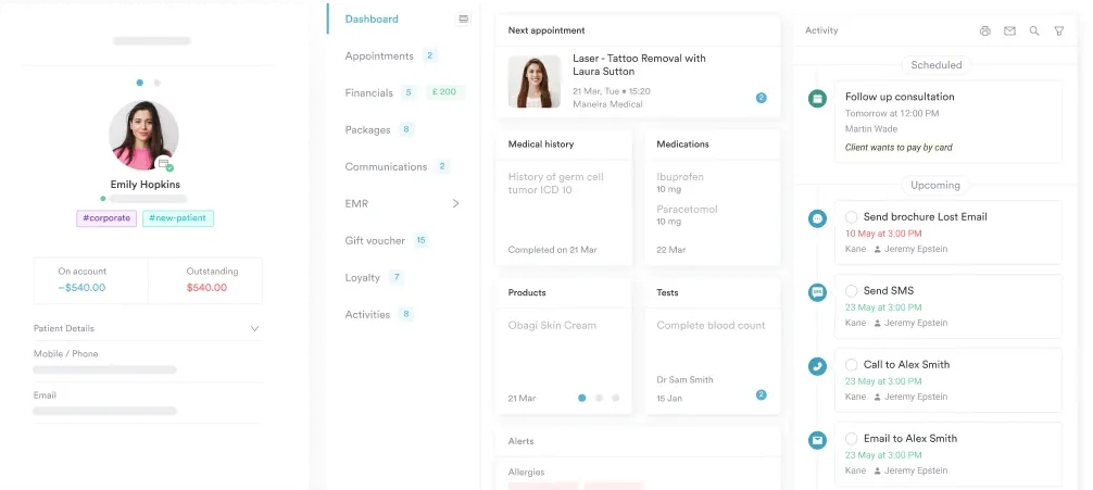
The digital forms feature lets you build an assessment form with a special test checklist inside it. Clinicians then work through the same evaluation for every relevant presentation instead of rebuilding a note from scratch.

Practices running Pabau as their physical therapy EMR can chart measurement changes across appointments and flag when a grade shifts. Assessment summaries then go to the referring clinician straight from the record, so nobody rekeys a number that already exists.

Pabau gives physical therapy and sports medicine practices customizable SOAP note templates, digital forms, and client records. Assessment findings then stay consistent across every clinician and every visit.

Ely’s test is quick, needs nothing but a table, and answers a question that the painful knee rarely answers on its own. It only rewards clinicians who run it the same way twice, though.
So treat the angle of onset as the finding, rather than the word positive. Compare legs, agree on one endpoint across your team, and confirm a positive with the Thomas test before you build a plan around it. The evidence supports acting on a positive result and staying skeptical of a negative one.
Consistent notes are what turn those angles into a trend your whole team can act on. Book a demo to see how Pabau keeps special test findings organized across a physical therapy caseload.

Suspect an Achilles rupture instead? Thompson test walks through the calf squeeze, the positive sign, and what to do when the result is equivocal.
Need an objective ankle dorsiflexion measure? Knee-to-wall test covers the setup, the distance thresholds, and how to track change between sessions.
Screening heel pain in a runner? Windlass test explains the plantar fascia mechanism and how to perform the weight-bearing version properly.
Working up a painful shoulder? O’Brien’s test sets out the two-position technique for labral pain and the accuracy you can expect.
They are close relatives and often confused. Both flex the knee in prone lying. The prone knee bend is usually described as a femoral nerve tension test. There, reproduced thigh or back pain matters more than a lifting pelvis.
No. Expect a stretch at the front of the thigh, not pain. Sharp knee pain, groin pain, or a pinch at the hip points to something other than muscle length. Stop the movement there and reassess before you push further.
Most plans start with prone quadriceps stretching, hip flexor mobility work, and glute strengthening to settle an anterior pelvic tilt. Where spasticity is the driver, tone management comes first. Retest at the same knee angle to see whether the program is working.
No. Reported sensitivity of 56% to 59% and specificity of 64% to 85% cannot carry a surgical decision alone. Gait laboratories use the Duncan-Ely test to support dynamic EMG findings rather than to stand in for them.
Retest at planned reassessment points rather than every visit, which usually means every four to six weeks in outpatient care. Muscle length changes slowly, so frequent testing produces noise instead of a trend you can act on.