Nursing narrative note template
A nursing narrative note template is a structured PDF that guides you through re...

3
Last Updated: Oct 21, 2026

An elastography test measures how stiff tissues are, with stiffer tissue indicating fibrosis, scarring, or disease
Four main types exist: strain elastography, shear wave elastography, transient elastography (FibroScan), and MRI elastography (MRE)
Results are reported in kilopascals (kPa), with values above 7-9 kPa often indicating significant fibrosis, depending on etiology and guideline
Pabau’s automated recall workflows and digital forms help practices schedule, document, and follow up on repeat elastography monitoring
Most liver conditions progress silently. By the time symptoms appear, fibrosis is already advanced. The elastography test gives clinicians a way to catch that progression early, without a needle, without sedation, and without the sampling errors that make traditional biopsies unreliable.
The World Health Organization estimates that 304 million people are living with chronic hepatitis B or C, a leading driver of the liver fibrosis that elastography is used to detect. Non-invasive monitoring tools like this one have become the standard of care, and hepatologists, gastroenterologists, and specialist radiologists now use elastography routinely across outpatient settings.
This guide covers what an elastography test measures, how each type works, how to interpret the results, and how practices can build it into their diagnostic workflow effectively.
An elastography test is a non-invasive imaging procedure that measures the stiffness of soft tissue. It works on a simple physical principle: diseased tissue is stiffer than healthy tissue. By quantifying that stiffness, clinicians can assess the degree of fibrosis, inflammation, or structural change without surgical intervention.
The liver is the primary focus, but elastography can also evaluate thyroid nodules, breast lesions, and musculoskeletal structures. For chronic liver conditions, it has largely replaced biopsy as the first-line assessment tool, which simplifies follow-up monitoring scheduling.
The Cleveland Clinic describes elastography as a test that uses “painless, low-frequency vibrations to check the elasticity of your body’s organs,” noting that stiff tissue “may signal disease, especially in the liver.” As the Clinic explains, “the most common reason for elastography is to check your liver for fibrosis.” It carries no ionizing radiation and requires no recovery time.
The test measures tissue elasticity, specifically how much a tissue resists deformation when a mechanical force is applied. Results are typically reported in kilopascals (kPa), a unit of pressure that reflects stiffness.
Healthy liver tissue is soft and elastic. As fibrosis develops, collagen deposits replace functioning liver cells and the tissue stiffens. The elastography test captures that change numerically as a liver stiffness measurement, letting clinicians track disease progression or regression over time.
Inflammation can also elevate stiffness readings temporarily, which is why results should always be interpreted alongside clinical context rather than in isolation. Tracking measurements over serial visits using measurements tracking software lets hepatology teams spot genuine progression versus transient changes.
The physics differ slightly by modality, but the core mechanism is consistent. A mechanical force (vibration, sound pulse, or magnetic gradient) is introduced into the tissue. The device measures how quickly the resulting shear waves propagate through it. Stiffer tissue transmits waves faster.
In ultrasound-based systems, the transducer both generates the acoustic push and measures wave speed. In MRI elastography, a pneumatic driver outside the bore produces low-frequency vibrations that travel through the patient’s body while the MRI captures the wave patterns.
The output is a quantitative stiffness value or, in the case of MRE, a full spatial map. Neither approach involves ionizing radiation, and both are well-tolerated by most patients. This is why the RSNA and ACR’s RadiologyInfo.org describes elastography as having “no known harmful effects.”
Four main modalities are in clinical use, and they fall into two families: ultrasound elastography (strain, shear wave, and transient or FibroScan) and MRI elastography. Each differs in equipment, technique, and the clinical scenarios where it performs best.
FibroScan is the most widely deployed standalone elastography device globally. It performs vibration-controlled transient elastography, sometimes written as a VCTE test. The probe emits a controlled vibration pulse and measures the resulting shear wave speed through liver tissue, reporting stiffness in kPa. It also captures the Controlled Attenuation Parameter (CAP) to quantify hepatic steatosis.
The procedure takes roughly 10-15 minutes and requires no sedation. Patients typically fast for 2-3 hours beforehand, as recent food intake can affect stiffness readings. For patients with elevated BMI, an XL probe is often required to achieve adequate signal depth. Results are available immediately after the scan.
Shear wave elastography (SWE) is integrated into modern diagnostic ultrasound platforms from manufacturers including Siemens Healthineers, GE HealthCare, Philips, and Canon Medical. It uses Acoustic Radiation Force Impulse (ARFI) technology to generate a localized push pulse and measures the resulting shear wave velocity.
Because elastography ultrasound runs on the same machine as a standard abdominal ultrasound, SWE is highly practical for hepatology departments. Point SWE gives a single-site kPa value, while 2D SWE generates a color-coded stiffness map of a region. Both provide quantitative outputs suitable for serial monitoring in patient care management workflows for chronic liver disease.
Magnetic resonance elastography combines standard MRI acquisition with low-frequency mechanical vibrations delivered via a passive driver placed on the abdomen. The MRI captures wave propagation patterns across the entire liver and produces an elastogram: a color-coded stiffness map showing spatial variation in tissue stiffness.
MRE is considered the most accurate non-invasive method for advanced fibrosis staging, performing well even in patients where obesity limits ultrasound quality. The trade-off is time (30-60 minutes total scan time) and cost. Standard MRI contraindications apply: patients with certain metal implants or severe claustrophobia may not be suitable candidates.
Liver fibrosis staging is the dominant clinical application. Clinicians use the elastography test to monitor patients with chronic hepatitis B, chronic hepatitis C, non-alcoholic fatty liver disease (NAFLD/MASLD), alcohol-related liver disease, and autoimmune hepatitis.
Beyond the liver, elastography has established uses in characterizing thyroid nodules (distinguishing benign from suspicious lesions), evaluating breast masses, and assessing musculoskeletal tissue integrity. Functional medicine practices running metabolic health assessments increasingly include liver stiffness monitoring as part of comprehensive metabolic panels. Weight loss clinic software can fold that liver-stiffness data into the same metabolic monitoring record patients already use for other markers.
The liver biopsy was the gold standard for fibrosis staging for decades. Elastography has not eliminated biopsy entirely, but clinical practice guidelines from EASL now position non-invasive testing, including elastography, as the preferred first-line approach for most patients with chronic liver disease.
Biopsy retains a role when elastography results are borderline, when a concurrent condition (autoimmune hepatitis, for example) needs histological confirmation, or when the clinical picture does not match the stiffness reading. Think of elastography as the filter that significantly reduces how many patients need to proceed to biopsy, not as a complete replacement in every scenario.
Schedule elastography tests in the morning and confirm patients have fasted for at least 2-3 hours beforehand. An unfasted FibroScan can overestimate liver stiffness by up to 20%, generating results that trigger unnecessary anxiety or referrals. Build the fasting reminder into your pre-appointment confirmation workflow so it reaches every patient automatically.
Preparation varies by modality, but a few rules apply broadly. Sending automated pre-appointment instructions ensures patients arrive prepared regardless of which type of elastography they are having.
Patients with ascites (fluid in the abdominal cavity) may obtain unreliable FibroScan results. In those cases, clinicians typically opt for MRE or defer the elastography test until the ascites is managed. This is worth flagging during the booking triage process so patients are not called in for a test that will not yield usable data.
For a FibroScan or ultrasound SWE, the patient lies on a couch with their right arm raised. The operator applies gel to the skin and positions the probe over the right lobe of the liver, typically between the ribs.
The device fires a series of vibration pulses, and patients feel a very mild tapping sensation. Ten valid measurements are taken, and the median value is reported. The whole appointment, including positioning, takes 10-15 minutes.
MRE takes longer: 30-60 minutes total, including standard MRI sequences. The patient lies in the scanner bore with a passive driver (a pad) placed against their abdomen. They may be asked to hold their breath briefly at intervals. Claustrophobic patients may find this more challenging than the ultrasound-based alternatives.
After either procedure, there is no recovery period. Patients can eat, drive, and resume normal activities immediately. Results from FibroScan are available straight away, while MRE results need radiologist interpretation and are usually ready within a few days.
A patient portal for results communication lets the specialist share a summary as soon as it’s ready, so patients get their results securely without an extra phone call or follow-up appointment.
Liver stiffness values are reported in kPa, and they correspond to fibrosis grades using the Metavir scoring system (F0 = no fibrosis, F4 = cirrhosis).
The thresholds below are based on EASL clinical practice guidelines for non-invasive liver disease assessment. They apply specifically to patients with chronic hepatitis C. Thresholds shift for NAFLD and cholestatic liver disease, so the treating specialist must apply the correct reference range.
For NAFLD patients, significant fibrosis thresholds are generally higher (F2 around 8-10 kPa), because fatty infiltration itself can elevate stiffness independent of fibrosis. Etiology-specific reference tables from AASLD and EASL should be consulted for each patient population. An isolated kPa number without clinical context is not a diagnosis. Reference norms matter in every domain — even a coordination screen like the alternate hand wall toss test is meaningless without age-matched scoring tables.
No test is perfect. Clinicians relying solely on the elastography test number without considering confounders risk acting on misleading data. The key factors that can falsely elevate stiffness readings include:
According to MedlinePlus (National Library of Medicine), accuracy can be affected by liver inflammation, tumors, obesity, and abnormal fluid buildup in the abdomen, so a specialist should interpret findings in the full clinical context. For borderline readings (F1-F2 boundary), combining the elastography test result with blood-based biomarkers (FIB-4 index, APRI score) is common practice per AASLD guidance.
Clinical references explain what elastography measures. They say far less about the logistics of running a high-quality elastography monitoring program at scale.
Chronic liver disease patients typically need elastography every 6-12 months to track progression. That creates a predictable, repeatable scheduling need. Without a structured recall system, patients fall through the cracks, results go undocumented, and clinicians lose the longitudinal data that makes elastography clinically meaningful. Patient recall software that links each test to a scheduled follow-up changes that dynamic.
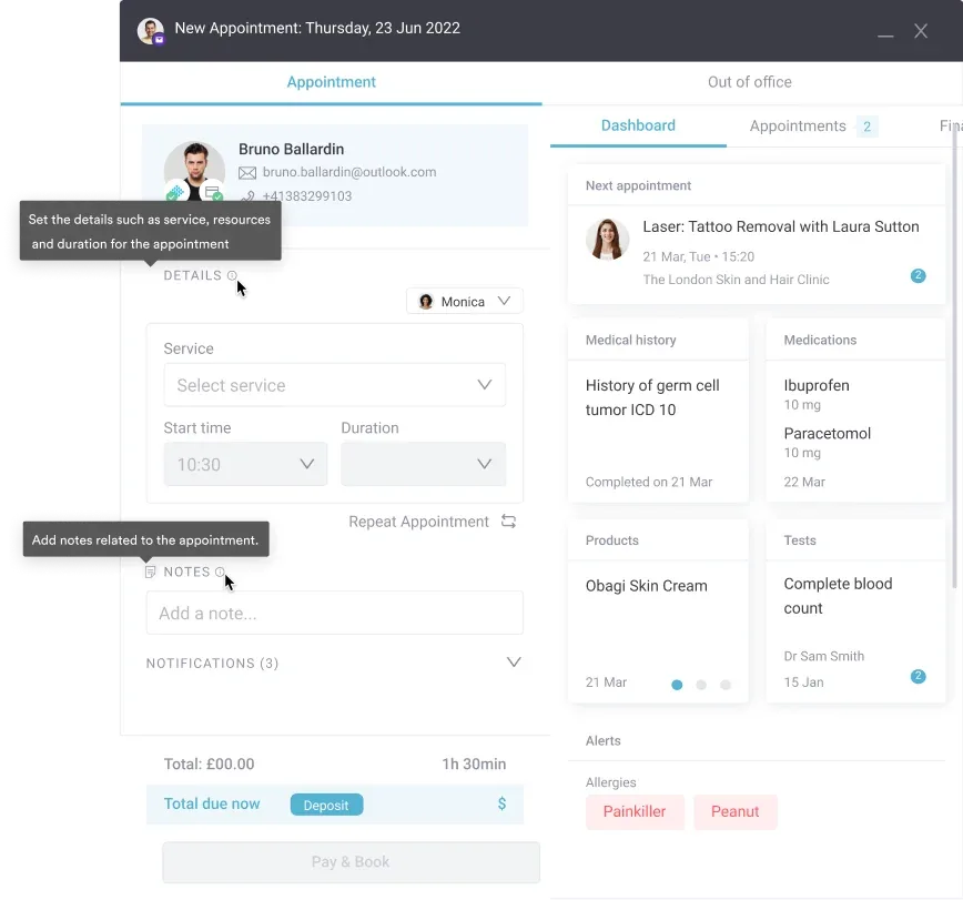
Practice management software like Pabau handles the scheduling side of this. Its automated recall workflows mean that once a clinician completes an elastography visit and documents the result, the system triggers the next monitoring appointment invitation automatically, with pre-set fasting instructions included in the confirmation. That removes the administrative burden from the clinical team while keeping the monitoring program consistent.
For multi-site hepatology services, patient engagement between monitoring visits matters too. Educational resources, lifestyle guidance, and check-ins via the patient portal can be automated to support behavior change between elastography appointments.
Secure patient data handling for diagnostic results is a compliance baseline any practice running structured diagnostic monitoring must address. Want to see how Pabau structures diagnostic follow-up pathways? Book a demo to walk through the workflow.
The elastography test has changed liver disease management by making serial, non-invasive fibrosis monitoring practical for outpatient settings. Choosing the right modality, preparing patients correctly, and interpreting kPa values against the right etiology-specific reference tables are the three areas where practice teams most commonly encounter avoidable errors.
Operationally, this test only delivers value if monitoring programs run consistently. Pabau’s record and recall tools give specialist practices the infrastructure to run elastography-based monitoring programs without manual follow-up burden. If your hepatology or gastroenterology service is expanding diagnostic capacity, see medical practice scheduling software options that integrate with diagnostic workflows.

Need a quick weight-related screening tool? Body mass index assessment explains how to read adult and pediatric BMI ranges and when to add waist circumference.
Preparing a patient for a metabolic workup? Glucose tolerance test guide covers preparation and how to interpret results for prediabetes risk.
Comparing software built for a weight loss practice? Top weight loss clinic software compares seven platforms built for clinical and engagement needs.
Tracking a patient’s glucose control over time? A1C conversion chart maps A1C percentages to estimated average glucose for diabetes management.
Monitoring cardiovascular risk between lab visits? At-home cholesterol test guide covers accuracy versus lab tests and how to interpret results.
An elastography test is a non-invasive imaging procedure that measures tissue stiffness, primarily in the liver, to detect and stage fibrosis (scarring). It uses sound waves or MRI technology to generate quantitative stiffness values in kilopascals (kPa) without needles, radiation, or sedation.
Elevated liver stiffness signals underlying fibrosis or inflammation, which can progress to cirrhosis if untreated. A single elevated reading requires clinical context before acting on it. Inflammation, recent meals, and obesity can all temporarily raise stiffness values, so a hepatologist should interpret results alongside blood tests and clinical history.
A FibroScan or ultrasound shear wave elastography test takes approximately 10-15 minutes from probe placement to result. MRI elastography (MRE) requires 30-60 minutes total. Neither modality requires recovery time, and patients can return to normal activities immediately.
For FibroScan in chronic hepatitis C patients, values below 6 kPa are generally considered normal (F0, no fibrosis). Values above 12.5 kPa suggest cirrhosis (F4). Thresholds shift for NAFLD and other etiologies, so results must be interpreted using the correct guideline reference for each patient’s underlying condition.
No. Standard ultrasound shows anatomy, while elastography measures stiffness. Shear wave elastography and strain elastography run on ultrasound hardware, but they use additional physics to quantify tissue mechanical properties that a conventional B-mode scan cannot detect. FibroScan uses dedicated equipment unrelated to standard diagnostic ultrasound.
Elastography is non-invasive and measures an average stiffness across a large tissue volume, while biopsy samples a tiny fraction (about 1/50,000) of the liver with procedural risk. Most hepatology guidelines now recommend elastography as the first-line assessment, reserving biopsy for cases where results are ambiguous or a concurrent pathology needs histological confirmation.
A doctor orders an elastography test to measure liver stiffness and stage fibrosis without a biopsy, most often to monitor chronic hepatitis B or C, fatty liver disease (MASLD), or alcohol-related liver disease. It is also used to check whether treatment is reducing liver scarring over time, and to decide which patients still need a biopsy.
The cost of an elastography test varies by modality, location, and insurance coverage. A FibroScan or ultrasound shear wave scan is generally far less expensive than MRI elastography, which uses scanner time and radiologist interpretation. Many insurers cover the test when it is ordered to stage or monitor chronic liver disease, so confirm coverage with your provider before booking.
FibroScan is one type of elastography test, not a separate procedure. It performs vibration-controlled transient elastography, a specific ultrasound-based method that reports liver stiffness in kilopascals alongside a CAP score for fat content. Shear wave elastography and MRI elastography are the other main types.