CRM for chiropractors: how to manage patients, bookings and marketing
A CRM for chiropractors is a patient relationship management system built around...

Last Updated: Oct 13, 2026

The alar ligament test assesses the integrity of the alar ligaments connecting the odontoid process (dens) of C2 to the occipital condyles, screening for upper cervical instability.
Normal mechanics: laterally flexing (or rotating) the head causes the opposite-side alar ligament to tighten, pulling C2 into prompt, immediate contralateral deviation. A positive test is an absent, delayed, or reduced-amplitude contralateral C2 response, suggesting ligament laxity or rupture.
Diagnostic accuracy studies against MRI report around 80% sensitivity and 69-77% specificity for the individual test variants (rising to roughly 85-91% specificity against supine MRI or when all three maneuvers are combined); a positive result is a contraindication to high-velocity cervical manipulation until imaging clears the craniocervical junction.
Practice management software like Pabau gives physiotherapy practices structured patient records and digital forms to document cervical assessment findings accurately within SOAP-based workflows.
Upper cervical instability is one of the most consequential findings a physiotherapist can encounter.
Miss it, and a high-velocity manipulation could cause serious neurological harm.
The alar ligament test is a key screening tool for detecting laxity or rupture at the craniocervical junction, yet its clinical utility depends entirely on understanding the anatomy, applying the technique correctly, and interpreting findings within the limits of its diagnostic accuracy. This guide covers the procedure, interpretation, diagnostic evidence, and how these findings integrate with your clinical documentation workflow.
The alar ligaments are short, strong bands that run bilaterally from the lateral aspects of the odontoid process (dens of C2) to the medial surfaces of the occipital condyles. They sit at the craniocervical junction, within the occipito-atlanto-axial complex.
Their primary biomechanical role is restraint: they limit contralateral rotation and side-bending. When you laterally flex the head to the right, it is the left, contralateral alar ligament that becomes taut, not the one on the side you flexed toward. That tension pulls on the dens, and with it the C2 spinous process, into contralateral deviation, meaning C2 moves to the left, opposite the direction of head movement. In an intact ligament, this coupling response is prompt and immediate, appearing essentially as soon as lateral flexion begins. This predictable contralateral coupling is the basis of the test.
Why does integrity matter? Damage to these ligaments, whether from trauma, inflammatory arthropathy (such as rheumatoid arthritis), or connective tissue disorders, removes the primary restraint against excessive atlantoaxial motion. The consequence is upper cervical instability, with the potential for cord compression or vertebral artery compromise during routine cervical movement.
Clinicians assessing compliance requirements for physiotherapy clinics treating post-traumatic or inflammatory cervical conditions will recognize the alar ligament test as a foundational pre-treatment screen.
The test is not routine for every cervical patient. It is indicated when upper cervical instability is a realistic possibility, either from the history or from clinical presentation.
When myelopathic symptoms raise that suspicion, the inverted supinator test adds a quick reflex check for cord compression in the neck. Understanding when to screen is part of broader patient care management workflows for musculoskeletal physiotherapy. If any of the above presentations apply, perform the alar ligament test before progressing to cervical joint mobilization or manipulation.
Correct technique is everything here. Operator variability is one of the test’s primary limitations, so a reproducible, standardized approach is essential.
Supine variation: The test can also be performed in supine with the head supported. The examiner stands at the head of the table, fixates C2 using a thumb-web contact on the posterior arch of C2, and applies lateral flexion passively. The same contralateral coupling criteria apply: a prompt contralateral C2 deviation is normal, and an absent, delayed, or reduced-amplitude response is positive. Some clinicians find the supine position allows better C2 stabilization, though evidence comparing the two positions is limited.
Physiotherapy teams setting up a physiotherapy clinic should include upper cervical instability screening within their initial assessment protocols, particularly if they see trauma or rheumatological caseloads.
Understanding what constitutes a positive result requires understanding normal coupling mechanics first.
Normal response: As the examiner introduces lateral flexion, the alar ligament on the contralateral side becomes progressively taut almost immediately. This tension transmits a rotational force to C2 through the dens, and the C2 spinous process deviates contralaterally, opposite the direction of head movement, promptly at or near the onset of lateral flexion. The coupling is discrete and follows a predictable, near-immediate timing.
Positive response: When the alar ligament is lax or ruptured, the normal tensioning mechanism is diminished or absent. The expected contralateral C2 movement either fails to appear, is noticeably delayed relative to the onset of head lateral flexion, or is reduced in amplitude compared with the opposite, uninjured side. A completely absent contralateral response on one side suggests probable rupture of that ligament.
A positive alar ligament test does not confirm atlantoaxial instability on its own. It raises clinical suspicion and should prompt imaging (typically MRI, which remains the gold standard for soft tissue integrity) and referral decisions. Reviewing guidance on writing safer clinical notes helps ensure these findings are documented with appropriate clinical context and caveats.
The most robust evidence for the alar ligament test comes from Osmotherly et al. (2019), which evaluated three variants of the alar ligament stress test, the side-bending stress test (SBST), the rotation stress test (RST), and the lateral shear test (LST), against MRI as the reference standard, using both upright 3T and supine MRI. The study did not evaluate the Sharp-Purser test or the transverse ligament test; those are separate maneuvers targeting a different ligament, covered in the comparison table below.
These figures come from a single validation study, and values shift depending on which maneuver is used and which MRI reference is applied, so treat them as a guide rather than a fixed number for “the” alar ligament test. Sensitivity is fairly consistent across variants at 80-86%, while specificity is more variable, from around 69% to 91%, and improves when all three maneuvers agree.
Key limitations to communicate in your clinical notes: the test cannot reliably differentiate between partial laxity and complete rupture; muscle guarding can mask a positive finding; C2 fixation quality is highly examiner-dependent; and a single negative result does not rule out instability in a high-suspicion patient. The physical therapy return-to-activity protocols framework is a useful parallel: a single negative screen is part of the picture, not the full clinical decision.
The alar ligament test is conceptually simple but easy to execute poorly. These are the most frequent sources of a misleading result.

Pabau helps physiotherapy practices capture structured cervical screening findings, build custom upper cervical assessment templates, and keep positive-finding follow-ups on track, all in one platform.

The alar ligament test is one of three primary manual tests for upper cervical instability. Each targets a different structure and has distinct procedural and interpretive characteristics. No single test is sufficient in isolation.
The three tests complement each other. The alar ligament test is most sensitive to rotational and side-bending instability; the Sharp-Purser and transverse ligament tests primarily assess anterior translation restraint. In clinical practice, perform all three when upper cervical instability is suspected. A cluster of positive findings carries more clinical weight than any individual result.
When assessing a post-whiplash patient with upper cervical symptoms, perform all three instability tests in sequence: alar ligament test, transverse ligament test, then Sharp-Purser test. Document each result individually and note whether findings are consistent across tests. Inconsistent results (positive on one, negative on two) warrant repeat testing on a separate visit, accounting for muscle guarding on the first assessment.
A positive alar ligament test changes the clinical pathway. The response must be proportionate to the degree of suspicion and the accompanying clinical picture.
The American Physical Therapy Association publishes clinical practice resources on cervical spine management, including guidance on pre-manipulative screening. Physiotutors also offers procedural video content for alar ligament test technique verification.
Refer to physical therapy clinic requirements in Arizona for a jurisdiction-specific example of how documentation and referral obligations are framed in regulatory guidance.
This is the content gap no competitor covers well. Performing the test correctly is only half the clinical obligation. What you write in the patient record shapes every subsequent decision, including who sees the patient next, what treatment is applied, and what your medicolegal position is if an adverse event occurs.
A well-structured SOAP note entry for the alar ligament test should include: the clinical indication for performing the screen, the exact findings (side tested, C2 response, timing and amplitude of contralateral coupling), interpretation relative to the contralateral side, and the clinical decision that follows. Vague entries such as “upper cervical instability screen performed, results normal” provide no evidence of what was actually assessed. Review guidance on structuring effective SOAP notes for frameworks applicable across clinical specialties.
The same discipline applies outside physiotherapy: SLPs administering an articulation test also rely on structured scoring fields to keep speech-sound error data consistent across visits.
Practices managing musculoskeletal caseloads benefit from digital intake and assessment forms that capture upper cervical screening data as structured fields rather than free text. This allows findings to be compared across visits, flagged when positive, and included in referral summaries without transcription errors. AI-assisted clinical note capture further reduces documentation burden by transcribing verbal assessments directly into structured patient records.

Practice management software like Pabau supports SOAP-based clinical documentation workflows through structured patient records, letting physiotherapy teams build assessment templates tailored to upper cervical screens. Practices using physical therapy EMR tools with customizable form fields can standardize how alar ligament test results are recorded, making documentation auditable and consistent across clinicians.
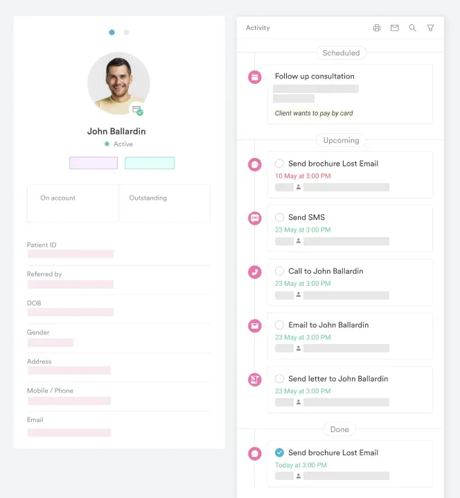
Good documentation also matters for physiotherapy clinic management software users integrating clinical findings with appointment workflows: linking a positive instability screen result directly to a follow-up or specialist referral task within the same platform reduces the risk of patients falling through the cracks between assessment and appropriate management. See also how teams approach managing medical forms across your practice for strategies on keeping assessment records accurate and retrievable.
Pabau helps physiotherapy teams capture structured assessment findings, build custom upper cervical screening templates, and link positive test results to referral workflows, all in one platform.
The alar ligament test is an essential tool in the upper cervical instability screen, but its value depends on technique precision, accurate interpretation of the contralateral coupling response, and integration with the broader clinical picture. Perform it alongside the Sharp-Purser and transverse ligament tests whenever the history raises suspicion. Treat a positive result as a flag, not a diagnosis, and respond with appropriate caution around cervical manipulation and timely imaging referral.
For physiotherapy practices that see trauma, rheumatological, or complex musculoskeletal caseloads, standardizing how these assessment findings are documented is just as important as the clinical skill itself. Book a demo to see how Pabau supports structured clinical documentation across physiotherapy workflows.

Doctor of osteopathy vs MD compares training, licensing, and practice rights between DO and MD providers.
The alar ligament test is a manual clinical examination used to assess the integrity of the alar ligaments at the craniocervical junction, helping to detect upper cervical instability associated with trauma, rheumatoid arthritis, connective tissue disorders, or other conditions that may compromise the dens-to-occiput ligamentous restraint system.
Normally, laterally flexing the head causes the opposite-side (contralateral) alar ligament to tighten, producing a prompt, immediate contralateral deviation of the C2 spinous process. A positive alar ligament test is an absent, delayed, or reduced-amplitude contralateral C2 response, indicating laxity or disruption of that ligament. An absent response suggests probable rupture. It raises clinical suspicion for atlantoaxial instability and warrants MRI referral; it does not confirm instability on its own.
A 2019 validation study (Osmotherly et al.) evaluated three test variants, the side-bending stress test, rotation stress test, and lateral shear test, against MRI. Against upright 3T MRI, sensitivity was around 80% across all three variants, with specificity ranging from about 69% to 77% depending on the maneuver, rising to around 85% when all three were positive together. Against supine MRI, the side-bending and lateral shear tests reached roughly 86% sensitivity and 91% specificity. These figures come from a single study population and should be interpreted alongside pre-test clinical probability, not applied as a universal accuracy figure for the test.
The alar ligament test assesses the alar ligaments by evaluating the contralateral C2 coupling response during lateral flexion or rotation, screening for rotational and side-bending instability. The Sharp-Purser test assesses anterior atlantoaxial instability by applying a posterior-directed force to the head in slight flexion, looking for symptom relief or a clunk, and targets the transverse ligament rather than the alar ligaments. The two tests are not interchangeable and are typically performed together as part of an upper cervical instability screen.
Yes, a positive alar ligament test is generally considered a contraindication to high-velocity thrust techniques in the upper cervical spine. Proceeding with manipulation in the presence of suspected ligamentous instability risks cord compression or vertebral artery injury. Imaging should be obtained before any decision to progress to manual therapy techniques involving the craniovertebral junction.
Yes. A supine variation exists where the examiner stands at the head of the table, contacts C2 posteriorly, and applies passive lateral flexion. Some clinicians prefer this position as it may allow more consistent C2 stabilization, though there is limited comparative evidence between seated and supine administration. The same interpretation criteria apply: a prompt contralateral C2 deviation is normal, and an absent, delayed, or reduced-amplitude response is positive.