GLP-1 diet plan
A GLP-1 diet plan is a structured eating framework for patients taking semagluti...

2
Last Updated: Oct 13, 2026

A radiology report is the radiologist’s written answer to the question a referring clinician asked when ordering the study.
Four sections carry the report: clinical history, technique, findings, and impression, and each one answers a different question.
Structured templates cut ambiguous wording, stop required fields being skipped, and make the report easier to bill from.
Practice management software like Pabau turns the report into a form, stores it on the client record, and tells the referrer it is ready.
A two-page A4 form with fields for patient details, clinical history and reason for exam, technique, findings, impression, and recommendations. Page two adds a critical-findings communication log and a sign-off checklist you run before you finalize.
Download templateA radiology report template exists for one reason. The referring clinician has to find the answer fast. They open the report, jump straight to the impression, and decide what happens to the patient next. If that impression does not answer the question they asked, the study cost time without changing care.
Get the structure right and the report reads the same way every time, whoever dictated it. Below you get the free template, what each section owes the reader, and how a report moves from the scanner to the referrer’s inbox.
A radiology report is a radiologist’s written interpretation of an imaging study, addressed to the clinician who ordered it. It stands as the record of the exam, and it drives the next decision about treatment.
According to the Radiological Society of North America, the report is written for the referring clinician rather than for the patient.
When two people read the report in practice, the referrer reads the impression and acts on it, and months later, another clinician reads the findings to compare them against the next study. This is why measurements and locations earn their place.
Every professional report follows the same four-part structure. Each section answers a different question, and each one fails in its own way.
The diagram below pairs each section with the question it owes the referrer, and with the error it tends to carry.
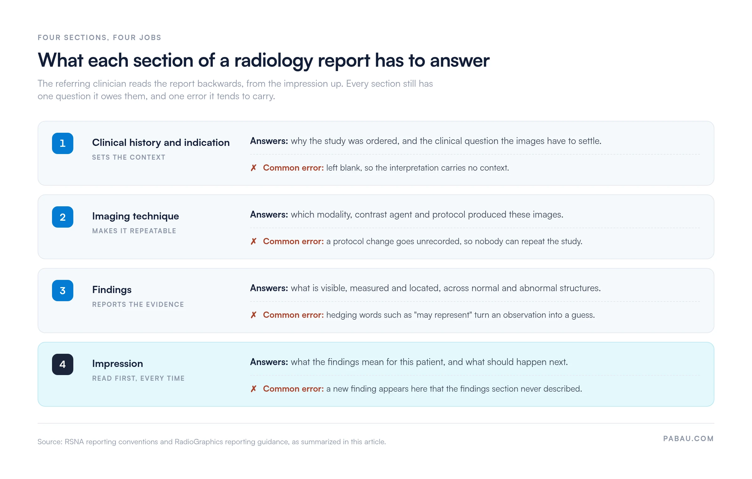
This section says why the imaging happened. Record the presenting symptom, the relevant history, any prior diagnosis, and the specific question the study has to settle.
Leave the field blank and the radiologist reads without context, which is how a technically correct report ends up answering the wrong question.
Name the modality, the equipment, and the protocol. Write it precisely enough that another radiologist could repeat the study. Multidetector CT with contrast, 3.0 Tesla MRI with and without gadolinium, and portable chest X-ray all work as phrasings. Record the contrast agent, the scanning parameters, and any change from your standard protocol.
Billing leans on this section too, because the code has to match what was performed. Contrast is the usual sticking point, so state whether it was given and by which route.
Findings are observations, not conclusions. Describe what you see with precise anatomic terms and measurements, working through the study by region or organ system.
Note the normal structures alongside the abnormal ones, so the referrer knows what you examined. Keep hedging language out of here. “May represent” belongs in the impression, where interpretation lives.
The impression answers the question from the history, in as few lines as it takes. State the diagnostic conclusion, the recommended follow-up, and any finding that needs a phone call rather than a report.
Nothing new belongs here. A lesion that appears in the impression but not in the findings makes the report contradict itself. The referrer then has to call you to sort it out.
Structured reporting wins for most practices, and the table shows where. Structured means the radiologist fills a template. Narrative means free text from a blank screen.
Hartung and colleagues make the same case in RadioGraphics. Structured reports read more consistently, and they leave less room for ambiguity than free text.
Narrative still has a place. An unusual case sometimes needs a paragraph that no template anticipated, so keep the option rather than forcing every study into fields.
A CT report and an ultrasound report do not ask for the same information, so one generic form will not serve both. The RSNA runs RadReport, a library of templates written by radiologists for each modality and body region.
If you report vascular studies, our Doppler ultrasound report carries the flow velocities and waveform fields a general form leaves out.
Structured client records keep each modality’s form beside the study it belongs to. Nobody hunts through a shared drive for the current version, and the wrong template stops turning up in finished reports.
A RADS category tells the referrer what to do, not just what you saw. The American College of Radiology publishes these reporting and data systems for specific screening and diagnostic settings.
Put the category in the impression, next to the finding it describes. A number on its own leaves the referrer guessing which lesion you meant, and that guess turns into a phone call.
The report is a workflow, not just a document. Here is the path it takes, and where practices lose time.
Most of the delay sits at step four. Forms built for clinicians can mark the indication and technique as required, so a half-filled report never reaches the signing queue in the first place.
Step seven is the one that gets written down last, usually after the call has already happened. Give that conversation a structure and it survives an audit, and our SBAR report template works well for the handoff.
Seven questions, and the report is ready to sign. The same list sits on page two of the template, so you can tick it on paper if that suits your team better.
One item deserves more than a checkbox. Communicate a critical finding as soon as possible, per your institution’s critical-results protocol. Use a phone call or a secure message rather than the report alone.
The ACR practice parameter on communicating imaging findings leaves the timing to local policy, so write yours down and hold to it.
Three questions come up again and again from practices tightening their reporting.
Why did the referrer call about a report that reads perfectly well? Usually the impression answered a different question than the one in the history. Check the indication field first, before you re-read the images.
Why do our reports vary so much between radiologists? Free text lets each person pick an order and a vocabulary. A template removes that choice for the parts that should never vary, and leaves it where judgment belongs.
Why does billing keep querying our technique wording? The code has to match the study performed, contrast included. Vague technique wording forces a coder to guess, and a guess is what a payer queries.
Assign one radiologist a week to review a random sample of finalized reports against your template. Score each on a single question: does the impression answer the indication? Feed the results back at your next team meeting. Peer review catches drift while it is still one person’s habit, before it becomes the house style.
Any practice that documents imaging findings can use it. That covers diagnostic imaging centers, hospital radiology departments, and radiologists in private practice.
It also suits practices with imaging on the side of another service. Sports medicine practices with an in-house ultrasound, and occupational health services running on-site X-rays, need the same structure as a dedicated center.
Three changes show up once the template is in daily use. Dictation gets shorter, because the radiologist stops deciding what order to work in. Clarification calls drop, because the impression sits where the referrer expects it. Audit and claims data gets cleaner, because the same fields are filled every time.
An audit asks one question. Can you show the record? A form that fills the same fields on every study keeps that answer short, and keeps it the same answer whoever is asked.
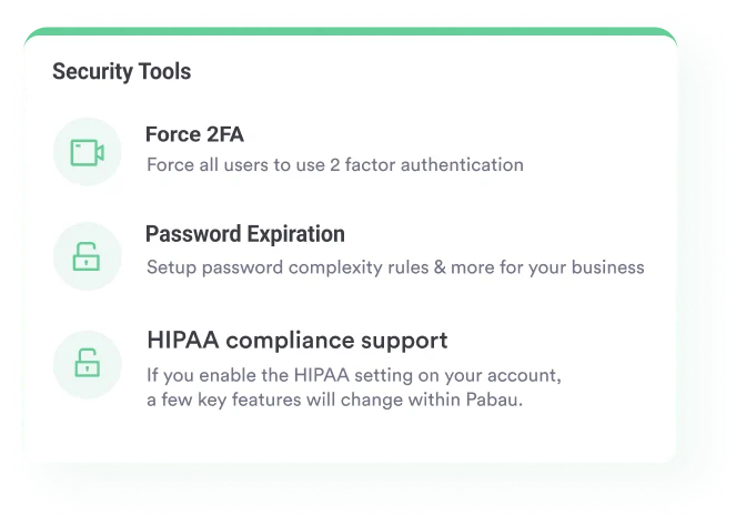
Most imaging practices keep the report in one place, the patient’s history in another, and the referrer’s details in a third. The report gets finalized, then somebody emails it. Practice management software like Pabau, our all-in-one platform for clinical and business operations, holds all three in the same record.
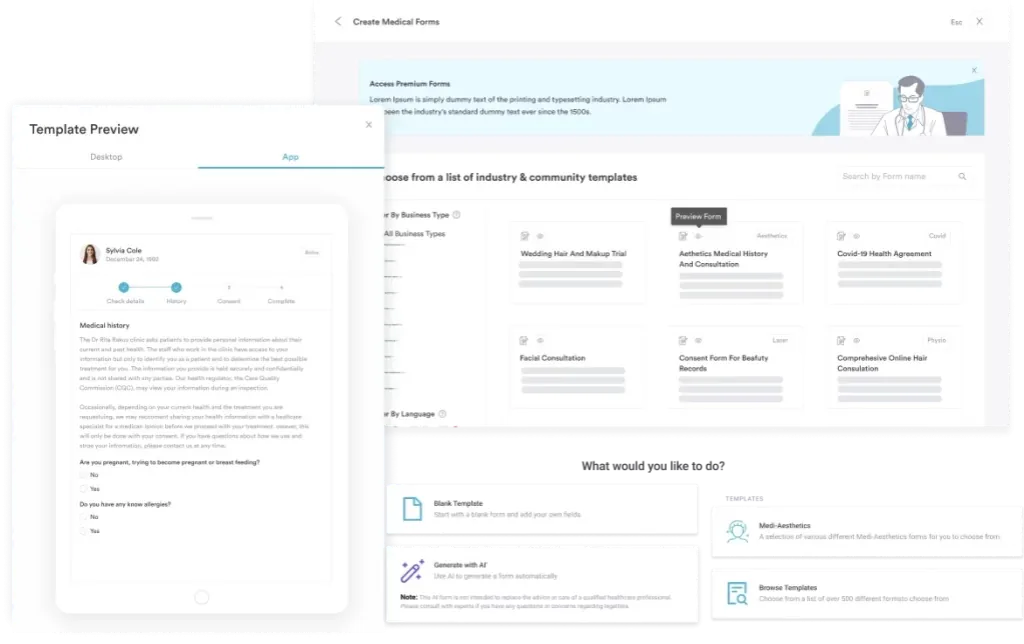
Build the report as a digital form and every field the template asks for becomes a field the software asks for. Mark the indication and technique as required, and an incomplete report cannot reach the signing queue. The finished report attaches to the client record, beside the imaging request and the prior study.
Delivery runs on the same record. Automated messages tell the referrer the report is ready, and each send is logged against the patient. Your audit trail then stops depending on somebody’s sent-mail folder.
Pabau Scribe, our AI scribe, drafts the clinical note from the consultation, which leaves the radiologist writing the report instead of retyping the history.
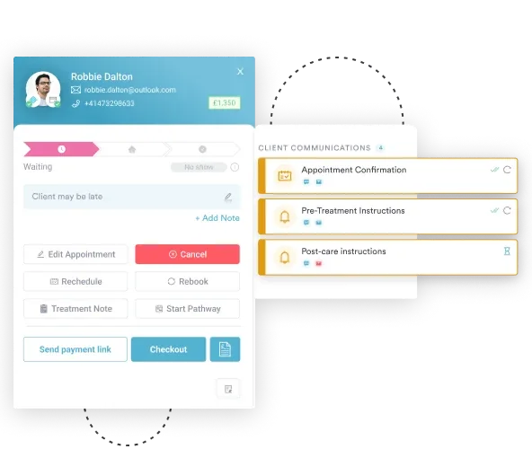

Pabau turns your radiology report template into a digital form and stores each finished report on the client record. Automated messages tell the referring clinician it is ready.

The template is the easy part. Holding to it is what changes how your reports read. That takes two decisions. Set a rule about the indication field, and name an owner for the unsigned queue.
So start narrow. Keep the modality sections you actually report, delete the rest, and put the sign-off checklist where the team will see it. A form nobody can find gets replaced by free text within a month.
One trade-off is worth remembering. A template constrains the unusual case, so leave room for a paragraph where the reasoning needs it.
If your reports are finished on time but still arrive late, the problem is distribution rather than dictation. Book a demo to see how Pabau moves a signed radiology report to the referring clinician without a manual email.

Reporting vascular studies? The Doppler ultrasound report template adds the flow velocity and waveform fields a general imaging form leaves out.
Need a structure for the critical-findings call? The SBAR report template gives that conversation a shape an auditor can follow.
Documenting procedures as well as imaging? Our operation notes template covers the fields a surgical record needs, from indication to post-operative plan.
Tracking a patient across visits? The clinical progress notes template keeps each follow-up entry consistent enough to compare month to month.
Moving off paper forms? Our guide to clinical documentation software explains what to look for before you commit a practice to one system.
Yes. Under the HIPAA right of access, patients can ask for their imaging records, including the report. Most practices release it once the referring clinician has reviewed the findings.
No. A preliminary read is a fast verbal or short written interpretation, often given out of hours. The signed final report remains the record of the study.
Add an addendum rather than editing the original. The addendum carries its own date, author, and reason, so the audit trail shows exactly what changed.
No. A radiologist interprets the study and signs the report. Technologists document positioning, dose, and protocol details, which feed the technique section.
Retention rules vary by state and by payer, and images often run on a different clock than reports. Check your state’s medical record retention rule before setting a policy.