Nitrates medication list
A nitrates medication list is a point-of-care reference that sets out the main c...

Last Updated: Oct 6, 2026
The pediatric assessment triangle is a rapid, equipment-free way to judge how sick a child is, and it takes 30 to 60 seconds. Ronald Dieckmann and colleagues published it in 1996. Its three equally weighted sides are appearance, work of breathing, and circulation to skin, and you read all three without touching the child.
Reading the three sides together gives you one of four clinical impressions. The child is stable, in respiratory distress, in shock, or in cardiopulmonary failure. That impression sets the pace and the priority of the primary assessment that follows.
Whether you work in emergency medicine, EMS, pediatric primary care, or PALS training, this is the first-line tool for sorting children by acuity. The guide below explains each side, how to read the findings together, and how to record what you saw. The template turns 30 seconds of observation into a note another clinician can act on.
A one-page form for recording a rapid pediatric assessment side by side. It carries tick-lists for the TICLS categories, the work-of-breathing signs, and the skin findings. There is a box for the clinical impression and space for the primary assessment notes that follow.
Download template
The pediatric assessment triangle takes 30 to 60 seconds, needs no equipment, and gives you a first read on how sick a child is.
Its three sides are appearance scored on TICLS, work of breathing, and circulation to skin. All three carry equal weight.
The three sides combine into one of four clinical impressions, running from stable through respiratory distress and shock to cardiopulmonary failure.
Recording the specific sign you saw, rather than the conclusion you reached, is what makes a PAT useful at handover and in audit.
Practice management software like Pabau stores PAT findings inside the patient record, so the next clinician reads them instead of starting again.
The pediatric assessment triangle (PAT) is a structured way of looking at a sick child before you go anywhere near them. It needs no equipment, no physical contact, and no more than 30 to 60 seconds. That is why it sits at the front of pediatric triage in emergency departments, in EMS systems, and in PALS certification training.
PAT names the three sides of the triangle. They are appearance, work of breathing, and circulation to skin. Each side stands on its own and each carries the same weight, so an abnormal finding on any one of them can signal serious illness. Because the whole assessment is visual, a new graduate and a 20-year attending run it the same way.
Validation studies in peer-reviewed journals report acceptable sensitivity and specificity for identifying seriously ill children. Emergency physicians, nurses, paramedics, and primary care clinicians all run it as the first step of a pediatric encounter. The diagram below puts the three sides and the four impressions they produce on one page.
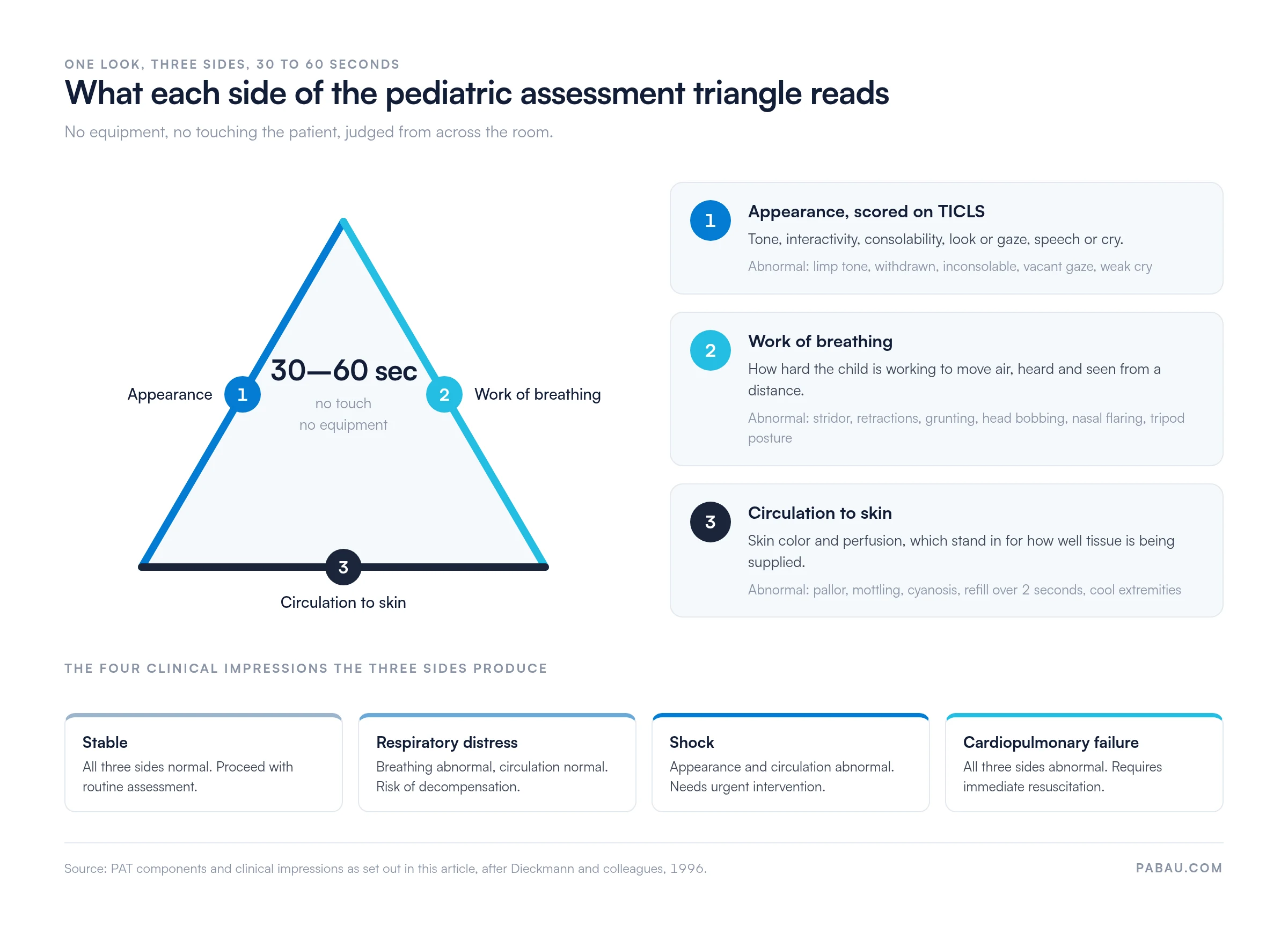
Each side reflects a different part of the child’s physiology. The table below sets out the normal and abnormal findings for all three.
Every finding in that table is visible or audible from a distance. An abnormality on any side raises the concern and changes how fast you move next.
Appearance is scored with the TICLS mnemonic, which breaks a child’s mental and physical status into five observable features. TICLS stands for tone, interactivity, consolability, look or gaze, and speech or cry.
An abnormality in any TICLS category points to possible serious illness. Record which category was abnormal rather than noting that appearance was poor, because the specific finding is what the next clinician needs.
Work of breathing shows whether the child is breathing comfortably or fighting for air. Abnormal effort points to inadequate oxygenation, inadequate ventilation, or an obstructed airway.
Any one of these warrants immediate attention and a closer look at oxygenation and ventilation.
Circulation to skin reflects perfusion and oxygenation. Abnormal findings here suggest shock, hypoxemia, or severe systemic illness.
Skin findings are the easiest side to under-record. Write down the sign you actually saw and the capillary refill time in seconds, not a summary word like “poorly perfused”.
The combination of findings across the three sides points to one of four clinical impressions. Each one sets a different urgency for the assessment and treatment that follow.
The impression drives the pace of the primary assessment. A stable child gets a complete, methodical evaluation. A child in respiratory distress or shock gets a rapid airway, breathing, and circulation check with intervention alongside it.
You run the PAT first, before you touch the patient, reach the bedside, or attach a monitor. The “across the room” label is literal. You form the impression from a distance, in the first 30 to 60 seconds, on observation alone.
The same 30 seconds does different work in different settings:
Practices that see a high volume of acute pediatric visits train every member of staff to spot an abnormal side and escalate. Reception and medical assistants often see the child a few minutes before the clinician does.
Once you have a clinical impression, you move into the primary assessment, also called the primary survey or ABCDE. The PAT decides how fast you work through it and which letter you reach for first.
The PAT sets the priorities and the primary assessment works through them. If your department records the whole encounter on one sheet, the emergency nursing assessment template covers the fields that come after the triangle.
The PAT has been validated in multiple peer-reviewed studies, and the American Heart Association builds it into PALS training. Research in peer-reviewed journals supports it as a reliable way of identifying seriously ill children.
Validation studies report acceptable sensitivity and specificity for detecting serious illness, consistent results across experience levels, and quicker identification of children who need higher-acuity care. HRSA’s Emergency Medical Services for Children (EMSC) program promotes the PAT as a standard assessment tool for EMS systems treating children.
Because it needs no equipment, the PAT travels. It runs the same way in a resuscitation bay and in the back of an ambulance. A primary care room with nothing but an otoscope on the wall works just as well.
Most practices record the PAT as free text, when they record it at all. The note says “appears well” or “increased work of breathing”, and the specific sign behind that judgment never reaches the chart. At handover, or months later during an audit, nobody can reconstruct what the first clinician saw.
Practice management software like Pabau replaces that free text with a structured form. Each side of the triangle gets its own fields. The clinician records the TICLS category, the breathing sign, and the skin finding in a few taps. It is the same builder behind Pabau’s intake forms for practices, so triage notes and intake records land in one chart.
AI clinical documentation turns a dictated assessment into structured text, which shortens the gap between the observation and the note. Structured patient records then carry the findings into every later handover, so the next clinician reads what you saw instead of re-assessing from scratch.
How you go about designing medical forms decides whether any of this sticks. A form with 40 fields gets skipped during a resuscitation. One with three tick-lists and a short free-text box gets filled in every time.
Train every member of staff to recognize an abnormal PAT side, not only the clinicians. Nurses, medical assistants and front-desk personnel often see the child first. Give them the template’s three tick-lists and a clear escalation route, and the clinician hears about respiratory distress in seconds rather than minutes.
See how Pabau’s digital forms and structured templates help pediatric practices standardize assessments and keep documentation retrievable. Book a demo to see how a PAT-aligned workflow would fit your practice.

Pabau captures each side of the pediatric assessment triangle as structured fields, then files the result in the patient record. Your team gets a consistent triage note and a clear audit trail with no extra admin.

The PAT earns its place because it costs nothing and runs before anything else can. Thirty seconds of looking tells you whether the next hour is a routine sick visit or a resuscitation.
The observation is the easy half. The record is the half that decides whether the next clinician starts from your findings or starts over. A structured form takes the same 30 seconds and leaves behind a finding somebody else can act on.
Download the template above and put it wherever your team performs triage. Book a demo to see how Pabau makes a PAT form a permanent part of the pediatric record.

Need the form that follows the triangle? Emergency nursing assessment template covers the primary survey fields you complete once the PAT has set the urgency.
Documenting an altered appearance? Level of consciousness assessment template gives you a structured way to record what an abnormal TICLS score actually looked like.
Want a faster consciousness check? AVPU scale template is the four-point scale most services use alongside the PAT in prehospital and triage settings.
Looking at how you store all of this? Clinical documentation software explains what to look for when assessment forms need to live inside the patient record.
The pediatric assessment triangle (PAT) is a rapid, equipment-free observational tool for judging a child’s clinical status in 30 to 60 seconds. It has three sides: appearance scored on TICLS, work of breathing, and circulation to skin. Together they point to one of four clinical impressions: stable, respiratory distress, shock, or cardiopulmonary failure.
TICLS stands for tone, interactivity, consolability, look or gaze, and speech or cry. Those five observable features are how you score the appearance side of the triangle.
You perform it first, before touching the patient or attaching any equipment. It takes 30 to 60 seconds of visual observation and produces the impression that sets the pace of your primary assessment.
The three sides are appearance, work of breathing, and circulation to skin. Appearance is scored with TICLS. Breathing is judged on stridor, retractions and grunting, and circulation on pallor, mottling, cyanosis and capillary refill.
Yes. The PAT is a core component of Pediatric Advanced Life Support training and certification from the American Heart Association. PALS teaches it as the foundation of the pediatric primary assessment.
An abnormal finding on any side points to possible serious illness and changes the urgency of what you do next. The particular combination across the three sides is what produces the clinical impression, which in turn directs your response.