OSHA respirator medical evaluation questionnaire
The OSHA respirator medical evaluation questionnaire is the mandatory health for...

3
Last Updated: Oct 15, 2026
A nursing pupil dilation chart records pupil size in millimeters, reactivity to light, and symmetry, so every shift measures the same way. One number matters more than the rest. A pupil wider than 8 mm that will not react to light is a neurological emergency.
Pupils shift faster than most neurological signs. An accurate size and a clear trend buy the medical team time before a patient deteriorates. The sections below set out the millimeter ranges, how PERRLA gets charted, and the findings that warrant a call.
Two printable pages. Page one carries a 1 to 9 mm pupil scale that prints at actual size, plus a timed PERRLA observation grid. Page two adds abnormal findings, drug effects, escalation criteria, and space for patient details.
Download template
Normal pupils measure 2 to 8 mm depending on the light, and 3 to 5 mm in average room lighting.
PERRLA is the standard framework nurses use to record size, symmetry, shape, light response, and accommodation.
A pupil above 8 mm with no light response is a neurological emergency and needs a phone call, not a handover note.
Pinpoint pupils of 1 to 2 mm usually point to opioids or Horner syndrome, so check the medication chart first.
Practice management software like Pabau keeps each pupil entry timestamped in the patient record, so the trend survives every handover.
A nursing pupil dilation chart is a one-page reference that pairs a millimeter scale with somewhere structured to write findings down. The scale runs from 1 mm, a pinpoint pupil, up to 9 mm at full dilation.
Next to the scale sit the PERRLA prompts and a short reference for the abnormal findings you are most likely to meet. Those are fixed and dilated pupils, pinpoint pupils, unequal pupils, and reactivity that has turned sluggish.
The chart belongs with the rest of the neurological observations rather than on a loose sheet. Filing it beside the intake forms for nurses and the treatment notes means the next clinician reads a trend, not a single entry.
Pupil changes often arrive before other signs do. A nurse who can measure, compare, and chart in under three minutes gives the medical team a head start. That matters most with raised intracranial pressure, brainstem herniation, and medication effects.
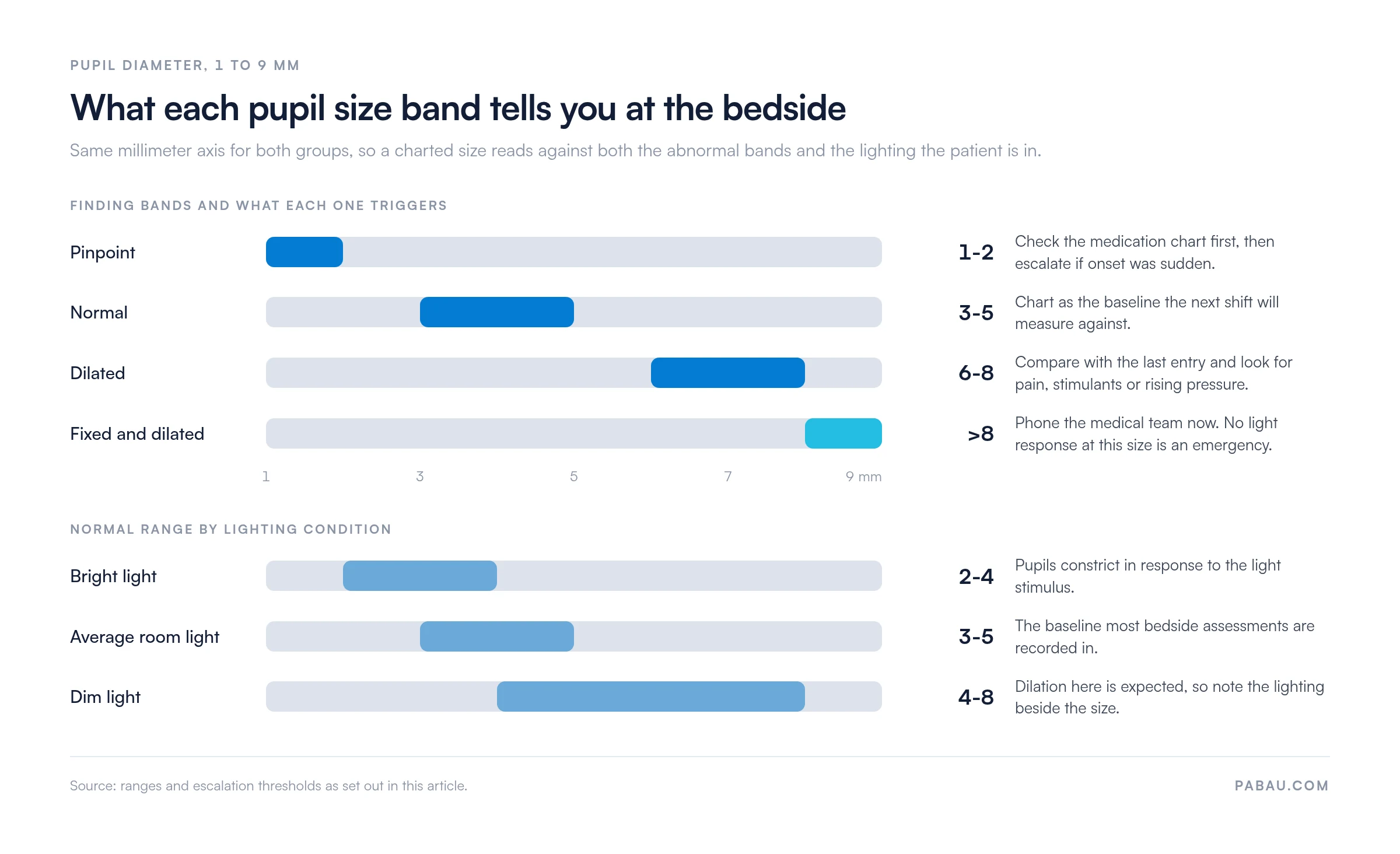
Normal pupil size depends on the light in the room and on the patient in front of you. Bright light constricts them to 2 to 4 mm. In dim light, they widen to 4 to 8 mm. Most adults measure 3 to 5 mm under average room lighting.
Measure with a pupil gauge held beside the eye rather than by eye alone. Record each pupil on its own line so anisocoria, a difference between the two, shows up at a glance.
Charting a pupil as medium or somewhat enlarged tells the next reader very little. Comparing it against the printed circles gives you an entry like “6 mm, round, brisk”. That line reads the same to the night nurse, the covering physician, and an auditor six months later.
The bands below sit on the same millimeter axis as the lighting ranges, so one chart answers both questions at once.
PERRLA stands for Pupils Equal, Round, Reactive to Light, and Accommodation. The mnemonic keeps five observations in the same order on every chart, which is what makes two entries comparable.
Chart PERRLA findings straight after the assessment, then read the previous entry. A sudden change, such as pupils that were equal this morning and now differ by 1 mm, goes to the medical team immediately.
A complete assessment takes two to three minutes and needs only a penlight and a gauge. Work through it during routine neurological observation, and always after a head injury, during post-operative recovery, or in a patient with a known neurological condition.
Use the table below to match a finding to its likely causes. Every abnormal result gets documented, and the ones marked for escalation get a phone call as well.
Knowing when to escalate is the skill the chart exists to support. These findings need urgent medical review, and usually a tighter monitoring frequency as well.
A pupil change rarely travels alone. Pairing it with a level of consciousness assessment gives the physician two data points instead of one, which shortens the conversation considerably.
When you find a red flag, write the exact time and measurement into the patient’s medical records. Then call the medical team directly rather than waiting for handover, and increase monitoring frequency per local protocol.
Several drug classes produce predictable pupil changes, and they turn up constantly in acute care. Knowing the pattern keeps you from escalating a side effect, or from dismissing pathology as one.
Cross-reference the medication chart every time you document an abnormal pupil. A patient on a fentanyl infusion with pinpoint pupils needs a different response to a patient whose pinpoint pupils have no drug behind them.
A pupil gauge is a card printed with circles from 1 mm to 9 mm. Hold it beside the eye in good light and match the pupil to the nearest circle. Direct comparison removes the estimating, which is what makes the number defensible later.
Here is how a complete entry reads after a routine round on a post-operative patient:
02:00, room light. Right 4 mm, left 4 mm, equal and round. Both brisk to light, direct and consensual. Accommodates. No change from 01:00. RN initials.
Every element there earns its place. The time and lighting explain the number, the two sizes make anisocoria visible, and the comparison line turns a snapshot into a trend.
Run the same five checks before you save the entry. They take less time than correcting a note later, and they catch the problems that show up in audit.
Three habits account for most of the pupil documentation that fails on review:
Pupils are one component of a wider examination, so the same round usually captures consciousness, motor and sensory findings too. A neuro checks nursing assessment sets out the full sequence and where pupils sit inside it.
A paper chart sits at the bedside and gets written on straight away, which is exactly what an observation round needs. The trouble starts at handover. Numbers stay on the ward while the patient record gets a summary, and the two drift apart.
Pabau takes the same fields and turns them into a digital form on the patient’s record. The mm fields, the reaction options, and the accommodation prompt are all there, so a nurse taps through the round on a tablet. Each entry is timestamped and signed automatically.
Because those entries land on the record rather than a clipboard, the trend follows the patient. A physician reviewing an escalation sees every prior measurement with the time and the nurse’s name against it. Handover and audit then read from the same line.
For a group running several sites, that also standardizes how neurological observations get recorded everywhere. No ward has to keep its own version of the form.

Pabau’s digital forms capture pupil size, reactivity and timing on the patient’s record, and every entry is timestamped and signed. The trend then follows the patient through handover and audit.

Pupil assessment earns its place because it is quick and it is sensitive. Two minutes with a penlight and a gauge can surface a change before the other observations do.
The chart is what turns that skill into a record somebody else can act on. Print it, keep it within reach, and measure against the last entry rather than against your memory of it.
One caveat is worth holding onto. A chart only helps when the numbers reach the person making the decision, and that is where a paper round tends to fall down. Book a demo to see how Pabau keeps every pupil entry on the patient’s record and visible to the medical team.

Need the wider neuro exam, not just pupils? Cranial nerve nursing assessment walks through all twelve nerves and the findings to record for each one.
Want a faster consciousness screen for the same round? AVPU scale covers the four-point check that takes about thirty seconds at the bedside.
Building a full admission assessment? Emergency nursing assessment template sets out ABCDE, SAMPLE history and triage categories in one printable form.
Losing detail between shifts? Bedside shift report structures the handover so trends like a widening anisocoria get spoken about rather than skipped.
Ready to move the paper round onto the record? Clinical documentation software sets out what to check before a practice changes systems.
A pupillometer measures size and reactivity with infrared light and reports a number, so two nurses get the same result. Penlight readings drift by 1 to 2 mm between observers. The units are costly, so most wards still chart with a gauge.
Yes. Dilating drops used for eye exams, such as tropicamide, can hold a pupil open for several hours. Write the drug and the time on the chart so the next nurse does not read it as a new finding.
Record the eye as prosthetic and assess the other one on its own. Cataract surgery, trauma and corneal scarring can leave a pupil irregular or hard to see. Write the reason rather than leaving the field blank.
Write right and left in full wherever your policy allows. OD, OS and OU sit on the ISMP list of error-prone abbreviations because they get misread as the ear abbreviations AD, AS and AU.
Eye color does not change pupil size, but a dark brown iris makes the pupil edge harder to see. Angle the light across the eye rather than straight at it, and hold the gauge close.