



GLP-1 diet plan
A GLP-1 diet plan is a structured eating framework for patients taking semagluti...

2
Last Updated: Oct 13, 2026

The sweep test detects small to moderate knee effusions by displacing fluid medially and watching for a returning bulge.
Grading runs from 0 (no effusion) to 3+ (spontaneous bulge before stroking), which sets the clinical urgency.
A positive result confirms effusion, not a diagnosis, so the underlying cause still needs investigating.
Two examiners agree on a sweep test grade far more often than on a patellar tap result.
Recording the grade, the side tested, and the clinical context gives the next clinician a usable baseline.
The sweep test is a bedside examination technique that detects small amounts of fluid, called effusion, inside the knee joint.
You displace fluid out of the medial compartment, then watch the medial side for a returning fluid wave. A wave or bulge means effusion is present, graded from 0 to 3+ by how quickly and how strongly it comes back.
According to Physiopedia, it is one of the most reliable bedside tests for identifying small to moderate knee effusions. It needs no equipment and takes under 30 seconds. This guide covers the technique, the grading scale, how to read a positive result, and when to image instead. It is written for physical therapists and clinicians running musculoskeletal knee assessments.
The sweep test is a physical examination technique that detects intra-articular knee effusion. Fluid is moved out of the medial compartment, and the examiner watches whether it returns when the lateral side is stroked downward. It is also called the brush test, stroke test, or bulge sign test. All four names describe the same maneuver.
Clinicians use it because effusions under 10 ml are invisible on inspection. Those small volumes can still signal significant pathology, from ligament injury to septic arthritis.
The test sits within the orthopedic knee special tests and usually runs during the initial musculoskeletal assessment. It requires no equipment and takes under 30 seconds, so it is a standard first pass before ordering imaging.
All healthy knees hold a small volume of synovial fluid, roughly 1 to 4 ml, produced by the synovial membrane to lubricate the joint. When the knee is injured or inflamed, that membrane produces excess fluid as part of the inflammatory response. The excess collects in the joint capsule and pools in the suprapatellar pouch and medial compartment, which have the most capacity.
Even 4 to 8 ml of extra synovial fluid produces no visible swelling. It still changes the mechanics of the joint and causes pain with full extension. The sweep test exists to catch effusions at that scale, before they become visually apparent. This anatomy explains both the technique’s logic and the limits covered later in the article.
Performing the sweep test correctly depends on precise hand placement and stroke direction. Small variations in technique account for most false negatives, so a standardized approach matters.
Record the finding straight after the test. Capturing the grade, the side tested, and any associated findings in a structured note keeps it searchable. Knee conditions evolve over weeks, so the next clinician needs to find it.

The sweep test grading scale runs from 0 to 3+ and reflects both the volume of effusion and its clinical significance. Documenting the grade rather than a bare “positive” or “negative” gives a more useful baseline for tracking change over time.
The volume figures above are approximate clinical estimates from examination literature rather than precise measurements. Individual anatomy affects how much fluid produces each visual grade.
Set side by side on one scale, the bands show how far apart a mild effusion and an imaging-worthy one really sit.
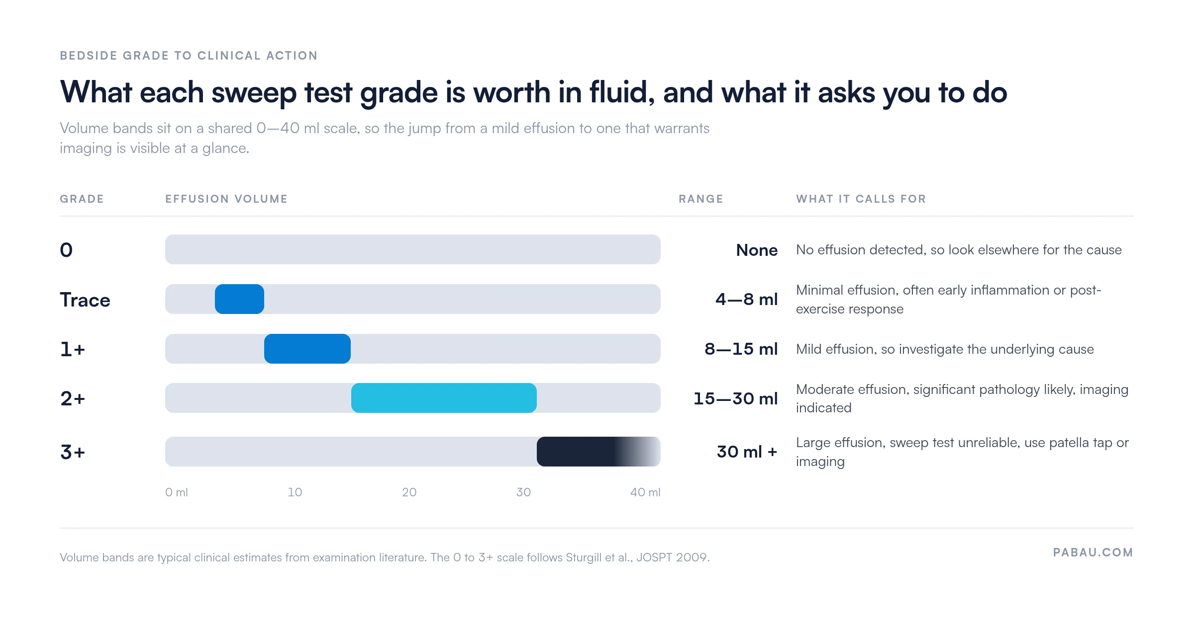
A positive sweep test confirms intra-articular effusion. It does not identify the cause. That distinction shapes how you explain the finding to the patient and what you write in the record.
Common causes of a positive sweep test finding include:
Septic arthritis and gout both need laboratory and imaging confirmation before treatment, and the sweep test cannot tell them apart. Tie the effusion grade to the referral decision in the same encounter note, so the clinical rationale stays visible and defensible. Practices on a physical therapy EMR can hold the grade, the reasoning, and the referral in one place.
The sweep test is the most reliable of the three common bedside effusion tests. Two trained examiners agree on the grade roughly 73% of the time.
That figure comes from Sturgill and colleagues (2009), who tested a five-point effusion scale on knee patients and reported a kappa of 0.75. The patellar tap test and the fluctuation test both scored far lower in the same body of work.
One point is often reported incorrectly, so it is worth stating plainly. No published study gives the sweep test a sensitivity or specificity figure against imaging or joint aspiration. What the literature measures is inter-rater reliability. That answers a different question. Do two clinicians grading the same knee land on the same number?
Kappa values are drawn from Sturgill LP, Snyder-Mackler L, Manal TJ, Axe MJ, “Interrater Reliability of a Clinical Scale to Assess Knee Joint Effusion”. It appeared in the Journal of Orthopaedic and Sports Physical Therapy, 2009;39(12):845-849 (PMID 20032559).
Kappa runs from 0, meaning agreement no better than chance, to 1, meaning perfect agreement. Values above 0.6 read as substantial, and anything under 0.4 is fair at best. That spread is the practical argument for the sweep test. When a colleague documents a 2+ sweep result, you can build a treatment decision on it.
Consistent grading is still not a diagnosis. The sweep test tells you that fluid is present and roughly how much. Combine it with patient history, range-of-motion testing, and other special tests whenever the result is ambiguous.
The sweep test is not the only option for detecting knee effusion. The patella tap test and the fluctuation test each suit different presentations. Picking the wrong one leads to under-detection of large effusions and false reassurance.
In practice, the sweep test is the logical first choice for any suspected effusion. If the result is equivocal and the picture suggests a larger collection, follow with the patella tap test. Running both in one assessment improves detection across the full range of effusion volumes. The Ottawa ankle rules offer a rule-in and rule-out model that translates well to knee assessment.
Technique errors are the most common reason a sweep test gives a misleading result. These are the pitfalls experienced examiners meet most often, and the ones new clinicians are rarely warned about.
Standardizing technique across a practice cuts variation between clinicians. Documented assessment protocols also reduce medico-legal exposure when a finding later supports a referral decision, which is part of wider physical therapy compliance obligations.
The sweep test performs well in the right context but has clear boundaries. Knowing them keeps your confidence in the finding proportionate.
When to escalate to imaging:
Ultrasound is the most accessible option for confirming effusion, and it quantifies fluid volume accurately. MRI remains the standard for identifying the structural source.
A sweep test grade written on paper or buried in free text stops being findable a week later. The next clinician re-examines the knee with no note of the previous grade, so there is no baseline to compare against.
Practice management software like Pabau keeps the grade in a structured patient record instead. Pabau’s patient record management holds the sweep test finding, the side tested, the referral decision, and the follow-up plan against one patient. All of it stays searchable across every visit.

Automated recalls then prompt the follow-up assessment for any patient with an active effusion grade, so the re-check does not fall between treatment episodes. Where several clinicians assess the same knee, physiotherapy practice management software keeps their grading documentation consistent.

The result is a knee that gets graded the same way by whoever sees it next. The reasoning behind each referral stays attached to the record months later.

Pabau gives physical therapy practices digital intake forms, clinical notes, and patient records in one place. Sweep test grades, referral decisions, and treatment plans stay connected across every visit.

Grade the sweep test every time, and write the grade down with the side tested. A bare “positive” throws away what the test is good for, which is showing whether an effusion is settling or growing.
The trade-off worth remembering is that reliable grading buys you a shared baseline, not a diagnosis. Two clinicians will usually land on the same number, which is exactly why that number should never travel alone. Pair it with the history, the mechanism of injury, and the range of motion.
Practices that standardize their assessment documentation get cleaner referral trails and better continuity of care. Book a demo to see how Pabau records effusion grades, referrals, and follow-up plans in one patient record.

Need a decision rule before ordering a knee radiograph? Pittsburgh knee rules sets out the criteria that decide when imaging is justified after an acute knee injury.
Suspect an ACL tear behind the effusion? Lateral pivot shift test walks through the rotational instability test and how to grade what you feel.
Tracking ankle mobility alongside the knee? Knee to wall test covers the dorsiflexion measure that often explains how a knee is being loaded.
Want a scored outcome measure for the knee? Knee outcome survey gives you a patient-reported questionnaire for tracking function between visits.
Planning rehab after a meniscal tear? Meniscus rehab exercises handout is a printable worksheet you can hand the patient at discharge.
The sweep test is a bedside examination technique used to detect intra-articular knee effusion. It picks up small to moderate amounts of excess synovial fluid inside the joint capsule. It runs as part of a routine musculoskeletal knee assessment, and it is most useful when effusion is suspected but not visually apparent.
Position the patient supine with the knee fully extended. Stroke firmly upward along the medial aspect of the knee to displace fluid into the suprapatellar pouch. Then stroke downward along the lateral side. A positive result is a fluid bulge or wave returning on the medial side within 1 to 2 seconds.
Grading runs from 0, meaning no fluid wave and no effusion, to 3+, a spontaneous bulge visible before stroking. Trace is barely perceptible fluid movement, 1+ is a small delayed bulge, and 2+ is an immediate distinct bulge. At 3+ the test becomes less reliable and imaging is warranted.
A positive sweep test confirms the presence of intra-articular effusion. It does not identify the underlying cause. Common causes include osteoarthritis, ACL injury, meniscal tear, gout, pseudogout, and septic arthritis. Patient history, additional clinical tests, and imaging are always needed to establish the specific diagnosis.
Yes. The sweep test, brush test, stroke test, and bulge sign test all describe the identical clinical maneuver. The different names reflect regional and institutional preferences rather than any variation in technique. All four terms cover the same medial-clearance and lateral-stroke procedure for detecting knee effusion.
The sweep test detects small to moderate effusions, Trace to 2+, by observing a returning fluid wave. The patella tap test detects moderate to large effusions, 2+ to 3+. It compresses the suprapatellar pouch so you can feel the patella ballotte against the femoral condyle. Using both together improves detection across the full range.






