



GLP-1 diet plan
A GLP-1 diet plan is a structured eating framework for patients taking semagluti...

2
Last Updated: Oct 13, 2026

The posterior interosseous nerve test assesses the deep motor branch of the radial nerve, checking for finger and thumb extension weakness without wrist drop.
The arcade of Frohse, the fibrous proximal edge of the supinator, is the most common site of PIN entrapment. It produces motor deficits with no sensory loss.
A positive test shows weak finger and thumb extension at the MCP joints, with wrist extension that deviates radially. Wrist drop is absent because ECRL is spared.
Practice management software like Pabau keeps MRC grades, Tinel’s findings, and investigation results in one patient timeline, so follow-up visits compare like with like.
The posterior interosseous nerve test is a clinical examination sequence that detects motor dysfunction of the posterior interosseous nerve (PIN).
The PIN is the deep motor branch of the radial nerve. A positive test reveals finger and thumb extension weakness at the metacarpophalangeal (MCP) joints, with preserved but radially deviated wrist extension. Because the PIN carries no sensory fibers, sensory function stays intact throughout.
That combination is what makes the test worth doing properly. PIN palsy looks like a hand problem, and a vague note makes the deficit disappear. The sequence below is written to be repeatable and gradable. It covers the anatomy that explains the findings and the four examination steps in order. It then sets out how to read the result against its differentials, and which investigations confirm it.
The radial nerve divides at the lateral epicondyle into a superficial sensory branch and the posterior interosseous nerve. The PIN descends into the radial tunnel, passing through or beneath the arcade of Frohse, the fibrous proximal arch of the supinator muscle. From there it enters the posterior forearm compartment.
One anatomical detail explains the whole clinical picture. The extensor carpi radialis longus (ECRL) receives its innervation proximal to the arcade of Frohse, before the PIN enters the supinator. When the PIN is compressed at or below the arcade, ECRL remains intact.
So wrist extension is still possible in PIN palsy. The wrist deviates radially because extensor carpi ulnaris (ECU) is weak while ECRL pulls unopposed. The map below shows where that dividing line falls.
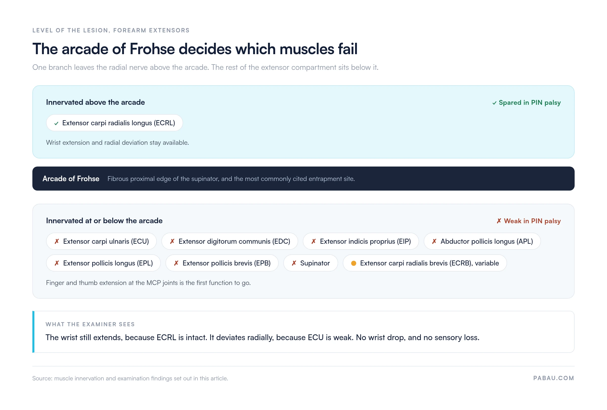
According to Orthobullets’ anatomical reference, the arcade of Frohse is the most commonly cited site of PIN entrapment. Repetitive pronation and supination loads that arch, which is why the presentation clusters in manual workers and racquet sports.
Knowing which muscles the PIN supplies is the foundation of every posterior interosseous nerve test. ECRL is innervated above the arcade and is therefore always spared in PIN palsy. All other posterior forearm extensors are PIN territory.
ECRB is the row worth reading twice. Its innervation varies between individuals, so it may sit above or below the arcade in any given patient. That variation is one reason wrist extension power grades inconsistently across cases that are otherwise identical.
PIN entrapment can be traumatic, compressive, or inflammatory. Knowing the cause guides both the examination focus and the investigation choice.
PIN syndrome presents as a pure motor neuropathy. That alone separates it from most other peripheral nerve lesions at the bedside.
Document whether the wrist deviates radially on active extension before any resistance is applied. Radial deviation at rest confirms ECRL activity with ECU weakness, and it is the fastest clinical marker separating PIN palsy from complete radial nerve palsy.
The test runs as a four-part examination sequence rather than a single maneuver. Each step isolates a different component of PIN function, so perform them in order and grade using the MRC scale (0-5) per muscle. Our printable MRC scale template gives you a one-page grading sheet to keep beside the plinth.
Position the patient with the elbow slightly flexed, the forearm pronated, and the wrist in neutral. Stabilize the wrist to remove its contribution. Apply downward pressure over the proximal phalanges of fingers 2-5 while asking the patient to extend at the MCP joints.
Positive finding: weakness or inability to hold MCP extension against minimal resistance. Document each finger individually where possible, since partial PIN lesions may spare some fascicles. Extensor digitorum communis (EDC) and extensor indicis proprius (EIP) are the primary targets here.
Ask the patient to extend the thumb at both the IP joint (EPL) and the MCP joint (EPB) against resistance. Stabilize the first metacarpal to isolate the movement.
Positive finding: weakness at the IP joint in particular indicates EPL involvement. Consider de Quervain tenosynovitis as a differential for localized first-ray symptoms. The Finkelstein test separates the two, because de Quervain produces pain rather than a true extension deficit.
Ask the patient to extend the wrist actively without stabilization. Observe the direction of deviation before applying any resistance. In PIN palsy the wrist extends but deviates radially. Then apply resistance and grade wrist extension power overall.
Positive finding: radial deviation on active extension, with ECU weak and ECRL intact. If the wrist drops entirely on attempted extension, the lesion sits at or above the spiral groove of the humerus rather than at the PIN.
Radial deviation is easy to miss by eye. A described bedside refinement is the window test. The examiner rests a hand along the ulnar border of the patient’s pronated forearm during active wrist extension. A visible window opening between the examiner’s forearm and the patient’s hand marks the radial drift.
Percuss over the radial tunnel, roughly 3-4 cm distal to the lateral epicondyle, along the course of the PIN through the supinator. A positive Tinel’s sign produces tingling or shooting pain into the dorsal forearm. Tinel’s sensitivity for PIN entrapment is limited, so a negative sign does not exclude the diagnosis.
Follow with resisted supination. Ask the patient to supinate the forearm against resistance with the elbow flexed to 90 degrees. Reproduction of lateral forearm pain or weakness on this maneuver supports radial tunnel involvement.
A clear positive test shows four findings together.
The most important differential to exclude is radial tunnel syndrome. The table below separates the three patterns that present around the radial tunnel.
Two further differentials sit outside the forearm. C7 radiculopathy produces sensory symptoms and may involve triceps weakness, and Spurling’s test helps separate a cervical root cause from a local one. Lateral epicondylitis is pain-dominant and produces no motor deficit at all.
Anterior interosseous nerve syndrome is the closest mimic in name only. It is a purely motor palsy of the volar forearm compartment rather than the extensor compartment. The pinch grip test is the bedside screen for it.
Clinical examination narrows the diagnosis substantially. Electrodiagnostic testing and imaging then confirm it and localize the lesion.
Electromyography (EMG): the gold standard for confirming PIN palsy and excluding radial tunnel syndrome. According to a peer-reviewed electrodiagnostic evaluation of posterior interosseous neuropathy, the key muscles sampled are EDC, EIP, ECU, and APL.
Denervation potentials and reduced MUAP recruitment in those muscles confirm a PIN lesion below the arcade of Frohse, provided ECRL and brachioradialis are normal. Radial tunnel syndrome by definition produces a normal EMG, which is what makes the study decisive between the two.
Nerve conduction studies (NCS): motor latency to EDC via the PIN can be prolonged or absent. Standard superficial radial sensory studies stay normal throughout, consistent with a pure motor neuropathy.
Ultrasound: dynamic imaging at the arcade of Frohse can show nerve enlargement and hypoechogenicity. It is particularly useful for detecting ganglia and lipomas as compressive causes.
MRI: reveals denervation edema and fatty infiltration in PIN-innervated extensor muscles. MRI is the most sensitive modality for identifying space-occupying lesions and quantifying muscle involvement, as confirmed by AJR sonography and MRI research on PIN syndrome.
Reference sources describe the examination well and stop at the finding. The note is where a PIN assessment either supports continuity of care or leaves a medico-legal hole, so it is worth specifying. A structured PIN test note should capture four elements.
Recording those four elements the same way at every visit is what makes a follow-up comparison meaningful. Software for physical therapists that carries a reusable assessment template removes the variation between clinicians and between appointments.

Management depends on the cause, the severity, and the duration of symptoms. The evidence base favors a stepped approach, as set out in clinical consensus sources including Physiopedia’s PIN syndrome reference.
Conservative management (first-line, 3-6 months):
Surgical decompression: indicated when an adequate conservative trial has failed, when imaging identifies a space-occupying lesion, or when neurology is worsening quickly. Decompression at the arcade of Frohse is the standard approach.
Prognosis is generally good when surgery happens within 12 months of symptom onset. Recovery timelines vary by case, so frame them for the patient as reported ranges rather than as a guarantee.
In most practices the PIN test lands in a free-text note. One clinician writes “finger extension weak”, the next writes MRC grades for three of the six muscles, and the third records only the Tinel’s response. Six weeks later nobody can say whether the patient improved.
Practice management software like Pabau replaces that free text with a form you design once. Build the four-step sequence as fixed fields, with a 0-5 grade per muscle, a deviation measurement, a Tinel’s site and response, and the investigation requested. The form attaches to the appointment and stays on the patient timeline.
The outcome is a set of visits you can read side by side. Grades line up against each other, and the imaging request sits next to the finding that prompted it. Any clinician picking up the case sees the same structure. Every Pabau subscription includes the full clinical records toolset, so this is a setup task rather than an upgrade.

Pabau’s digital forms and client records let musculoskeletal and physiotherapy practices capture MRC grading, Tinel’s findings, and investigation results in one structured patient timeline. See it in action.

PIN palsy is quiet. There is no wrist drop and no numbness to prompt a referral. The deficit sits at the fingers and thumb until someone tests for it deliberately. The four-step sequence takes under five minutes and needs no equipment.
The trade-off worth remembering is that this examination is only as useful as the record it leaves. A grade per muscle turns a subjective impression into a number you can retest in six weeks. A vague note means starting the assessment again from scratch.
Build the sequence into your assessment template once and it runs the same way for every clinician who uses it. Book a demo to see how Pabau keeps MRC grades, Tinel’s responses, and investigation requests together on one patient timeline.

Screening a hand for more than one nerve? Hand nerve tests walks through the median, ulnar, and radial screens you can run in the same appointment.
Need to grade strength consistently? Manual muscle testing covers positioning, resistance, and how to apply the MRC scale without drifting between clinicians.
Suspect a neural rather than muscular cause? Upper limb tension tests gives you a printable sequence for testing neural mechanosensitivity in the arm.
Ruling out a compression at the wrist? Phalen’s test explains the median nerve screen for carpal tunnel syndrome and how to interpret a positive result.
Reviewing the software behind your notes? Physiotherapy clinic management software compares the platforms that handle clinical records and scheduling together.
The posterior interosseous nerve test is a clinical examination sequence that checks for motor dysfunction of the PIN, the deep branch of the radial nerve. It involves four steps: resisted finger MCP extension, resisted thumb extension, assessment of wrist extension deviation, and Tinel’s sign with resisted supination provocation. A positive test reveals finger and thumb extension weakness with radially deviated (not dropped) wrist extension and no sensory loss.
No. PIN palsy does not cause wrist drop because extensor carpi radialis longus (ECRL) is innervated proximal to the arcade of Frohse and remains intact. The wrist extends but deviates radially due to ECU weakness. Wrist drop indicates a more proximal radial nerve lesion, at or above the spiral groove of the humerus.
PIN syndrome is a motor neuropathy: it produces measurable weakness of finger and thumb extension. Radial tunnel syndrome is a pain syndrome: it produces lateral forearm pain without motor deficit and with normal EMG findings. Both involve the radial tunnel and arcade of Frohse region, but they are clinically distinct entities requiring different management.
Supinator syndrome is an alternative name for PIN entrapment at the arcade of Frohse. It refers to compression of the PIN as it passes through the supinator muscle. It is used interchangeably with posterior interosseous nerve syndrome in much of the surgical and rehabilitation literature. The clinical presentation and examination findings are identical.
Order EMG and nerve conduction studies when the diagnosis stays uncertain despite a suggestive clinical picture. They also exclude radial tunnel syndrome, which by definition produces a normal EMG. Order them to quantify lesion severity and set a prognostic baseline. They are also warranted when symptoms persist for 3-4 weeks with no obvious structural cause. EMG should sample EDC, EIP, ECU, and APL as the primary PIN-innervated muscles.
The PIN innervates extensor digitorum communis, extensor indicis proprius, extensor carpi ulnaris, and abductor pollicis longus. It also supplies extensor pollicis longus, extensor pollicis brevis, the supinator, and extensor carpi radialis brevis variably. ECRL is spared because it receives innervation proximal to the arcade of Frohse.






