



GLP-1 diet plan
A GLP-1 diet plan is a structured eating framework for patients taking semagluti...

2
Last Updated: Oct 13, 2026

The Phelps test is performed prone: hip abduction is measured with the knee extended, then again with the knee flexed to 90 degrees.
A positive Phelps test is an increase in hip abduction after the knee is flexed, which points to gracilis tightness rather than adductor contracture.
The gracilis crosses both the hip and the knee, so flexing the knee slackens it and lets the hip abduct further.
Record both angles and the difference between them, because a single positive or negative label loses the measurement a later examiner needs.
Practice management software like Pabau stores both angles, the delta, and your interpretation as structured fields in the patient record.
The Phelps test is a prone orthopedic special test that identifies gracilis tightness as the cause of restricted hip abduction.
You measure passive hip abduction twice: first with the knee extended, then again with the knee flexed to 90 degrees. If abduction increases in the second phase, the result is positive and the gracilis was the limiting structure. If the range does not change, the restriction sits elsewhere, usually in the mono-articular adductors, the hip capsule, or the bony architecture.
That distinction sets the treatment plan. A tight gracilis has to be lengthened across the hip and the knee together, while an adductor contracture is worked at the hip alone. According to the American Physical Therapy Association (APTA), special tests for the hip and thigh are a core part of orthopedic assessment. This guide covers the procedure, the anatomy behind it, the clinical indications, and how to document the result so it survives a handover.
The Phelps test assesses whether restricted hip abduction in a prone patient comes from gracilis tightness rather than a contracture of the deeper adductor muscles. That single distinction changes the treatment pathway. A patient with gracilis tightness benefits from interventions that address the hip and the knee at the same time, because the gracilis spans both joints. A patient with adductor contracture needs an approach focused on the hip in isolation.
The test belongs in physical therapy and orthopedic assessment when a patient presents with limited passive hip abduction in prone. It appears regularly in assessments of patients with neuromuscular conditions, post-injury hip restriction, and hip adductor spasticity.
Its usefulness comes from changing one variable at a time. Knee position selectively slackens or tensions the gracilis while the adductors stay under a consistent stretch. If abduction improves with the knee flexed, the gracilis was the limiting structure. If it stays the same, the restriction is elsewhere.
The gracilis is a long, slender muscle running along the medial aspect of the thigh. It crosses two joints, and that is what makes it identifiable through this maneuver.
Because the gracilis runs from the pubis to the tibia, it is at its longest when the hip abducts and the knee stays extended. Flexing the knee to 90 degrees shortens the distance the muscle has to cover at its distal end, so overall gracilis tension drops. If gracilis tightness was limiting abduction, that drop in tension lets the hip travel further. This is the biomechanical logic the test rests on.
The pure adductors (adductor longus, adductor magnus, adductor brevis, and pectineus) are mono-articular or primarily hip-crossing. Flexing the knee does not meaningfully alter their length. So if the restriction does not change when the knee is flexed, the gracilis is not the primary culprit.
The procedure follows a two-phase sequence. Standardizing each step reduces examiner variability and makes findings comparable across repeat assessments.
A goniometer improves reliability. Estimating by eye introduces examiner bias, particularly when the difference between the two phases is small. Where the same patient will be reassessed over several appointments, consistent measurement technique is what makes the comparison worth anything.
A positive Phelps test is an increase in hip abduction range after the knee is flexed to 90 degrees. It indicates gracilis tightness: the muscle was limiting abduction with the knee extended, and slackening it at the knee end allowed more hip abduction.
A negative result is an abduction range that does not change between the two phases. The gracilis is then unlikely to be the limiting structure. The restriction usually sits in the adductors, the hip capsule, or the bony architecture of the joint.
No threshold in degrees is universally agreed. Most clinical texts treat any consistent increase in abduction as meaningful. In practice, a difference of fewer than 5 degrees may sit inside measurement error, particularly without goniometry. The diagram below follows the difference between the two phases through to the finding and the next clinical step.
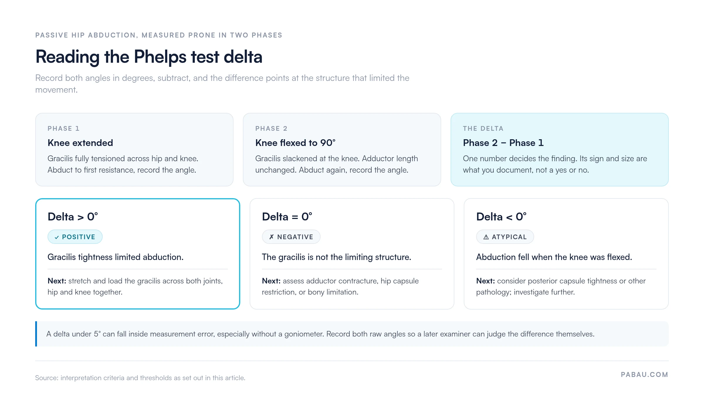
The diagnostic question here is whether restricted hip abduction originates in the gracilis or in the mono-articular adductors. The distinction matters because the two present similarly and are managed differently.
Gracilis tightness produces hip abduction restriction that depends on knee position. The restriction is worse with the knee extended and eases with the knee flexed. Patients may also report medial knee discomfort during combined hip abduction and knee extension, as in lateral lunges or side-step running.
Adductor contracture produces a restriction that holds steady whatever the knee is doing, because adductor longus, brevis, and magnus do not cross the knee. The limitation feels equally firm in both phases. Groin pain with resisted adduction points the same way, and an adductor squeeze test will usually reproduce it.
A negative result alongside persistent hip abduction restriction should prompt you to consider hip joint capsule tightness, labral pathology, or bony hip morphology. The test isolates one variable. It does not rule out co-existing pathology.
The test is indicated when a patient presents with restricted passive hip abduction in prone and you want to know whether the gracilis is contributing. Several patient populations appear consistently in the clinical literature.
The test is safe across most patient populations. It uses passive motion within a comfortable range, and the examiner controls the force throughout. Contraindications are not widely reported, but avoid it where there is acute hip or knee injury, suspected fracture, or significant pain on any passive movement. Documenting why you selected the test is standard practice in most jurisdictions.
Record the Phase 1 angle before you flex the knee. Without that baseline, the second measurement has nothing to be compared against, and the result cannot be called positive or negative. Both angles belong in the note, in degrees.
Published psychometric data is limited. The Thomas test and the Ober test have both been studied heavily. Formal sensitivity and specificity studies for this one are sparse in peer-reviewed literature. Treat it as a biomechanically rational screen rather than a high-accuracy diagnostic tool.
Its clinical value rests on anatomy. The gracilis crosses two joints, and the maneuver isolates the tension relationship between its proximal and distal ends, which gives the test strong face validity. Where older texts cite psychometric figures, those usually reflect expert consensus or case series rather than blinded controlled studies.
According to the Journal of Orthopaedic and Sports Physical Therapy, evidence quality for many hip special tests is rated low to moderate. Small samples and heterogeneous patient populations account for much of that. The Phelps test fits that pattern. Treat a positive result as one piece of evidence to be read alongside symptom history, other special tests, and imaging where it is available.
Reliability between examiners improves with standardized goniometry and clear documentation protocols. Inter-rater reliability for passive abduction measured in prone is generally acceptable, per guidance from the Chartered Society of Physiotherapy. That holds where the protocol specifies the stabilization point and the measurement landmark.
Clear documentation supports clinical continuity, referral quality, and medicolegal protection. A complete record captures the measurements and the interpretation, not just the verdict.
A well-structured entry names the tested limb and both angles, with the knee position recorded against each one. It also carries the difference between the two and your interpretation. Recording both angles against a dated hip range of motion chart lets a colleague judge the difference for themselves rather than trusting your label.
Free-text notes are where this data goes to die. The angles get written into a paragraph, the delta is never calculated, and the next clinician re-measures from scratch because the baseline cannot be found. Structured fields keep all three numbers retrievable, which matters most for multi-disciplinary teams treating cerebral palsy or post-surgical hip restriction.

This test sits within a broader battery of hip and thigh special tests. Knowing how it relates to the adjacent ones helps you pick an assessment sequence and avoid redundant or conflicting findings.
The Ober test and this one both examine thigh musculature, but they target different structures. The Ober test looks at the iliotibial band and tensor fasciae latae. This one looks at the gracilis. They can be performed in the same session without interfering with each other. The Thomas test, performed supine, covers the hip flexors. Ely’s test, performed prone, covers rectus femoris, and together the four round out a thorough assessment.
For clinicians managing return-to-sport pathways, running all three biarticular thigh tests together gives a fuller picture than any one of them alone. A structured return-to-running protocol will typically weigh findings from all three when deciding readiness for full training loads.
In many practices, a Phelps test is recorded as one line of free text in the visit note. Six weeks later, nobody can tell whether abduction has improved, because the baseline angle was never written down as a number anyone can query.
Pabau, our practice management software for physical therapy and musculoskeletal practices, handles this with structured assessment forms. You build the form once, with numeric fields for the Phase 1 and Phase 2 angles and a field for your interpretation. Every clinician who runs the test then fills in the same fields, in the same units, in the same place in the record.
Those values then plot against a timeline, so you can show a patient or a referrer how abduction has changed across a course of treatment. Pabau Scribe, our AI scribe, drafts the narrative around them, which means the note reads properly without anyone retyping the numbers into a letter.
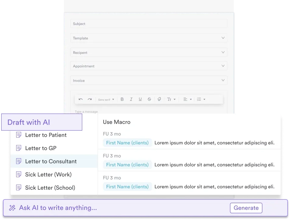

Pabau gives physical therapy and musculoskeletal practices structured assessment forms, range of motion tracking across appointments, and referral-ready notes. See how it fits the way your team already works.

The Phelps test answers one narrow question well: is the gracilis what is holding hip abduction back? Read alongside a thorough history and the neighboring special tests, it points treatment at the right structure instead of the obvious one.
The evidence base is thin, and that is the trade-off worth remembering. The number you record carries more weight than the label you attach to it. Write down both angles and the difference, every time, and the next clinician inherits something they can act on. Software for physical therapists should make that the path of least resistance rather than an extra step. Book a demo to see how Pabau structures musculoskeletal assessment data for your team.

Assessing rectus femoris tightness in the same prone position? Ely’s test covers the procedure and what a positive heel-to-buttock finding means.
Need the hip flexor half of the picture? Thomas test template gives you a printable recording sheet for iliopsoas and rectus femoris length.
Building a full hip assessment sequence? Hip examination sets out the order to work through observation, range of motion, and special tests.
Looking for a clinical documentation system for your musculoskeletal team? Physiotherapy clinic management software explains what to look for when choosing a system for physical therapy practices.
Running a physical therapy practice and need guidance on compliance? Mandatory compliance for physiotherapy clinics covers the regulatory requirements practice owners need to meet.
The Phelps test assesses gracilis muscle tightness as a cause of restricted passive hip abduction. You compare hip abduction range with the knee extended and again with the knee flexed to 90 degrees. The comparison shows whether the gracilis, which crosses both the hip and the knee, is limiting the movement.
Position the patient prone and stabilize the pelvis. Passively abduct the hip with the knee fully extended and record the range in degrees. Then flex the knee to 90 degrees and abduct the hip again, recording the second measurement. An increase in abduction during the second phase indicates a positive result.
A positive result is an increase in hip abduction range after the knee is flexed to 90 degrees, compared with the knee-extended baseline. The increase shows that the gracilis was the primary limiting structure during Phase 1. A negative result, where abduction is unchanged, suggests the restriction comes from adductor contracture or another structure.
It uses the fact that the gracilis crosses two joints. Flexing the knee reduces tension in the gracilis at its distal end, which allows more hip abduction if gracilis tightness was the limiting factor. The mono-articular adductors are unaffected by knee position, so a restriction caused by adductor contracture does not change between the two phases.
Yes, and it is one of the test’s most established applications. In patients with spastic diplegia or hemiplegia, hip adductor spasticity can involve both the gracilis and the pure adductors. The test helps distinguish dynamic gracilis spasticity from fixed adductor contracture, which is a key distinction when planning conservative management or considering surgical referral.
Three tests are commonly paired with it. The Ober test assesses iliotibial band and TFL tightness, performed side-lying. The Thomas test assesses hip flexor tightness, performed supine. Ely’s test assesses rectus femoris tightness, performed prone. Together they cover the biarticular thigh muscles in a hip and thigh assessment battery.






