Nitrates medication list
A nitrates medication list is a point-of-care reference that sets out the main c...

2
Last Updated: Oct 6, 2026
The interlocking fingers test is a rapid bedside screen for parietal lobe dysfunction. The examiner demonstrates four interlocking finger figures, and the patient copies each one in turn. Every figure copied correctly scores one point, so the total runs from 0 to 4. A score of 2 or below points to constructional apraxia and warrants a full cognitive assessment.
The test was first described by Moo et al. in the Journal of Neurology, Neurosurgery and Psychiatry in 2003. It takes under two minutes and needs no form, no pen, and no training beyond the four figures. Neurology, geriatrics, and primary care teams use it to decide who needs a longer battery.
Four versions have been published since. Only the largest of them, ILFT 15, carries cut-offs validated in a patient population.

The interlocking fingers test screens for parietal lobe dysfunction using four imitated finger figures, scored out of 4. It does not diagnose dementia on its own.
Failure to reproduce the figures is associated with constructional apraxia, which turns up in Alzheimer’s disease, stroke, and Parkinson’s disease with cognitive involvement.
A 2022 study tested four variants of the original. ILFT 15 uses five figures and a three-point scale, and it performed best in Parkinson’s disease.
Reported sensitivity runs from about 39% to 79%, depending on the cut-off used, so a pass never rules out early disease.
The test complements the MoCA or MMSE rather than replacing them. A positive result should always trigger a full cognitive assessment.
The interlocking fingers test is a bedside cognitive screen built on imitation. The examiner demonstrates four interlocking finger figures, and the patient copies each one without physical help.
Its clinical value rests on the parietal lobe’s role in visuospatial processing and constructional praxis. Damage to that region leaves patients unable to copy spatial arrangements, even when vision and motor strength are intact.
The original paper positioned the test as a rapid flag for parietal lobe lesions during a hospital neurological examination. It needs no printed materials, no scoring sheet, and no training beyond familiarity with the four figures. It has since spread to geriatric wards, memory services, and primary care rooms where a full battery is impractical.
Memory services rarely sit on their own. Where cognitive screening runs alongside talking therapies and psychiatric review, unified therapy practice management keeps the score and the clinical notes in one record. The next clinician reads the result without hunting for a loose sheet.
The interlocking fingers test evaluates three cognitive-motor domains at once. Each one contributes to whether a patient can imitate the examiner’s hand posture.
Beyond those three domains, the test targets constructional apraxia, a difficulty assembling parts into a whole despite intact limb power. Constructional apraxia is a hallmark of parietal impairment, and it turns up often in memory care caseloads. Teams that log the finding in a mental health EMR can compare today’s score against the last one.
Administering the test correctly means following the same protocol every time. The set contains four figures, and variation in how they are demonstrated is a known source of inter-rater unreliability.
The four figures were published as drawings alongside the original paper. The descriptions below are a working translation of that figure set rather than a replacement for it. Keep a printed copy of the figures in the room, so every examiner demonstrates the same four configurations.
One practical point. If a patient has problems in both hands, document that before you administer. A failed test alongside significant hand pathology cannot be read as evidence of parietal dysfunction.
Clinicians who record these details in structured patient record systems can tie the examination findings to the assessment outcome for cleaner tracking over time.
A 2022 study in Brain and Behavior tested four variants of the original against each other. Two of them change the number of figures, and two change the scoring. They are labeled ILFT 4, ILFT 5, ILFT 12, and ILFT 15.
ILFT 4 is the original: four figures, one point each, maximum score 4. ILFT 5 adds a fifth figure on the same one-point scale. ILFT 12 keeps the four original figures but scores each one out of three.
In that three-point scheme, each figure earns up to three points:
ILFT 15 combines both changes, using five figures on the three-point scale.
The four versions differ on two axes only, and the table below sets them side by side.
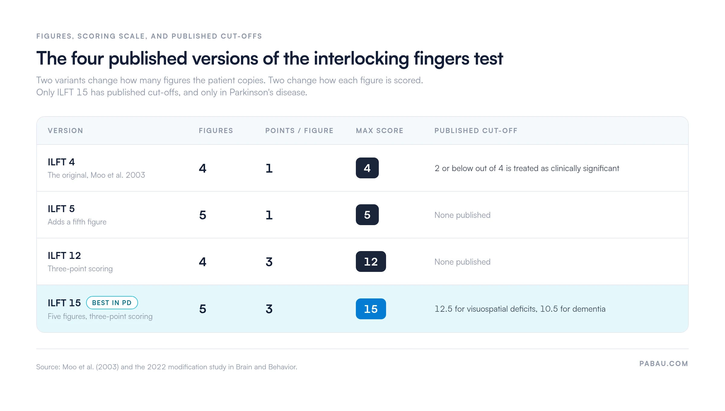
ILFT 15 performed best in the study’s Parkinson’s disease cohort. Its cut-off was 12.5 for visuospatial deficits and 10.5 for dementia. A modified version is worth the extra minute where documentation has to be systematic, such as in research or across a multi-clinician team.
One point is worth stating plainly, because it causes regular confusion. No published version of this test uses three figures. A three-posture bedside task described as the interlocking fingers test is almost always a mix-up with Luria’s fist-edge-palm sequence. That one assesses motor sequencing rather than constructional praxis.
The original version awards one point for each of the four figures the patient imitates correctly, giving a total between 0 and 4. Only the interlocking finger component counts, so the non-interlocking fingers and the posture of the arms are ignored. The modified variants score each figure out of three instead.
In the original four-figure version, a total of 2 or less out of 4 is treated as clinically significant. The modified versions use cut-offs that vary by population, such as 10.5 out of 15 for dementia in Parkinson’s disease.
Apply the threshold validated for your patient group. Recording the error type, not just the total, gives you far more to work with when you plan follow-up.
Interpreting a result means pairing the score with the patient’s broader clinical picture. A failed result is a signal to investigate further, not a diagnosis. The Oxford Academic review of IFT clinical utility reports sensitivity of roughly 39% to 79%, depending on the cut-off and the population. It reaches 79% at a cut-off below 4 out of 4, and falls to 39-42% at a stricter cut-off below 3 out of 4.
The table below maps result patterns to likely clinical correlates, based on published research.
The conditions most often associated with failure are Alzheimer’s disease and other dementias, stroke involving the parietal cortex, and Parkinson’s disease with cognitive involvement. In each case the result argues for further assessment rather than confirming a specific diagnosis.
Document the error type alongside the score out of 4. A mirror reversal on figure 1 carries different clinical weight than random finger placement on figure 4. Being specific in your notes shapes the referral pathway and hands the receiving specialist something usable.
The interlocking fingers test has specific research validation in Parkinson’s disease, which makes it a useful addition to routine PD cognitive monitoring. PD affects motor control and visuospatial processing through dopaminergic disruption in the basal ganglia. Parietal cortex involvement adds to that as the disease progresses.
Two practical considerations apply when you administer it to PD patients. First, tremor and rigidity affect fine motor execution independently of cognition. A patient with advanced motor PD may fail because of motor impairment rather than parietal dysfunction, so note the mechanism of failure explicitly.
Second, the 2022 modification was validated in a Parkinson’s disease cohort. ILFT 15 is therefore the version to reach for when PD is the primary clinical concern. Rehabilitation teams often fold it into regular review cycles, using a standardized template to track change across visits rather than reading one score in isolation.
The interlocking fingers test does not replace the Montreal Cognitive Assessment (MoCA) or the Mini-Mental State Examination (MMSE). It covers a narrower domain, much faster. Knowing where the differences sit helps you choose between a first-pass triage and a full battery.
Serial cognitive assessment works best when every tool has a template in the same system. Practice management software like Pabau can hold an IFT, MoCA, or MMSE form inside the patient record. Results then sit next to appointment history and clinical notes. If you want the MMSE on paper first, our MMSE screening template is free to download.

No bedside screen is free of constraints. Know where this one stops before you use it as a triage tool.
The American Academy of Neurology recommends reading any cognitive screening result alongside clinical history, collateral information from caregivers, and functional assessment. The interlocking fingers test fits that framework as an efficient first filter rather than a conclusive evaluation.
The interlocking fingers test produces a data point that has to land somewhere useful. In many practices it lands on a handwritten note that never reaches the patient’s longitudinal record. Repeat assessments then lose their comparative value, and the follow-up never gets booked.
Pabau handles that in four ways relevant to teams running regular cognitive screens:
For practices running serial cognitive monitoring, structured digital forms and one integrated record cut the admin work of tracking outcomes over time. Pabau is rated 4.7/5 on Capterra from 600+ verified reviews, with clinical documentation and multi-practitioner functionality cited most often.

Pabau lets neurology and geriatrics teams record structured assessment results directly in the patient record. Follow-up assessments get scheduled automatically, and standardized screening templates keep every clinician consistent.

The interlocking fingers test earns its place at the bedside. It returns something usable in under two minutes, with nothing in your hands but your hands. It will not replace the MoCA or the MMSE, and it cannot stand alone as a diagnosis. Use it to decide who needs the longer assessment, and record the error type alongside the score.
The trade-off worth remembering is the sensitivity floor. At a strict cut-off the test misses a large share of parietal dysfunction, so a clean 4 out of 4 settles nothing on its own. Pair it with the clinical history and a collateral account before you reassure anyone.
A second test is only worth doing if the first one is still findable. Keeping the score, the error type, and the follow-up in one record is what makes that true. Book a demo to see how Pabau keeps screening results and follow-up tasks on the same patient timeline.

Building out the wider bedside examination? Mental state examination covers the structured observation that sits around a screen like this one, domain by domain.
Explaining a dementia trajectory to a family? 7 stages of dementia chart lays out the progression in a format you can hand to a caregiver during the consultation.
Looking for follow-up activities after a positive screen? Dementia worksheets template provides structured cognitive exercises you can use between review appointments.
The interlocking fingers test is a rapid bedside screen for parietal lobe dysfunction. The patient imitates four interlocking finger figures demonstrated by the examiner. Failure to reproduce the figures may indicate constructional apraxia, which is commonly associated with Alzheimer’s disease and other dementias. It is a triage tool rather than a diagnostic instrument, and a failed result should trigger a full cognitive assessment such as the MoCA.
Four. The original test, described by Moo et al. in 2003, uses a standardized set of four interlocking finger figures. Each figure scores one point, for a total out of 4. The 2022 modified versions use either the same four figures with a three-point scale (ILFT 12) or five figures (ILFT 5 and ILFT 15). No published version of the test uses three figures.
Seat the patient facing you and instruct them to watch and copy each hand figure. Demonstrate the four interlocking finger figures one at a time, holding each for about three seconds. Let the patient imitate each one without touching your hands. Award one point per figure copied correctly, for a total out of 4, and note the specific error type for any figure the patient fails.
Failing two or more of the four figures, which is a score of 2 or less out of 4, suggests clinically significant parietal lobe dysfunction. Common causes include Alzheimer’s disease, stroke with parietal involvement, and Parkinson’s disease with cognitive impairment. Hand pathology such as arthritis, contracture, or weakness can also produce failure without reflecting brain dysfunction, so record physical status alongside the result.
Sensitivity for detecting parietal dysfunction runs from roughly 39% to 79%. The figure depends on the cut-off used and the population tested, according to the Oxford Academic clinical utility review. It reaches 79% at a cut-off below 4 out of 4, and drops to 39-42% at a stricter cut-off below 3 out of 4. The test should therefore be used as a first-pass triage screen rather than a definitive rule-out tool.
Yes, the interlocking fingers test is used in Parkinson’s disease cognitive monitoring, and the 2022 modified versions were validated in PD populations. ILFT 15, which uses five figures and a three-point scale, performed best in that cohort. Account for motor impairment such as tremor and rigidity when you interpret the result, because motor deficits can cause failure independently of parietal dysfunction.
The interlocking fingers test takes under two minutes and requires no materials. The MoCA takes 10-15 minutes and covers six cognitive domains, and the MMSE takes 7-10 minutes. The IFT targets visuospatial and constructional praxis specifically, while the MoCA and MMSE assess memory, language, orientation, and attention more broadly. Use the IFT for rapid triage and the MoCA or MMSE for a full cognitive profile.
The interlocking fingers test cannot diagnose any specific condition. Reported sensitivity of 39% to 79% means it misses a substantial share of parietal dysfunction at stricter cut-offs. It also produces false positives in patients with hand disabilities. Comprehension barriers from language differences or low education level can affect results too. It does not assess memory registration, executive function, language, or orientation, so a normal result does not rule out impairment in other domains.