



GLP-1 diet plan
A GLP-1 diet plan is a structured eating framework for patients taking semagluti...

2
Last Updated: Oct 13, 2026
A depth perception test measures stereoacuity, the smallest depth difference your two eyes can detect together. Results come back in arc seconds, where a lower number is better, and 40 arc seconds or better counts as normal.
Most people never see that number until a school screening, a driving medical, or a military entrance exam flags it. At that point the useful questions are what the score means and whether it can improve.
The sections below cover each test, how to read a result, what pushes a score down, and which treatments work.

A depth perception test measures stereoacuity, your ability to judge distance using both eyes together.
Scores are reported in arc seconds, and 40 arc seconds or better is the normal adult benchmark.
The Randot stereotest is the clinical default, while the Lang and fly tests suit children who cannot wear glasses.
Amblyopia and strabismus cause most reduced scores, and both respond best to treatment before age seven.
Online and at-home checks screen for an obvious problem but cannot produce an arc-second score.
Depth perception is your visual system’s ability to see in three dimensions and judge how far away objects are. No single organ produces it. Your brain combines a slightly different image from each eye, then checks that result against cues in the scene itself.
Reach for a coffee cup and you can watch the system work. Each eye sends a marginally different view of the handle. Your brain measures that difference, decides how far away the handle sits, and stops your hand in the right place.
The strongest binocular cue is retinal disparity. Each eye views a scene from a slightly different angle, and your brain reads that offset as distance. Take one eye out of the equation and the offset disappears, which changes how you judge close distances.
Your visual system draws on two kinds of cue. Binocular cues need both eyes and carry the fine depth judgments at close range. Monocular cues work with one eye and take over at longer distances, where retinal disparity becomes too small to read.
Binocular cues operate only when both eyes are open and aimed at the same target.
Monocular cues let you estimate depth with one eye. They carry most of the load at longer distances, where retinal disparity falls below the level your brain can use. That is why losing an eye reduces depth perception without erasing it.
A depth perception test is a clinical measure of stereoacuity, the finest retinal disparity you can detect and use to judge depth. The result is a single figure in arc seconds. A lower figure means finer depth discrimination, so lower is better.
Optometrists, ophthalmologists and orthoptists all run these tests. Stereopsis testing usually sits inside a broader eye physical examination, alongside visual acuity, eye movement and pupil checks. It also appears on its own in occupational health screening, pediatric vision assessments and military entrance processing.
In practice, the admin around the test decides whether it runs smoothly. Before the patient sits down, staff need four things on file.
Miss one of those and a five-minute test becomes a repeat visit.

Which test a clinician picks depends on your age, the referral reason, and whether you can follow instructions in a chair. Most appointments use two of them. One measures stereoacuity, the other looks for the misalignment behind a poor score.
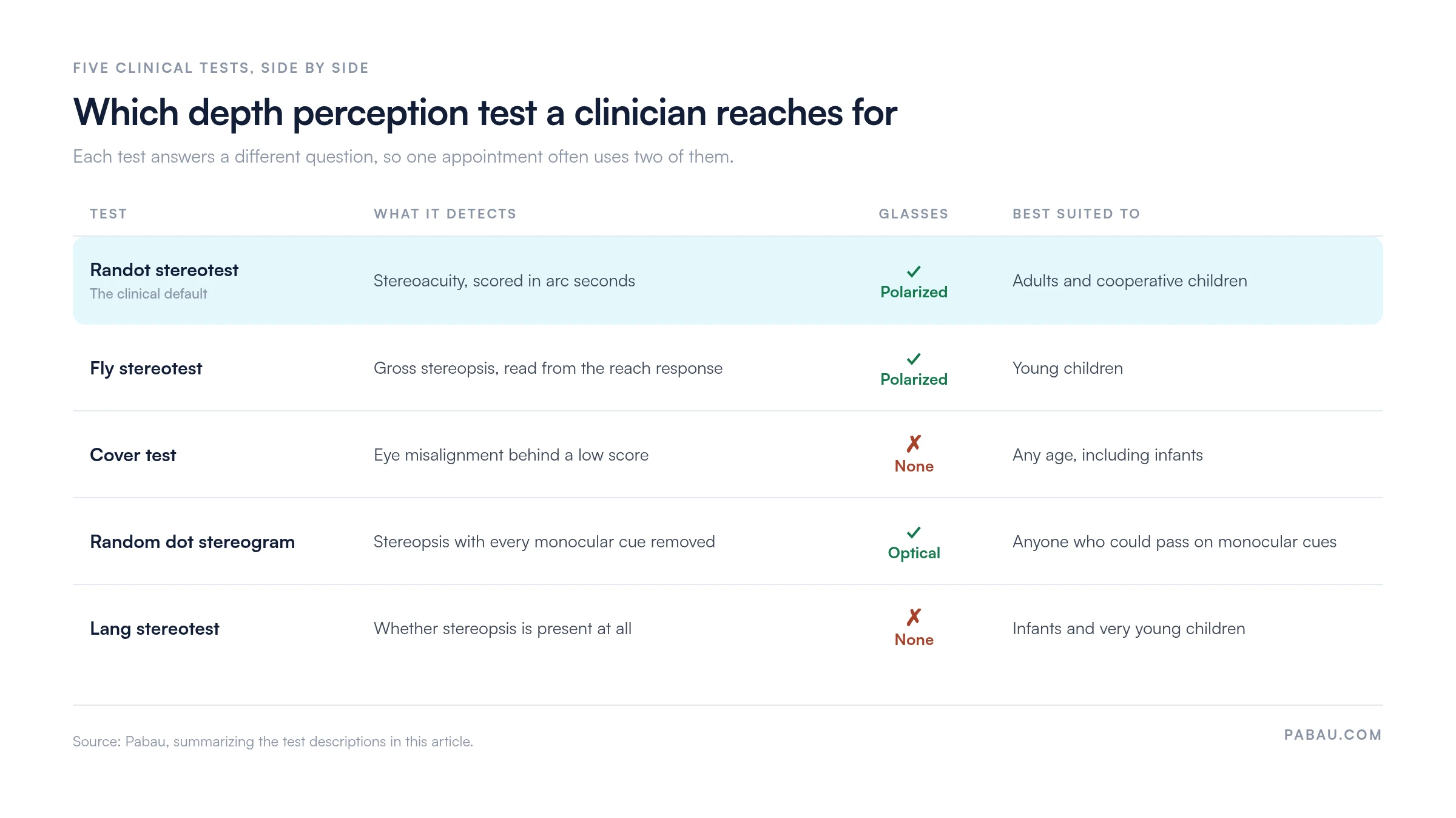
The Randot stereotest is the most common depth perception test in clinical use. You wear polarized glasses and look through a booklet of images, where certain shapes float above the page when both eyes work together.
The examiner records the smallest disparity you can still identify correctly. That figure is your stereoacuity, and 40 arc seconds or better is normal for an adult.
The fly stereotest turns the measurement into a game, which is why it works with young children. The child wears polarized glasses, looks at a large picture of a fly, and is asked to grab its wings.
A child with normal stereopsis reaches above the page, because the wings appear to hover. A flat reach onto the paper points to reduced or absent stereopsis.
The cover test targets alignment. It finds the strabismus that most often explains a poor stereo score, which a stereoacuity figure on its own cannot localize. The examiner covers one eye while you fixate on a target, then watches the uncovered eye. Movement to take up fixation confirms a misalignment, and the sequence is repeated on the other side.
Many practices pair it with an eye movement test in the same appointment, since tracking problems and alignment problems often travel together.
A random dot stereogram shows two panels of apparently random dots. Viewed through the right optical system, a shape emerges, but only for someone with working stereopsis.
Every monocular cue has been stripped out of the image, so a patient cannot guess from shading, size or perspective. That makes it the strongest choice when a pass has to be objective, in children and adults alike.
The Lang stereotest needs no glasses, which makes it practical for infants. A lenticular screen separates the image reaching each eye, so the card does the job the glasses would normally do. The child simply looks at it. The examiner watches for interest, pointing, or a reach toward a shape, any of which indicates stereopsis is present.
A home check can catch an obvious problem, and that is where its usefulness ends. It produces no arc-second score, so it cannot stand in for a clinical test.
The pencil version takes five steps:
Online depth perception tests using red-green anaglyph images give a rougher indication still. Screen calibration, room lighting and individual color sensitivity all shift the result. Treat either version as a reason to book an eye exam rather than as a measurement.
Stereoacuity is reported in arc seconds of angular disparity, and lower is better. A result of 40 arc seconds means you can detect the depth difference produced by 40 arc seconds of horizontal retinal offset. That is the benchmark for normal adult binocular depth perception.
The table below sets out the interpretation ranges in common clinical use, together with the step each one usually triggers.
A worked example makes the table easier to use. Say a patient scores 140 arc seconds on the Randot stereotest, and the cover test shows a small inward drift in the left eye.
That combination reads as a moderate deficit with a probable cause. The next step is a full binocular vision assessment rather than a watch-and-wait review.
Scores only stay comparable if every visit records them the same way. A stereopsis test template gives you fixed fields for the test name, the version, the arc-second result and the viewing distance.
Document stereoacuity scores in arc seconds with the test name and version used (e.g. Randot Stereotest, TNO random dot test). Vague entries like ‘stereopsis normal’ are not reproducible across appointments or practitioners. Precise records allow a meaningful comparison when a patient’s binocular vision changes over time, or before and after treatment.
Reduced stereoacuity almost always comes from a condition that disrupts binocular fusion, the brain’s ability to merge the two eyes’ images into one. The causes below run roughly in the order clinicians meet them:
Sudden change is the signal worth acting on. Depth perception can drop after a head injury or a stroke. A neurological eye exam checks the cranial nerves and eye movements a standard sight test leaves alone.
Routine clinical testing and occupational testing answer different questions. Three settings apply their own standards, and each can flag a score that a regular eye exam would pass without comment.
The US Military Entrance Processing Station includes depth perception in the vision screening battery for every enlistee. The test is usually an anaglyphic stereopsis test presented at a fixed distance.
Pass thresholds and waivers vary by branch and by military occupational specialty. Aviation roles, including Air Force pilot applicants, work to tighter stereoacuity standards than ground-based roles.
Monocular applicants generally fall short of the standards for combat arms roles. Some support and administrative positions allow a waiver instead.
Verify current MEPS and branch vision standards with the Department of Defense before making any application decision.
Commercial licensing in the United States sets no stereoacuity threshold in arc seconds. FMCSA regulations require at least 70 degrees of field in the horizontal meridian in each eye.
Drivers must also recognize standard red, green and amber traffic signals. Depth perception gets assessed informally during the physical.
Commercial drivers are checked against that standard during the DOT eye test, and the examiner records the outcome on the medical examination report.
Some employers go further still. Crane operators, pilots and certain surgical specialties work to internal standards that exceed routine clinical norms.
Treatment follows the cause, so the diagnosis matters more than the score itself. Catching a problem before age seven improves the odds considerably, because the visual system is still neurologically plastic at that stage.
Treatment runs over months, across several appointments and often more than one clinician. The record has to hold the original arc-second score, the referral letter, the therapy or surgical notes and every re-test, in order. Paper charts and separate letter folders are where that sequence usually comes apart.
Book an appointment with an optometrist or ophthalmologist if any of the following applies to you or your child.
Home and online checks are prompts, not diagnoses. Only a clinical assessment identifies the cause and points to a treatment. In childhood, the sooner that happens, the more the treatment can do.
Vision practices rarely lose a depth perception result. They lose the thread after it. The arc-second score sits in a free-text field, and the referral letter lives in a separate folder. The recall for a re-test then depends on someone remembering to set one.
Practice management software like Pabau keeps that sequence intact. Digital intake forms capture the prescription and the history before the patient arrives. Structured clinical forms record the test name, version and arc-second result as fields you can compare between visits.
Automated recall covers the part practices most often drop. A patient flagged for a re-test in six months gets the reminder automatically. Nobody tracks the date by hand, and more screening flags turn into a booked follow-up.
The outcome is a record a second clinician can read in order, from the first score through treatment to the most recent re-test. That matters most in pediatric cases, where the treatment window is short and the history decides what happens next.

Pabau helps optometry practices capture test results in structured forms and link them to referrals and treatment notes. Automated recall brings patients back for a re-test without anyone tracking the date by hand.

A depth perception score is a number with a cause behind it. The arc-second figure tells you how well the two eyes work together, and the cover test tells you why. Neither one is much use on its own.
If your result came back above 40 arc seconds, the useful next move is an appointment rather than another home check. For a child under seven, that appointment is genuinely time-sensitive, because treatment gets harder as the visual system loses plasticity.
For the practices running these tests, the admin around the result decides whether a flag turns into treatment. Book a demo to see how Pabau keeps the score, the referral and the recall on one patient record.

Want the form that goes with this test? Stereopsis test template gives you fields for the test version, the arc-second result and the viewing distance.
Trying to tell refractive blur from pathology? Pinhole test explains how a pinhole occluder separates the two in about a minute.
Checking how the eyes focus up close? Accommodation eye test covers the near-focusing checks that sit alongside a binocular vision work-up.
Recording pupil reactions properly? PERRLA eye exam form sets out the fields for pupil size, reactivity and accommodation.
No. Visual acuity and stereoacuity measure different things. You can read the bottom line of a chart with each eye and still score poorly on a stereo test. Stereopsis depends on the two eyes working together, not on the sharpness of either one.
Yes. Stereoacuity tends to decline gradually from middle age onward. Cataract, age-related macular changes and reduced contrast sensitivity all degrade the image each eye sends, which weakens fusion. A drop that happens suddenly is a different matter and warrants an examination.
They should improve them. Stereo tests are run with your habitual correction in place, because a blurred image in one eye lowers the score. Wear your usual glasses or lenses to the appointment. Mention it if the two prescriptions differ sharply, since that can affect fusion.
Only a few minutes. A Randot or fly stereotest is quick once the patient is seated, and a cover test adds a minute or two. Most of the appointment goes on history, correction checks and explaining the result.






