HIPAA compliant texting: What you can send patients & how to communicate securely
HIPAA compliant texting is possible when a text holds no protected health inform...

Last Updated: Oct 16, 2026

An ultrasound test uses high-frequency sound waves rather than radiation, so it is safe to repeat and safe in pregnancy.
Only abdominal and pelvic scans need patient preparation, and skipping it can obscure the organs you scanned for.
Seven scan types cover most of what a practice orders, from abdominal and obstetric through to musculoskeletal and FAST.
A defensible record needs labeled images, measurements in the report, a written interpretation, and a signature within about a day.
The free template below carries fields for patient information, preparation, examination details, findings, interpretation, and signature.
A printable clinical form with fields for patient information, preparation, examination details, findings, final interpretation, and the signing clinician’s credentials. Print it for the exam room, or attach it to the patient record after every scan.
Download templateAn ultrasound test builds a live picture of soft tissue using sound waves, not radiation. That is why it is the one scan you can repeat as often as the clinical question needs. The freedom is also the trap. A scan that adds no radiation dose still costs a room, a sonographer, and an appointment slot.
And a repeat usually traces back to a step the practice controls. Preparation instructions the patient never received, say, or a report too thin to stand up later.
The sections below work through the seven scan types you’ll order most, the preparation each one needs, and what your record must contain.
An ultrasound test is a noninvasive imaging procedure that uses sound waves of roughly 2 to 18 megahertz to picture structures inside the body. A handheld transducer, or probe, sends those waves into tissue and catches what bounces back. A computer turns the returning echoes into a moving image on the monitor.
Clinically, “ultrasound” and “sonography” mean the same thing. “Sonogram” usually refers to the image or the report that comes out of it. The American Institute of Ultrasound in Medicine (AIUM) publishes the practice parameters most US practices work to.
The physics is simple enough to walk through in five steps:
Because it runs live, ultrasound shows movement a still image cannot, from blood tracking through a vessel to a valve closing mid-beat. That is also why it works well as a guide during needle procedures.
Different organ systems need different approaches, and the choice follows the question you are trying to answer. Seven types cover most of what a general practice orders.
Evaluates the liver, gallbladder, pancreas, spleen, and kidneys. Ordered for suspected gallstones, hepatic disease, abdominal pain, or abnormal liver function tests. Needs 4 to 6 hours of fasting to settle bowel gas.
Assesses the uterus, ovaries, and surrounding pelvic structures. The transvaginal approach places the probe closer to the anatomy, so it resolves smaller detail than a transabdominal scan. Both are used to work up cysts, fibroids, and other gynecologic findings.
Monitors fetal development, measures growth, and screens for anomalies. First-trimester scans at 11 to 14 weeks assess nuchal translucency. Second-trimester scans at 18 to 22 weeks cover anatomy in full, and third-trimester scans check position and placental function.
Shows the heart chambers, the valves, and wall motion. Adding Doppler measures the speed and direction of blood through each valve. Used to work up valve disease, heart failure, and congenital defects.
Assesses flow through arteries and veins, most often for deep vein thrombosis, carotid stenosis, and peripheral vascular disease. Because the findings are numeric as well as descriptive, a structured Doppler ultrasound report keeps velocities and waveform notes in a consistent place.
Evaluates muscles, tendons, ligaments, and joints. Frequently used to diagnose soft tissue injury and to guide injections into a joint space or tendon sheath.
Focused Assessment with Sonography in Trauma is a bedside protocol for detecting free fluid. It looks in the abdomen, chest, or pericardium after blunt or penetrating injury. It takes 2 to 5 minutes and needs no preparation, because the patient never moves.
Ultrasound is strongest on soft tissue and fluid, and weakest wherever bone or gas sits between the probe and the target. Within that boundary it does six jobs well:
Across those jobs, ultrasound identifies or helps assess a wide range of conditions:
Three limits are worth stating plainly. Bone reflects nearly all the sound, so the adult brain and spinal cord stay out of reach. Bowel gas scatters it, which is exactly why fasting improves an abdominal study. And depth costs resolution, so a deeper structure returns a softer image.
Ultrasound is also operator-dependent in a way that CT is not, since two sonographers can produce different images of the same anatomy. That is an argument for protocol and for documentation, and both are covered further down.
Only abdominal and pelvic scans ask the patient to prepare beforehand. Obstetric, cardiac, vascular, and musculoskeletal studies are walk-in ready, and a FAST exam happens wherever the patient already is. The grid below sets the rules against the scan types, so your front desk knows which bookings need a reminder.
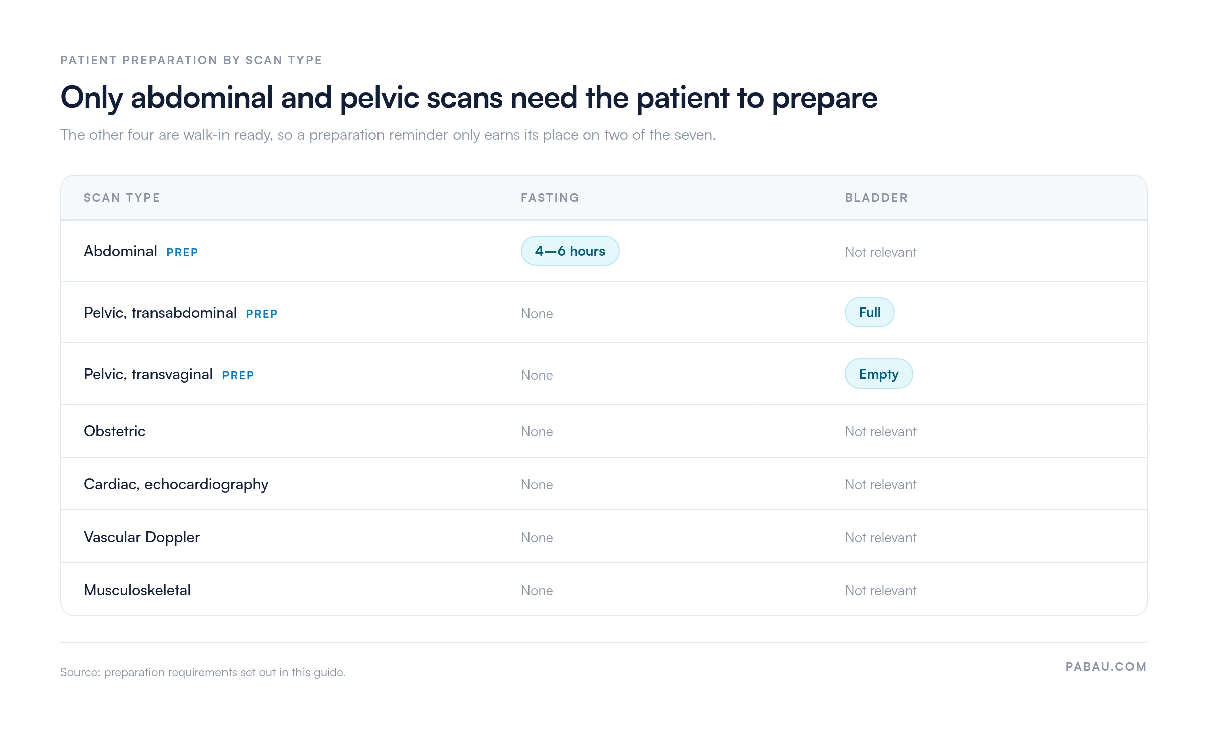
For abdominal work, patients fast for 4 to 6 hours, taking no food and no drink. Fasting settles the bowel gas that otherwise hides the pancreas and the common bile duct. Transabdominal pelvic scans want the opposite of an empty patient: a full bladder acts as an acoustic window onto the uterus and ovaries.
Ask those patients to drink water one to two hours ahead and hold it. Transvaginal scans reverse that again, because an empty bladder lets the probe sit closer to the anatomy. Most medications continue as normal, though a diabetic patient facing a fasting scan is worth flagging to the ordering clinician.
Consent sits on that list because it is the step people backfill. A general medical consent form covers a routine transabdominal scan. Transvaginal and transrectal work needs its own wording, since the patient is agreeing to an internal examination.
Most appointments run 20 to 45 minutes, depending on the scan type and how much measuring the protocol calls for. The sequence rarely changes:
Two habits separate a smooth study from a repeated one. Warm the gel, because a cold probe makes patients tense the abdominal wall and tense muscle scans poorly. And label each stored image as you capture it, since reconstructing orientation after the patient leaves is guesswork.
An ultrasound report carries three things: descriptive findings, measurements, and a clinical impression drawn from both. Results usually come back within 24 hours, reviewed by a radiologist or another qualified clinician.
In obstetric work, a handful of parameters do the dating and growth assessment:
Those figures get compared against standardized growth charts to catch growth restriction, dating errors, or anomalies. Where an early measurement and the menstrual dates disagree, first-trimester crown-rump length is the more reliable of the two.
Does a normal report mean nothing is wrong? No. It means the structures examined looked normal on that day, through that approach. A negative FAST exam does not exclude injury, and an early pelvic scan can miss a very early pregnancy. Read the impression alongside the clinical picture, not instead of it.
Yes. Ultrasound uses sound rather than ionizing radiation, and no harmful tissue effects have been documented at diagnostic power levels. MedlinePlus and the American College of Radiology both affirm its safety across all populations, pregnancy included.
Equipment is regulated under the ALARA principle, short for As Low As Reasonably Achievable. Operators use the least power needed for a diagnostic image, and the FDA sets the equipment standards that make that possible.
The residual risks are practical rather than biological. Scan quality varies with the operator, and a false-positive finding sends a patient into follow-up imaging they never needed. Keepsake fetal videos sit outside diagnostic use, and the FDA advises against them for that reason.
Five steps, and four of them are administrative. Knowing where each one can stall is usually more useful than knowing more about the physics.
Step one is where most of the friction sits. A standing medical referral form captures the indication, the requested scan, and the relevant history. That saves a phone call later, because the sonographer already knows which question to answer.
A defensible ultrasound record is a permanent one, holding labeled images, a written interpretation, and a signature. The AIUM practice parameter for documentation sets out what that means in practice, and accreditation surveyors work from the same list.
Store images of every relevant area, normal findings included, in a retrievable electronic format. Each stored image carries five labels: patient identification, facility identification, exam date, image orientation, and the anatomic structure shown. Miss the last two and a later comparison becomes guesswork.
The final report is generated, signed, and dated by the interpreting clinician, in line with state and federal requirements. Aim to have it available within 24 hours of the study, or by the next business day for non-urgent work.
None of that is hard to fix on a single scan. It gets hard across a full week’s list and several sonographers. That is where clinical documentation software earns its keep, holding every record to the same shape.
The usual setup splits this across three places. Consent lives on paper in a folder. Preparation instructions get read out over the phone. Images sit in the ultrasound machine’s own archive until someone remembers to export them. Each handoff is a chance for a record to go missing.
Practice management software like Pabau pulls those three into one patient record. You build the ultrasound consent and the exam form once in patient intake software. Attach them to the appointment type, and the patient signs on their phone before arriving. Preparation instructions travel with the booking confirmation.
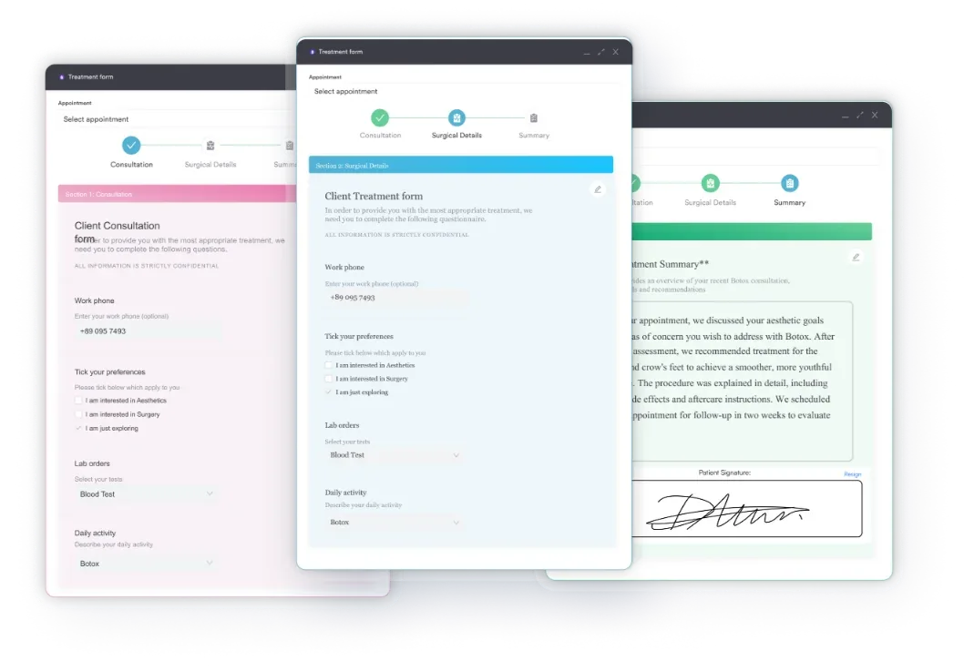
Findings, measurements, and images then file against the same record as the appointment and the invoice. The whole study sits in one place at audit time. Your sonographer stops chasing paperwork, and the record is complete before the patient has left the building.

Pabau’s digital forms, automated preparation reminders, and secure patient records keep ultrasound consent, instructions, and findings on one file. No record waits on an export.

Ultrasound earns its place because it is cheap in risk and rich in information. The limits are practical rather than clinical. Bone and gas block it, depth softens it, and the operator matters more than with any other modality.
So the leverage sits in the parts you control. Send the right preparation instruction to the right booking, label images as you capture them, and sign the report the same day. Do that consistently and repeat scans become rare, because the first study was usable.
If your consent forms, preparation reminders, and scan records currently live in three separate places, that is the workflow worth fixing first. Book a demo to see how Pabau keeps ultrasound consent, instructions, and findings on one patient record.

Running vascular studies as well? Doppler ultrasound report template gives you a structured place to record velocities, waveforms, and the comparison against the contralateral side.
Standardizing records across several sonographers? Clinical documentation software explains how a shared template library keeps findings, measurements, and signatures consistent from one study to the next.
Need written consent before an internal scan? Medical consent form for adults covers the wording, the signature fields, and the capacity checks a routine imaging consent needs.
Sharing images outside the practice? HIPAA authorization form sets out what a patient signs before their imaging leaves your system for another provider or an insurer.
Writing up the follow-up visit? Progress note template keeps the interval history, the imaging result, and the plan in one structure your next reviewer can read quickly.
Not usually. You feel the gel and steady pressure from the probe, which can be uncomfortable over a tender area or a full bladder. Transvaginal and transrectal scans feel much like an internal examination.
A sonographer usually performs the scan and captures the images. A radiologist or another qualified clinician then interprets them and signs the report. In point-of-care settings, one clinician does both at the bedside.
2D produces the flat grayscale image used for most diagnostic work. 3D assembles several 2D planes into a surface view, and 4D adds motion to that surface. Diagnosis still rests on the 2D images.
Point-of-care ultrasound, or POCUS, is a focused bedside scan run by the treating clinician to answer one question. A FAST exam is the best-known example. It supplements a formal study rather than replacing it.
Retention comes from state law and your accrediting body, not from one national rule. Many practices align imaging retention with their wider medical records policy, and pediatric records generally have to be kept longer.