Best chiropractic billing software in 2026: Top 5 compared
Chiropractic billing software automates insurance claims, CPT coding, eligibilit...

Last Updated: Oct 8, 2026
A spinal nerve diagram maps the 31 pairs of spinal nerves that leave the spinal cord. They divide into eight cervical, 12 thoracic, five lumbar, five sacral and one coccygeal pair.
Each pair supplies a predictable strip of skin and group of muscles. That pattern lets you trace a symptom back to its root. Numbness on the top of the foot usually points to L5, while a numb little finger points to C8.
This guide walks through each region, the test point for every key root, and the red flags to act on. It also includes a free one-page form for recording your findings.
A blank one-page form with name and date fields, plus Findings and Additional notes boxes for recording the nerve levels you tested. It carries no printed artwork, so pair it with the key sensory points chart in this guide.
Download template
The 31 spinal nerve pairs split into five regions: eight cervical, 12 thoracic, five lumbar, five sacral and one coccygeal.
Each root supplies a predictable skin area, or dermatome, so one test point per level is enough for a quick sensory screen.
L5 and S1 are among the most commonly compressed roots, and both can cause sciatica down one leg.
Saddle numbness or new bladder or bowel changes with leg symptoms need a same-day referral.
The free download is a blank findings form for recording the levels you test, not a labeled illustration.
A spinal nerve diagram shows how the peripheral nervous system branches off the spinal cord, level by level. In adults, the cord itself stops around the L1–L2 vertebrae.
The nerves keep going and leave the spine through small openings called intervertebral foramina. Each one carries motor signals out to muscles and sensory signals back about pain, temperature and touch.
A good diagram labels five layers of information:
Start with the simplest layer, which is how the 31 pairs divide by region.
The thoracic spine holds the most pairs, while the coccyx has just one. Each region also has its own clinical pattern.
There are eight cervical pairs but only seven cervical vertebrae. The extra pair comes from how they exit. C1 to C7 leave above their matching vertebra, while C8 leaves below C7. From T1 down, every nerve exits below its own vertebra.
C1 to C4 form the cervical plexus, which supplies the neck and shoulder. C5 to T1 form the brachial plexus, which supplies the whole arm.
Compression here usually shows up as neck stiffness, shoulder pain and symptoms running down the arm. Check this region first when a patient has tingling or weakness in the hand after whiplash.
The thoracic nerves supply the chest wall, rib cage and upper back muscles. Most of them don’t join a plexus. Instead, they run between the ribs as intercostal nerves. These nerves are vulnerable to rib dysfunction, herniated thoracic discs, shingles, and postural dysfunction.
Their band-like layout makes thoracic dermatomes easy to read. A shingles rash, for example, usually follows one band around one side of the trunk. Mapping the band also helps you separate local musculoskeletal pain from pain referred from an organ.
The lumbar nerves carry innervation to the lower back, abdomen, and legs. They merge into the lumbosacral plexus, which gives rise to the major nerves of the leg. These include the femoral nerve (front of the thigh), the obturator nerve (inner thigh) and the sciatic nerve, the largest nerve in the body.
A herniated disc or spinal stenosis can compress these roots and cause sciatica. Patients describe pain, numbness or weakness running down one leg.
Upper lumbar roots behave differently and refer pain to the front of the thigh. When you suspect one of those higher levels, the femoral nerve tension test stresses the L2 to L4 roots.
The sacral nerves (S1–S5) continue the lumbosacral plexus. They supply the buttocks, back of the thigh, calf, foot and pelvic floor. L5 and S1 are among the most common sites of lumbosacral nerve root compression, causing sciatica and functional loss.
When a disc herniation is the suspect, the crossed straight leg raise test adds specificity to your exam. The single coccygeal pair (Co1) rarely matters in assessment. S2 to S4 matter a great deal, though, because they carry bladder, bowel and saddle-area sensation.
A dermatome is the area of skin supplied by a single spinal nerve root. If a patient reports numbness along the outer leg and the top of the foot, L5 becomes your first suspect. Targeted testing then confirms it or rules it out.
Dermatome boundaries vary slightly between sources. The classic Keegan and Garrett maps differ from Lee’s more recent mapping, and neighboring dermatomes overlap. Pick one reference and stick with it, so findings stay comparable between visits.
For a fast screen, use the key sensory points from the International Standards for Neurological Classification of Spinal Cord Injury (ISNCSCI). The chart below gives one test spot for each key root.
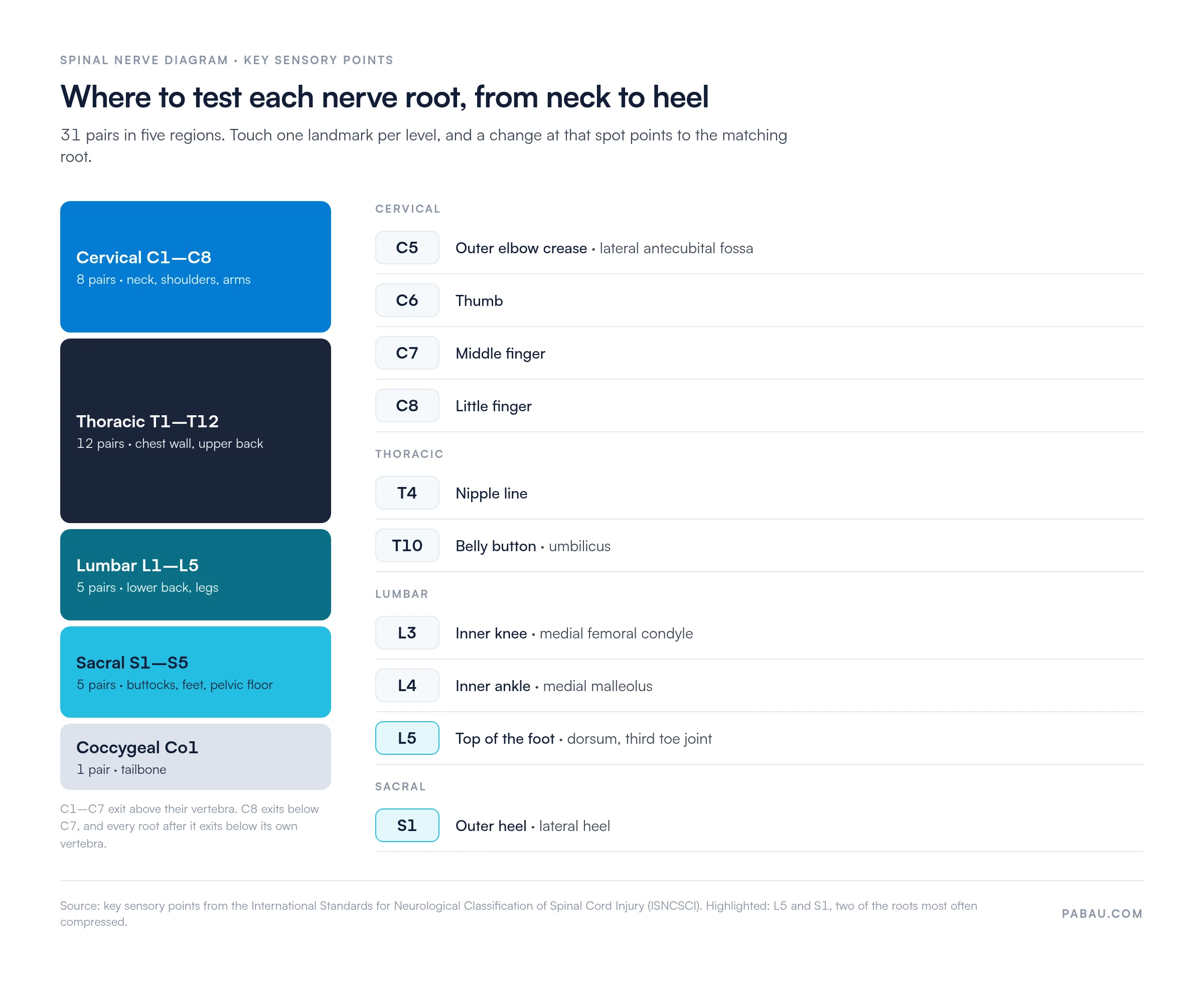
The findings form on this page has no printed map. Note the dermatome reference you used in the Additional notes box, then record the level and side under Findings.
Once you know the landmarks, the diagram fits into a five-step routine.
Reflexes are often the tiebreaker between two neighboring levels. A structured deep tendon reflex exam checks the biceps (C5–C6), triceps (C7), patellar (L4) and Achilles (S1) reflexes the same way at every visit.
Run through this quick checklist before each assessment:
Most level errors come from a handful of habits. Watch for these five:
Inside the spine, a cross-section of the cord shows two zones:
Damage to the cord interrupts those tracts, so sensation and movement change below the injured level. Root compression behaves differently, because it only affects the strip that one root supplies. After a cord injury, the ASIA Impairment Scale grades how complete the loss is, from A to E.
A spinal nerve diagram supports your clinical judgment, but it can’t make a diagnosis on its own. Refer the same day if you find any of these:
The first three can point to cauda equina syndrome, which is a surgical emergency. When in doubt, refer first, then record what you found and when.
Paper findings forms do the job, but they get scanned late, filed in the wrong place, or never compared with the last visit. A digital record keeps every level you test next to the patient’s history and treatment plan.
Pabau, our all-in-one practice management system, lets physical therapists record assessment findings, treatment notes, and digital forms in each patient record. In Pabau’s physical therapy EMR, you can rebuild this findings form as a digital form, so each visit’s results sit on one timeline.
Pabau Scribe, our AI scribe, can help practitioners record detailed nerve-distribution assessments during evaluation. That keeps your hands on the patient instead of the keyboard between tests.
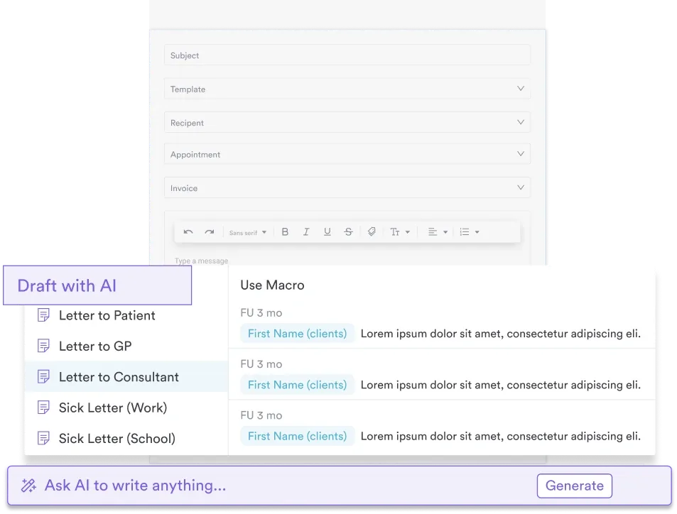

Pabau stores assessment findings, treatment notes and digital forms in each patient record, so every nerve-level result is ready at the next visit. Pabau Scribe drafts the note while you examine.

A spinal nerve diagram earns its place when it changes what you write down. Name the root, the side and the tests that confirmed it. Your next visit then starts from a baseline rather than a guess.
Keep one dermatome reference across the practice, and treat sensory findings as a lead until muscles and reflexes back them up. Suspected cauda equina always gets a same-day referral.
Download the findings form, keep the landmark chart next to your treatment table, and record every level the same way. Book a demo to see how Pabau keeps nerve-level findings, notes and referral letters in one patient record.

Mapping where the pain sits? Back pain location charts help patients mark their symptoms before you test levels.
Need a function score alongside the levels? Oswestry Disability Index template tracks how low back pain limits daily activity.
Testing the hand in more depth? Hand nerve tests covers the median, ulnar and radial nerve exams.
Screening above the spinal cord too? Cranial nerve examination checklist walks through all 12 cranial nerves.
Choosing software for your practice? Best physical therapy practice management software compares the leading options.
It shows the 31 spinal nerve pairs by level, the plexuses they form, the skin each root supplies, and often a cross-section of the cord. The free download on this page is a blank findings form, so use it alongside the chart above.
A dermatome is the skin area one nerve root supplies. Its muscle counterpart, the myotome, is the group of muscles that root controls. Testing both, plus reflexes, gives you a more reliable level than sensation alone.
In most adults, the cord tapers into the conus medullaris around the L1–L2 vertebrae. Below that, the lumbar and sacral roots continue down the canal as a bundle called the cauda equina.
The S2 to S4 roots carry the nerve supply for bladder, bowel and sexual function, plus saddle-area sensation. That’s why saddle numbness with new bladder changes needs urgent referral.
Mostly, yes. Pinched nerve is the everyday term, and radiculopathy is the clinical one. Both describe a compressed or irritated nerve root causing pain, numbness or weakness along its path.
S1 is the usual cause when pain runs down the back of the thigh and calf into the outer foot. L5 tends to cause pain along the outer leg and the top of the foot.