Healthcare risk management: A practical guide for private practices
Healthcare risk management is the process of finding, assessing, and controlling...

Last Updated: Oct 5, 2026
A pediatric rash chart is a clinical reference that helps clinicians identify and triage common rashes in infants and children. It matches what you see, such as morphology, body location, and age of onset, to a short list of likely conditions.
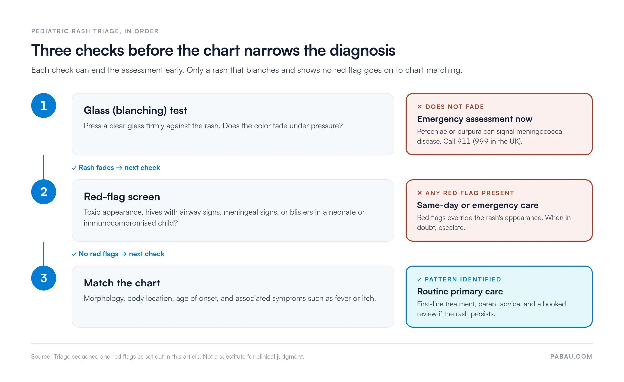
The chart works best in a fixed order. Run the glass (blanching) test first, because a rash that does not fade under pressure needs emergency assessment whatever else the chart suggests. If it blanches, screen for red flags, then narrow the diagnosis by morphology, location, and associated symptoms.
This guide covers the newborn, diaper, infectious, and inflammatory rashes that primary care teams see most often. It also sets out the red-flag features that override the chart and send a child for urgent care.
A two-page printable chart for your practice. Page one is a reference table you complete with lesion sites, sizes, notes, and the source checked, then sign off. Page two is a dated log for recording each rash’s site, size in millimeters, and who assessed it.
Download template
A pediatric rash chart sorts common childhood rashes by morphology, location, age of onset, and clinical features, so assessment follows the same steps every time.
The glass (blanching) test comes first. A non-blanching rash, such as petechiae or purpura, can signal meningococcal disease and needs emergency referral.
In diaper rash, satellite pustules and skin-fold involvement point to candida rather than irritant dermatitis, which decides between antifungal and barrier cream.
Red flags, such as a toxic appearance, hives with airway signs, or fever with a non-blanching rash, mean same-day or emergency assessment.
A pediatric rash chart is a structured reference, usually a table or decision tree. It links what a rash looks like to likely diagnoses and first-line management. Clinicians use it both to train new staff and as a quick decision aid during a consultation.
Its main job is sorting. The chart helps a busy primary care team decide whether a rash is benign and self-limiting, such as erythema toxicum, milia, or heat rash. It also flags rashes that need treatment, such as eczema, diaper rash, or impetigo.
A third group needs more than treatment. Measles, scarlet fever, and chickenpox are communicable, so the child may need isolation and the case may need reporting to public health.
Work through the same five steps at every assessment:
The blanching test is your safety gate. Press a glass or clear tumbler firmly against the rash and look through it. A rash that fades completely is blanching, as in most viral exanthems, measles, fifth disease, roseola, and erythema toxicum.
A rash that stays visible under pressure is non-blanching. Petechiae and purpura fall into this group, and they can signal meningococcal disease. Treat a non-blanching rash as a red flag that needs immediate emergency assessment.
The order matters more than any single step, and the flow below shows why. Each of the first two checks can end the assessment before chart matching begins.
The PDF has two pages. The first is a reference table your practice completes with lesion sites, sizes, notes, and the source you checked. A clinical lead then signs and dates it, so every clinician works from the same approved values.
The second page is a recording log. Each entry captures the date, time, lesion site, size in millimeters, and who recorded it. Measuring the same lesion at each visit shows whether a rash is spreading, fading, or holding steady.
Standard morphology terms let clinicians and parents describe a rash precisely, and they are the first input the chart asks for. Learn them before you start matching rashes to conditions.
Newborns often develop benign, short-lived rashes. Recognizing them as self-limiting reassures parents and avoids unnecessary tests. A structured newborn exam is the natural place to record them.
Erythema toxicum neonatorum appears as red macules and papules on the trunk and face within 24 to 48 hours of birth. It peaks at 24 to 72 hours and clears on its own within one to two weeks. No treatment is needed, and parents can be told it is a normal newborn response.
Milia (milk spots) are tiny white papules clustered on the nose, cheeks, and forehead. They come from blocked sebaceous glands and fade within two to four weeks without treatment.
Neonatal heat rash (miliaria) develops where skin rubs or overheats, such as neck folds and the diaper area, when sweat ducts are blocked. It improves with air exposure and lighter clothing.
Diaper rash is one of the most common skin complaints in infant primary care. Careful assessment of its pattern guides first-line management and cuts unnecessary antifungal prescribing.
Where a rash sits narrows the differential quickly. A rash confined to the face points to different conditions than one in the skin folds or across the whole trunk.
Spotting an infectious rash early drives three decisions: Isolation, public health notification, and treatment. Fever is often the first clue, so log it on a fever temperature chart alongside the rash findings.
Atopic dermatitis (eczema) in infants appears as an itchy, red, sometimes oozing rash on the face and the outer surfaces of the limbs. In older children it moves to the flexures, such as elbows, knees, and neck. Management combines emollients, topical steroids matched to site and severity, and avoiding triggers such as irritants and allergens.
Hives (urticaria) are itchy wheals that each last under 24 hours. They are often linked to allergic triggers or viral infections. Antihistamines such as cetirizine are first-line, and recurrent hives call for a look at allergen exposure. Hives with lip or tongue swelling or breathing difficulty need emergency care immediately.
These features call for same-day or emergency assessment, whatever the rash looks like. Abnormal vital signs strengthen the case, so check them against a pediatric vital signs chart. When in doubt, escalate.
Most rash consultations end with a follow-up decision, and paper notes make that decision easy to lose. Pabau, the practice management platform we build, keeps it attached to the child’s record. Digital intake forms collect onset date, fever, and exposures before the visit, so the clinician starts with the history in hand.
During the consultation, the clinician records morphology, distribution, and the blanching test result in Client records. Dated photos, taken with the parent’s consent, sit alongside the notes. At the review, a second photo shows whether the rash has spread or faded, without relying on memory.
Reviews are booked before the family leaves, and automated reminders bring them back on time. For a candidal diaper rash, that means a review after about seven days of antifungal treatment. With follow-up-ready GP software, family physicians and GPs can run these reviews at volume without a separate recall list.

Pabau links intake forms, rash photos, and consultation notes to one patient record, then sends review reminders automatically. Fewer children miss the follow-up that shows whether a rash is settling.

A rash chart earns its place only when it changes what happens next. Describe the morphology first, then let the red-flag criteria decide whether the child goes home or to emergency care.
The trade-off is speed against certainty. A chart narrows the options quickly, but it never replaces examining the whole child. When a presentation fits no single row, document what you see and book a review.
Practices that record rash findings in a structured way spot a worsening course sooner. Book a demo to see how Pabau keeps rash assessments, photos, and follow-up reviews in one patient record.

Assessing a sick child beyond the skin? Pediatric assessment walks through the structured evaluation that puts a rash finding in its clinical context.
Need a head-to-toe exam sequence? Pediatric physical examination sets out an age-appropriate exam order, so no skin area or system gets skipped.
Want to capture symptoms that come with the rash? Pediatric review of systems gives you a ready-made checklist for fever, feeding, and other associated findings.
Checking whether a rash comes with abnormal vitals? Pediatric vital signs chart lists normal ranges by age, so you can spot the warning signs that change triage.
Erythema toxicum neonatorum shows red macules and papules, often with small yellow-white centers, and clears within one to two weeks. Neonatal pustulosis (pustular melanosis) shows pustules without surrounding redness, which rupture and leave brown macules lasting weeks. Both are benign and need no treatment. The key distinction is the redness around the lesions and the brown marks left behind.
Perform the blanching test on any red or purple rash. Press a clear glass firmly against the rash for a few seconds and watch whether the color fades or stays. A non-blanching rash needs immediate emergency assessment. A blanching rash can usually be managed in primary care unless other red-flag features are present.
Candidal rash involves the skin folds, such as the groin, gluteal cleft, and thighs, and has satellite pustules beyond the main red area. It is often beefy red and may look shiny. Irritant rash spares the deep folds and has a sharper edge at the diaper margin. Candidal rash needs antifungal cream, while irritant rash responds to frequent changes and zinc oxide barrier cream.
Seek emergency care for non-blanching petechiae or purpura, fever with a toxic appearance, or meningeal signs such as a stiff neck and dislike of light. Widespread hives with airway swelling and a blistering rash in a newborn also need urgent assessment. When in doubt, escalate rather than reassure.
Scarlet fever needs oral antibiotics, usually penicillin or amoxicillin, to prevent rheumatic fever. Widespread or rapidly spreading impetigo may need oral antibiotics. Localized impetigo responds to topical treatment, with hydrogen peroxide 1% cream first-line and topical fusidic acid if that is unsuitable. Most viral rashes, including chickenpox, roseola, and HFMD, need only supportive care.
Record the child’s age, the date the rash started, its morphology, and its distribution. Add associated symptoms such as fever, itching, or contact exposure, the blanching test result, and your clinical assessment. Photograph the rash if the parent consents. Finally, record the triage decision, from routine follow-up to emergency referral, and the first-line management plan.