CRM for chiropractors: how to manage patients, bookings and marketing
A CRM for chiropractors is a patient relationship management system built around...

Last Updated: Oct 13, 2026

Craig’s test measures femoral anteversion by finding the angle of hip internal rotation with the patient prone.
Normal femoral anteversion in adults sits between 8 and 15 degrees. Values outside that range may point to biomechanical dysfunction.
Two examiners rarely agree on the same hip, so record who measured and compare left with right.
Read the number as a clinical estimate. It does not replace CT or MRI when you need a structural diagnosis.
Practice management software like Pabau keeps each measurement, interpretation, and follow-up plan in one structured client record.
A ready-to-use form covering patient positioning, examiner technique, and angle measurement. It also carries normal-value reference ranges and space for your clinical interpretation.
Download templateCraig’s test measures femoral anteversion, the inward twist of the femur. Also called the trochanteric prominence angle test, it needs only a treatment table and a goniometer. Adults normally measure between 8 and 15 degrees.
Physical therapists, sports medicine clinicians, and orthopedic teams use it to screen in-toeing gait, hip pain, and knee mechanics. Agreement between two examiners is poor with the standard technique, so how you record the result matters as much as the number itself.
Femoral anteversion is the forward twist of the femoral neck relative to the knee. Everyone has some. It becomes clinically interesting when it sits well outside the normal range and matches the patient’s complaint.
The test estimates that twist without imaging. You palpate the greater trochanter, rotate the hip internally, and read the angle off the lower leg with a goniometer. It costs nothing beyond two minutes of table time, which is why it stays in the standard hip screen.
Reach for it in these presentations:
For a fuller picture.
Follow the same sequence every time. Consistency is what makes repeat measurements comparable.
Technique tip: Move slowly and pause at end range so the soft tissue can relax. Abrupt movement triggers guarding and gives you a false reading. Keep your hand placement and goniometer alignment identical on the second side.
Pairing this scale with digital measurement tracking turns individual scores into longitudinal trend lines your whole team can read.
Use this table to read your measurement and flag anything that needs a closer look:
One caveat: The result is a clinical estimate, not a measurement of bone. Always read it alongside the patient’s history, movement pattern, and pain provocation tests. Order imaging when a structural diagnosis will change the plan.
Craig’s test repeats well for one clinician and travels badly between two. That single fact should shape how much weight you give any number you inherit.
Intra-examiner reliability is good. A reliability study of healthy adults reported intraclass correlation coefficients, or ICC, of 0.72 to 0.86 for the same examiner repeating the test. Your own follow-up measurements are therefore worth comparing.
Inter-examiner reliability is the weak point. Across methods, the same study found ICC values from 0.25 to 0.62. The standard goniometer technique described above scored 0.25, which counts as poor agreement.
That has a practical consequence. Treat a single absolute number with caution when a colleague took the baseline. Compare the patient’s left side with the right, flag asymmetry above 5 degrees, and note who performed the test.
Validity against imaging is contested. A 2019 validity study of healthy adults found weak agreement with CT. Anteversion measured on CT and the patient’s sex together explained only 23% of the variation in Craig’s test values.
Earlier surgical work was more favorable, reporting clinical estimates within roughly 4 degrees of the angle measured at operation. Either way, use the test to screen, and use CT or MRI when you need a structural diagnosis.
None of this makes the test useless. It is free, quick, and repeatable.
Craig’s test rarely stands alone. These assessments fill in what an anteversion angle cannot tell you:
Ankle and calf findings feed the same picture.
A battery only helps if every result lands in the same clinical record.
Record these five elements every time you run the test:
AI-assisted documentation can draft that note while you work.
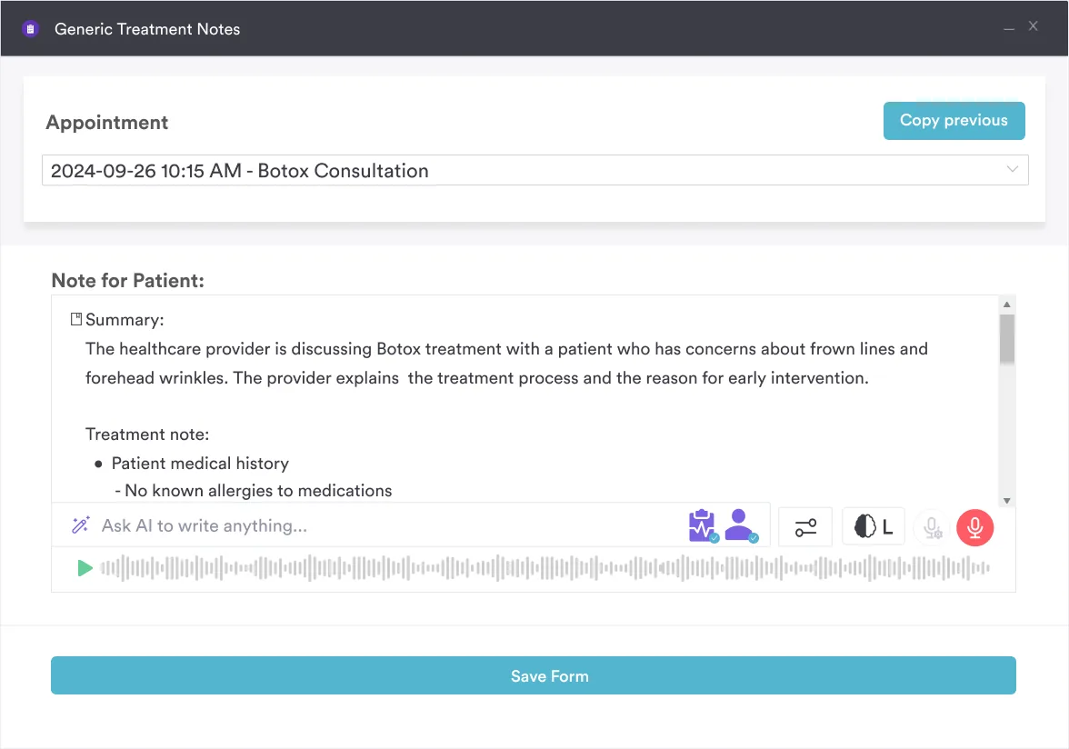
Most practices still capture Craig’s test in free text. The angle ends up buried in a paragraph, the examiner’s name never makes it in, and nobody can compare February’s reading with June’s.
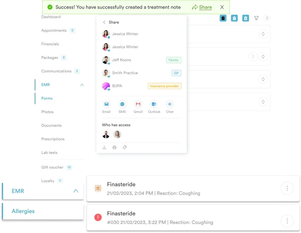
Pabau replaces that with structured assessment forms. Each measurement has its own field, normal-range flags fire automatically, and the interpretation template travels with the reading. Your team fills in the same fields in the same order, whoever is on shift.
The result is a client record you can track over time. Pull a patient’s hip history in one view, see how the numbers moved, and show them the trend rather than describing it. Pabau Scribe handles the narrative note, so structured data costs you no extra typing.

Pabau’s digital assessment forms capture each Craig’s test measurement, examiner, and interpretation in the client record. Your team records the same fields every time, so follow-up readings stay comparable.

Craig’s test earns its place on speed and cost rather than precision. Read the number as a screen and weigh it against the patient in front of you. Reach for imaging when the plan depends on the answer.
The bigger win is discipline around the record. Name the examiner, record both sides, and use the same fields every visit. Poor inter-examiner agreement then shows up as something you can spot and manage. Download the assessment form above and give your team one way of doing it. A rehab practice reporting outcomes to a payer needs consistent measures, not narrative summaries.
Book a demo to see how Pabau keeps hip measurements, examiners, and follow-up plans in one client record.

Assessing balance in a frail or elderly patient? The sitting balance scale template scores postural control across 11 functional items.
Hearing a grinding sound at the shoulder? This guide covers the snapping scapula syndrome test exam sequence, differential, and imaging rules.
Assessing hip pain in an athlete? The Stinchfield Test template guides hip flexion testing and captures pain patterns during assessment.
Assessing the shoulder next? The Kim test template walks through positioning and scoring for posteroinferior labral tears.
Treating neck pain as well? Neck pain exercises gives you a progression you can hand to patients between visits.
Normal femoral anteversion in adults is 8 to 15 degrees. Below 8 degrees suggests retroversion, and above 15 degrees indicates increased anteversion. Children naturally sit higher, around 20 to 40 degrees, remodeling toward the adult range by skeletal maturity.
Not reliably enough to replace it. The same examiner repeating the test scores an ICC of 0.72 to 0.86. Agreement between two examiners drops to 0.25 with the standard goniometer technique. A 2019 study also found only weak agreement with CT-measured anteversion. Use the test to screen, not to diagnose structure.
A reading above 15 degrees means increased inward twist of the femur. It commonly travels with in-toeing gait, anterior impingement symptoms, and altered hip loading. Correlate it with symptoms, movement analysis, and other hip tests before blaming anteversion for the presentation.
Yes, provided you use age-specific reference ranges. Children typically show 20 to 40 degrees of anteversion, remodeling toward adult values by ages 16 to 18. Where in-toeing persists beyond age 4, the test helps screen, but imaging guides any surgical decision.
Craig’s test measures the femoral anteversion angle specifically. The Ober test assesses iliotibial band tightness, and the Thomas test checks hip flexor length. Patrick’s test screens the hip joint and the sacroiliac region. Together they cover different structures in one battery.
Yes, and name the examiner while you are at it. Record both sides in degrees, your interpretation, the patient’s response, and how it links to the complaint. That is what makes the next clinician’s measurement comparable to yours.