Newborn assessment
A newborn assessment is a structured head-to-toe check of a baby’s health,...

5
Last Updated: Oct 9, 2026
A cervix length during pregnancy chart records cervical length in millimeters against gestational age. It shows whether a reading sits inside the normal range for that week, scan after scan. Cervical length usually falls as pregnancy advances, and a measurement under 25mm is commonly treated as a short cervix that needs closer review.
Most readings are taken by transvaginal ultrasound, often at the 20-week anatomy scan. A number only means something once it is plotted against a reference range and recorded the same way each time.
ACOG treats a length of 20mm or less before 24 weeks as the trigger for vaginal progesterone. SMFM goes further and recommends transvaginal screening for all singleton pregnancies at about 18 to 24 weeks.
A printable chart with one row per scan, covering gestational week, measurement in millimeters, percentile reference and operator. Threshold and follow-up columns flag any reading under 25mm and leave space to sign off the plan.
Download template
A cervical length under 25mm is the commonly used threshold for a short cervix.
ACOG treats 20mm or less before 24 weeks as the trigger for vaginal progesterone.
SMFM recommends transvaginal screening for singleton pregnancies at about 18 to 24 weeks.
Readings are plotted against singleton or twin reference data, never a mix of the two.
Record the week, the measurement, the reference set and the operator on every scan.
A cervix length during pregnancy chart is a structured record that plots measured cervical length against gestational age. It gives the team one consistent way to compare a reading with what is typical for that week.
The cervix normally holds the pregnancy closed until term. When it shortens early, the risk of preterm birth rises, which is why serial measurement and clear documentation matter in obstetric and midwifery care.
Length before labor and dilation during labor are separate measurements. Dilation belongs on a cervical dilation chart, which keeps the two records apart and easy to read.
Use the chart as a standing record for each scan. Complete the steps in the same order every time so readings stay comparable across visits.
Bladder filling and probe pressure can change a reading, so note those conditions where they apply. Repeat measurements should follow the same technique wherever possible.
Many practices file this chart with the patient’s blood pressure chart, so one antenatal visit leaves one set of records.
Cervical length normally decreases as gestation advances, and reference ranges differ between singleton and twin pregnancies. Always check the percentile source your practice uses, because published values vary between studies.
The 20-week anatomy scan is the most common point at which cervical length is measured in routine care. Guidance from ACOG and SMFM supports screening between roughly 18 and 24 weeks, in high-risk women and, per SMFM, universally in singletons.
Your reference percentile decides what counts as typical. Record the exact value, the reference used, and any clinical reason the reading needs follow-up.
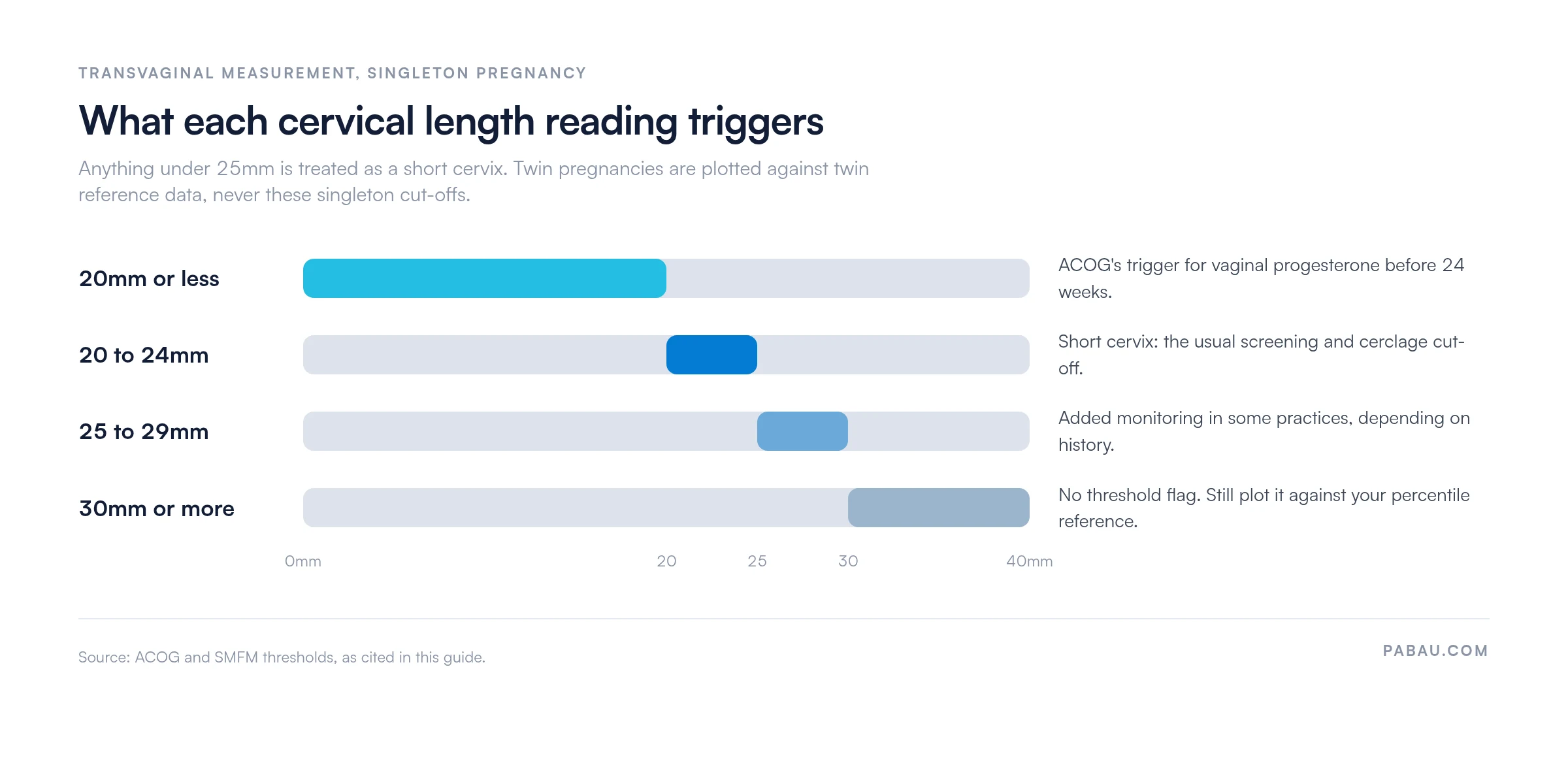
A short cervix is defined from a transvaginal ultrasound measurement. Under 25mm is the commonly used threshold, and ACOG treats 20mm or less before 24 weeks as the progesterone trigger. Both are population-level cut-offs rather than a forecast for one pregnancy.
A reading between 25mm and 29mm may warrant extra monitoring, depending on local practice and the patient’s history. The thresholds sit on one scale, and each band changes what happens after the scan.
A percentile chart shows where a measurement falls compared with a reference population at the same gestational age. The 5th, 50th and 95th percentile columns show whether a reading is low, typical or high for that week.
Twin reference ranges are shorter than singleton ranges. Applying a singleton threshold to a twin pregnancy can misclassify the result, so always confirm which dataset you are using.
A short cervix often causes no symptoms, so the scan result matters more than what the patient reports. Some patients do describe warning signs that prompt an urgent review.
Any of these symptoms should be assessed the same day, even if a previous scan was reassuring. That assessment often pairs a repeat scan with fetal monitoring, recorded on a fetal heart rate template.
Management depends on gestation, the measured length and the patient’s history, and a specialist should decide it. The options below appear in ACOG and SMFM guidance and do not replace individual assessment.
Patients are often advised to limit heavy lifting, strenuous exercise and intercourse. Evidence of benefit is limited, and ACOG and SMFM report that activity restriction and bed rest are not proven to prevent preterm birth. The treating team decides which restrictions apply to a particular pregnancy.
Document the advice given, the patient’s understanding and the review date, so the plan is clear at the next visit.
On paper, each scan result lands somewhere different. The report goes into one system, the printed chart sits in the notes, and the follow-up date depends on someone remembering it.
Pabau, the women’s health practice software we build, holds the chart as a digital medical form inside the patient record. Each reading carries its week, its value and the clinician who took it, so the series reads in order.
Automated reminders then chase the repeat scan after a flagged reading, and the record shows who was contacted and when. Your team opens the same history at every visit, so a borderline measurement stays in the plan instead of falling out of it.

Pabau stores the chart as a digital medical form in the patient’s file, and automated reminders chase the repeat scan after a flagged reading. Your team reads the whole series without hunting through scanned paper.

A cervical length reading is only as useful as the record around it. The number settles nothing on its own, because the week, the reference set and the technique all shape what it means.
Agree the details before the next scan rather than after it. Decide which percentile reference your practice plots against, who measures, and what happens at 25mm and at 20mm. Follow-up then stops depending on memory.
Download the chart above for the paper version, and keep the same fields in your patient record. Book a demo to see how Pabau stores serial cervical length readings and schedules the follow-up scan.

Charting labor rather than pregnancy? Dilated pregnancy chart maps cervical dilation from 0 to 10 cm through each phase of labor.
Documenting an invasive test? Amniocentesis test documentation covers consent, sampling details and the results your record needs.
Recording pain through labor? Pain scale for giving birth gives you a scoring sheet to use at each stage.
Tracking cycles before conception? BBT pregnancy chart logs basal body temperature so ovulation patterns are easy to read.
Planning care after delivery? Postpartum checklist sets out the checks and follow-ups for the weeks after birth.
A normal cervical length depends on gestational week and pregnancy type, so there is no single number. Plot the reading against the singleton or twin percentile reference your practice uses.
A reading under 25mm is generally classed as a short cervix. It raises the population-level risk of preterm birth and usually prompts specialist review.
Most measurements are taken between about 18 and 24 weeks, often at the 20-week anatomy scan. Patients with risk factors may be scanned earlier or more often.
It is usually measured by transvaginal ultrasound, which gives a clearer view than transabdominal scanning. Keep bladder filling and probe pressure consistent between scans.