



GLP-1 diet plan
A GLP-1 diet plan is a structured eating framework for patients taking semagluti...

2
Last Updated: Oct 13, 2026

The transverse ligament stress test screens for atlantoaxial instability at C1-C2 by applying a controlled anterior shear force to the occiput.
A positive result shows a soft or empty end-feel, neurological symptoms such as paresthesia or nystagmus, or muscle guarding instead of a firm bony stop.
The test should not be performed on patients with confirmed or suspected atlantoaxial instability, and must stop immediately if neurological symptoms appear.
Pabau’s digital forms and client record tools help physical therapy and manual therapy practices document upper cervical assessment findings, flag contraindications, and maintain audit-ready records.
Upper cervical instability is one of the highest-stakes findings in manual therapy. The transverse ligament stress test gives clinicians a structured, hands-on way to screen for atlantoaxial instability before any cervical treatment begins. Missing this screen on a patient with an undetected ligament injury is the kind of error that can lead to serious neurological harm.
The transverse ligament stress test, also called the anterior shear test, evaluates the integrity of the transverse ligament of the atlas (C1). This ligament holds the dens of the axis (C2) firmly against the anterior arch of C1. When it is damaged or lax, the dens can translate anteriorly into the spinal canal, compressing the spinal cord. This article covers the anatomy, indications, technique, result interpretation, diagnostic accuracy, and safety precautions, plus how the test fits into an upper cervical screening battery.
The transverse ligament of the atlas runs horizontally behind the dens of C2, anchoring it against the anterior arch of C1. It forms the posterior boundary of the atlantodental interval. It is the strongest ligament in the upper cervical spine and the primary restraint against anterior translation of the atlas on the axis.
The alar ligaments and apical ligament of the dens provide secondary restraint. This is why a battery of upper cervical tests, rather than one test alone, gives the most defensible clinical picture.
The transverse ligament stress test is indicated as a pre-treatment screen whenever atlantoaxial instability is clinically plausible. Several patient groups warrant routine consideration. Physical therapists must show sound clinical reasoning behind upper cervical screening decisions, under standards such as the HCPC standards for physiotherapists in the UK. Documenting that reasoning matters as much as the test itself. Structured digital intake forms that flag relevant history before the patient reaches the treatment table reduce the chance of an unstated contraindication slipping through.
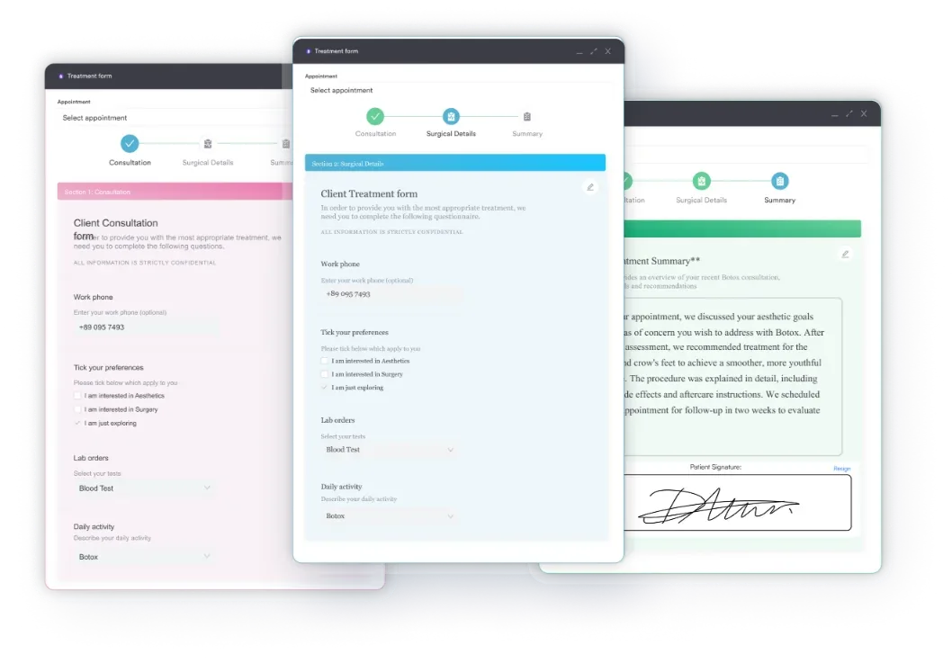
Practices managing patients across these groups also need systems that flag contraindications consistently across appointments. Keeping a physiotherapy compliance standard at the practice level means individual practitioners do not carry the full cognitive load alone.
The technique is consistent across the authoritative sources. Position and force direction matter: a misapplied force or an unsupported occiput can introduce error and risk.
The patient lies supine with the head at the edge of the treatment table, cervical spine in a neutral position. The head must not be in flexion or extension at the start of the test. Neutral alignment allows the examiner to apply a pure anterior shear force without simultaneously loading the anterior or posterior cervical muscles.
Stand at the head of the table. Cradle the occiput in both hands, with the fingers interlaced or spread across the base of the skull. The web of one hand, or the pads of both index fingers, contacts the posterior arch of C1. This provides a stabilizing contact point without gripping the neck.
The key clinical question is: what do you feel as the movement approaches end range? A firm bony end-feel is normal. The absence of that firm stop is the hallmark of a positive test.
Result interpretation is binary in concept but nuanced in practice. The examiner is assessing both the mechanical feel of the test and any patient-reported symptoms during the procedure.
A positive transverse ligament stress test does not confirm atlantoaxial instability. It indicates the need for further investigation. Imaging with MRI, CT, or dynamic flexion-extension radiographs is required for definitive diagnosis. Continue any cervical treatment only after imaging has excluded significant instability.
The most robust diagnostic accuracy data for the anterior-shear transverse ligament stress test comes from Kaale et al. (2008). The study compared clinical stress tests against MRI findings in patients with suspected upper cervical ligament injury. Against MRI as the reference standard, the anterior shear test showed roughly 65% sensitivity and 99% specificity. The positive likelihood ratio was about 51, and the negative likelihood ratio was about 0.35. These figures come from a single study with a modest sample size, so treat the point estimates as directional rather than exact. No large-scale replication in a general clinical population has been published.
The practical takeaway is to use the transverse ligament stress test as part of a screening battery rather than a standalone diagnostic. Its negative likelihood ratio means a negative result cannot rule out ligament injury on its own, especially in RA, Down syndrome, or significant trauma history. Urgent imaging is still warranted whenever those risk factors are present. The APTA neck pain guideline (Blanpied et al., 2017) recommends this same approach. It combines patient history, red flags, and physical examination findings before deciding on treatment.
The test involves applying a shear force to the upper cervical spine of patients who may already have compromised ligamentous support. Getting the safety parameters wrong can cause direct patient harm.
Practices using chiropractic practice software or physical therapy platforms can build a pre-treatment contraindication checklist into the patient intake workflow. This reduces the risk of a clinician encountering a contraindicated patient without warning. A chiropractic intake form template that captures trauma history, RA diagnosis, and known spinal conditions gives the treating practitioner relevant safety context before they start.
Document every upper cervical screen result, including negative findings, in the patient record immediately after assessment. If a patient later develops symptoms and the test was never recorded, reconstructing the clinical reasoning retrospectively is difficult. A structured field in your EMR for upper cervical screening status protects both the patient and the practitioner.
No single upper cervical test provides sufficient certainty on its own. A full screening battery combines the transverse ligament stress test with at least one other test, selected based on clinical suspicion. Clinicians working in sports medicine practice settings will often encounter these tests together in pre-participation screening for contact sports athletes with cervical risk factors. Reviewing physical therapy return-to-sport protocols that include upper cervical clearance criteria can contextualize how these tests are sequenced in practice.
How this screen fits into a physical therapy or chiropractic session is not fixed. What happens after a positive result is where clinical decision-making diverges most sharply between practitioners.
Pre-treatment screening before cervical manipulation. Complete the transverse ligament stress test, along with vertebral artery testing where indicated, before any high-velocity, low-amplitude (HVLA) thrust technique to the cervical spine. A positive result is a clinical stop sign for manipulation until imaging has cleared the patient.
Documentation. Record these details after every screen:
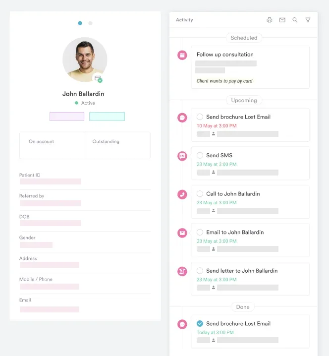
Safer clinical documentation practices help ensure that upper cervical screening notes hold up to clinical audit. Connecting those records to a structured client record keeps the screening status visible to any practitioner who sees that patient later. It is not limited to the one who performed the original assessment.
Referral pathway after a positive result: a positive transverse ligament stress test warrants urgent medical review before any cervical treatment continues. In practice, this means referring the patient to their primary care physician or directly to orthopedics or neurosurgery, depending on symptom severity. If the patient reports acute neurological symptoms during the test, treat it as a medical emergency. Do not continue manual therapy, position the patient safely, and arrange urgent medical assessment.
Practices managing larger patient volumes benefit from having a clear referral protocol embedded in their clinical workflows. Physical therapy practice software with structured clinical note templates and automated recall flagging helps prevent this. A patient with an unresolved positive screen is less likely to return for a later appointment without the flag being noticed. Patient care management workflows that link screening outcomes to follow-up actions keep an unresolved positive screen from being missed at the next visit.
Practices planning to formalize their upper cervical screening procedures should build clear protocols in from the start. Opening a physiotherapy practice with assessment protocols already in place is considerably easier than retrofitting them into an existing culture. Pabau’s digital forms and physical therapy EMR tools support the documentation, intake, and follow-up workflows behind this test. Together, they turn a single clinical screen into a defensible, auditable patient safety system.

Pabau helps physical therapy and manual therapy practices build structured assessment documentation, flag contraindications at intake, and keep audit-ready records from first visit to discharge.

The transverse ligament stress test is a low-tech, high-stakes screen. Diagnostic accuracy data exists for it, but it comes from a single study. Clinicians should treat the numbers as a guide rather than a certainty. Its practical value is as a safety gate. It gives clinicians a reason to pause, investigate, and refer before applying any technique that loads the upper cervical spine. Performed correctly with clear contraindications in mind, it protects patients and practitioners alike.
Pabau’s patient care management and digital documentation tools help musculoskeletal practices turn sound clinical protocols into consistent, recorded practice. To see how Pabau supports physical therapy and manual therapy workflows, book a demo.

Need a compliance framework for your physiotherapy practice? Mandatory compliance for physiotherapy clinics covers the regulatory requirements UK physiotherapy practices must meet.
Looking to formalize clinical documentation standards? Safer clinical notes provides a practical guide to writing clinical records that hold up to audit and protect practitioners.
Setting up or scaling a musculoskeletal practice? Opening a physiotherapy clinic walks through the operational and regulatory steps for launching a compliant practice.
The transverse ligament stress test is a manual orthopedic screen. It assesses the integrity of the transverse ligament of the atlas, which holds the dens of C2 against the anterior arch of C1. By applying a controlled anterior shear force to the occiput, the examiner determines whether the ligament is providing adequate resistance to anterior C1-C2 translation. An absent or soft end-feel suggests ligamentous insufficiency and raises concern for atlantoaxial instability.
The patient lies supine with the head at the edge of the treatment table in a neutral cervical position. The examiner cradles the occiput in both hands and applies a slow, gentle anterior-to-posterior shear force through the head and C1, relative to C2. The test begins with minimal force and is stopped immediately if any neurological symptoms appear. A firm bony end-feel indicates a normal finding. A soft or empty end-feel, muscle guarding, or neurological symptoms indicate a positive result.
A positive transverse ligament stress test can show a soft or empty end-feel, which is the absence of the expected bony resistance. Other signs include protective muscle guarding, paresthesia or numbness in the limbs or face, nystagmus, dizziness, or patient apprehension during the anterior shear force. A positive result does not confirm atlantoaxial instability. It indicates the need for urgent imaging, such as MRI, CT, or dynamic radiographs, before any cervical treatment continues.
The transverse ligament stress test applies an anterior shear force to the occiput and C1 to assess ligamentous resistance. The Sharp-Purser test starts with the cervical spine in flexion and applies a posterior, or reduction, force to C1. This tests whether the head relocates onto C2, indicated by a clunk or relief of symptoms. Both test C1-C2 stability but use opposite force directions and starting positions. The Sharp-Purser test is particularly associated with rheumatoid arthritis screening.
The alar ligament stress test assesses the lateral, or rotational, stability of C1-C2 provided by the alar ligaments. These ligaments run from the dens to the occiput and limit rotation and lateral flexion. The transverse ligament stress test assesses anterior-posterior stability provided by the transverse ligament. Used together, a positive result on both tests substantially increases clinical concern for upper cervical instability and strengthens the case for urgent imaging.
The transverse ligament stress test should not be performed on patients with confirmed atlantoaxial instability on imaging. Patients with known rheumatoid arthritis affecting the cervical spine, Down syndrome, or a history of C1-C2 surgery require specialist review before any upper cervical assessment. If neurological symptoms appear during the test, the procedure must be stopped immediately and the patient assessed for ongoing neurological signs before any further intervention.






