Bear hug test: Procedure, interpretation and free template
The bear hug test is a quick shoulder special test that screens for subscapulari...

2
Last Updated: Oct 23, 2026

The Thessaly test looks for a meniscal tear while the knee carries body weight, the position in which most tears become painful.
You run it at 5 and 20 degrees of knee flexion, rotating the patient’s body three times in each direction.
A positive result means joint line pain, catching, or locking during rotation. On its own it is never enough to diagnose a tear.
Independent studies report far lower accuracy than the original 89% sensitivity figure published by the test’s designers.
Practice management software like Pabau keeps special test findings, treatment notes, and referral decisions in one patient record.
Your patient stands on one leg, twists twice, and winces. The Thessaly test needs no equipment and adds barely a minute to a knee examination. It also loads the joint the way walking does, which a supine test cannot.
The trouble starts with what you do next. A positive result feels definitive, but joint line pain under load has several possible sources. Independent studies also report accuracy well below the 89% sensitivity of the original 2005 paper. Treat a positive as proof of a tear and you will refer patients who do not need imaging.
Start with what the maneuver is loading, because that explains both its strength and its failure modes.
The Thessaly test checks whether a meniscus produces symptoms when it is compressed and twisted under body weight. Karachalios et al. 2005 described the maneuver in the Journal of Bone and Joint Surgery. Other meniscal tests keep the patient supine or prone. Single-leg stance is what makes this one different.
The reasoning is biomechanical. A torn meniscus often stays quiet during passive handling and speaks up under load. Ask the patient to rotate their trunk over a flexed, weight-bearing knee. That recreates the compression and shear that hurt them on the stairs at home.
Reach for the test when the history and the joint line both point at the meniscus. Twisting injury, joint line pain, swelling after activity, and catching or locking are the classic set. It earns its keep when weight-bearing function matters, such as clearing an athlete for return to sport.
Consider the test when these are present:
Some patients should not be asked to try it. Skip the test if the patient cannot balance on one leg, has an unstable knee, or cannot bear weight at all. The same applies to a hot, swollen knee in the first days after injury. In that knee, pain limits rotation before the meniscus is ever loaded properly.
The test also cannot stand alone. An NIHR Health Technology Assessment (HTA) review of meniscal examination tests concluded that its findings belong inside a wider examination.
The protocol is the same across the original description and later clinical references. Test the good leg first. That gives you the patient’s baseline balance and shows them what you are asking for.
The 5 degree trial is a screen, not the test. It lets the patient rehearse the movement and picks up pain at minimal load. Most symptomatic tears stay silent at this angle.
At 20 degrees the posterior horn takes compression from the femoral condyle while the joint rotates. That extra load is what provokes a torn meniscus. The two-angle structure also gives you a useful contrast. Pain at 5 degrees but not at 20 suggests something other than the meniscus is complaining.
Technique explains much of the disagreement about this test. Five errors are worth watching for, in yourself and in anyone you are teaching.
Each of these produces a result you cannot defend later.
A positive test means the rotation reproduced medial or lateral joint line pain, catching, or locking. Where the symptom appeared, and in which direction, points you at a compartment.
Pain at the medial joint line during internal rotation suggests the medial meniscus. That meniscus is firmly anchored to the capsule and moves less, which leaves it exposed to rotational shear.
Pain at the lateral joint line during external rotation raises the lateral meniscus instead. The lateral meniscus moves more freely, yet compression during external rotation can still provoke a tear. Younger patients after a sports injury are the common picture.
Not every positive finding is meniscal. Patellofemoral pain, fat pad impingement, and ligament laxity all produce joint line pain under loaded rotation. Catching on its own, with no joint line pain, is the weakest finding of the set. Bilateral or migratory joint pain belongs on a different pathway, closer to rheumatoid arthritis testing than to a meniscal work-up.
Write down the compartment, the direction of rotation, and the exact symptom while the patient is still in front of you. That record drives your imaging decision later. Digital assessment forms with a field per finding keep the detail from evaporating between the treatment room and the note.

A positive Thessaly test earns your trust only when the answers below all hold.
Accuracy depends heavily on who ran the study. The original figures are high, independent replications are much lower, and the two are not always measuring the same thing.
Those first two rows answer different questions. Sensitivity and specificity describe how often the test catches tears, and how often it correctly clears healthy knees. Diagnostic accuracy is the share of all results that were right. It blends both measures together, and it moves with how common tears are in the sample.
The size of the drop matters more than any single number. Konan and colleagues reported 61% accuracy for medial tears and 80% for lateral ones, measured against arthroscopy. Originator studies tend to outperform independent ones across diagnostic test research. Treat the 89% figure as a ceiling rather than an expectation.
Five limitations shape how much weight a result deserves.
Because the result is probabilistic, your note has to carry the reasoning as well as the finding.
Record the flexion angle, the direction of rotation, the joint line involved, and the symptom you provoked. ‘Thessaly test positive’ on its own will not hold up in a records review. Aim for something like ‘Thessaly positive at 20 degrees, medial joint line pain on internal rotation’.
No single maneuver settles a meniscal diagnosis, so the useful question is what each one adds. These four tests differ in patient position, in how they load the joint, and in the kind of error they make.
McMurray gives you more specificity for posterior horn tears, and it still works when a patient cannot weight-bear. Joint line tenderness is the opposite trade. It catches most tears and flags plenty of knees that do not have one, which makes it a good opening screen. The Thessaly test adds the loaded, functional picture neither supine test can give you.
Use the Thessaly test fourth, after history, palpation, and McMurray. By that point you already have a pre-test probability, and the loaded rotation either raises it or leaves it where it was. When the patient cannot balance on one leg but can manage a full squat, Ege’s test applies loaded rotation with both feet on the floor.
Referral is where the test’s value gets cashed in. If imaging confirms the tear, the diagnosis lands on a code, and a right medial tear becomes S83.241. Patients who move on to arthroscopic meniscectomy are billed under 29881. Keeping that pathway inside the record stops a positive finding from stalling in someone’s inbox.
The test has a second life in discharge decisions. Loaded rotation that no longer provokes symptoms is one signal the knee is ready. Pair it with a return to running progression and a patient-reported measure such as the Global Rating of Change.
Consistency across a team is the harder problem. Practices running physical therapy EMR software with configurable examination templates get the same fields from every clinician. That makes a second opinion possible months later. Good patient care management depends on the record as much as on the hands doing the test.
Right now a knee examination probably lives in three places. The findings go into a note, the referral goes into an email, and the follow-up sits in someone’s head. Two months later, nobody can reconstruct which angle produced the pain.
Practice management software like Pabau puts those pieces in one patient record. You build the knee examination once as a template. The flexion angle, the compartment, and the direction of rotation each get their own field.
Pabau Scribe, our AI scribe, drafts the note from what you said during the session. The structured patient record then ties that finding to the treatment plan and tracks it across visits.
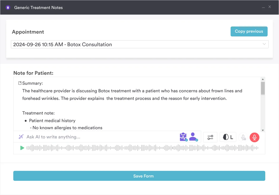
The benefit compounds across a team. When every clinician documents into the same sports medicine software template, a colleague covering your list finds the same fields you filled in.
Nobody is deciphering handwriting or guessing what was tested. That consistency is what practices shopping for physiotherapy clinic management software tend to ask about first.

Pabau gives physical therapy and sports medicine practices structured patient records, configurable examination forms, and AI-assisted notes. Your team spends less time documenting and more time treating.

The Thessaly test is worth keeping in your examination, as long as you hold it to what it can do. It moves your clinical probability. A diagnosis needs more than that, and the independent accuracy figures make the point plainly.
What changes your results is the routine around the maneuver. Standardize the technique, test the good leg first, and write the angle and the compartment into the note every time. Do that and your positives start meaning something consistent.
None of that survives a busy list without somewhere reliable to put it. Book a demo to see how Pabau captures special test findings, notes, and referrals in one patient record.

Comparing systems for a sports medicine practice? 7 best sports medicine software platforms weighs the options against clinical documentation and scheduling needs.
Need a patient-reported measure for the knee? Knee outcome survey gives you a scored template for tracking function through rehab.
Documenting a mobility-limited patient’s care plan? Impaired Physical Mobility Nursing Care Plan A ready-to-use care plan template covers NANDA-I diagnosis, related factors, defining characteristics, and mobility documentation.
Treating a patient with lateral knee pain? ITB syndrome exercises handout The handout explains iliotibial band tightness and inflammation, with exercises addressing overuse and hip stabilizer weakness.
It should not be, unless the meniscus is torn. The maneuver itself is a twist on a slightly bent knee. Warn the patient that pain is the finding you are looking for, and stop as soon as they report it. Anyone guarding through the rotation gives you an unreliable result anyway.
No. Sensitivity in independent studies is too low to rule anything out on one maneuver. A patient with a convincing history, joint line tenderness, and mechanical symptoms still deserves imaging after a negative result. Treat the negative as one data point, not permission to stop looking.
No. It targets the meniscus, and a positive result says nothing reliable about the ACL. Use Lachman and the pivot shift for cruciate integrity. Run the ligament tests first, because an unstable knee makes single-leg loaded rotation unsafe and hard to read.
It is not designed for self-testing. The patient needs someone holding their hands, and the flexion angle has to be controlled. A positive result also means little without joint line palpation and a history to set against it. Home attempts on an unstable knee risk a fall.