CRM for chiropractors: how to manage patients, bookings and marketing
A CRM for chiropractors is a patient relationship management system built around...

Last Updated: Oct 13, 2026
The pronator teres syndrome test checks for median nerve compression in the proximal forearm, at the level of the pronator teres muscle. It is a provocative maneuver. You hold the forearm in resisted pronation for 30 to 60 seconds and watch for the patient’s own symptoms.
The test is positive only when the patient’s familiar paresthesia returns in the median nerve distribution. That means the thumb, index, middle, and radial half of the ring finger. Forearm ache on its own does not count. Two companion maneuvers, the lacertus fibrosus test and the FDS middle finger test, then narrow down which compression site is involved.
Pronator teres syndrome is uncommon and often mistaken for carpal tunnel syndrome (CTS). A 2025 PMC case report on diagnostic pitfalls in pronator teres syndrome describes exactly that pattern. That overlap is why a structured physical examination, rather than electrodiagnostic testing, usually settles the diagnosis.

The pronator teres syndrome test uses resisted forearm pronation to reproduce median nerve symptoms in the proximal forearm, not at the wrist.
A positive test means reproduction of paresthesia or aching pain in the median nerve distribution during resisted pronation, not simple discomfort.
Nocturnal symptoms are typically absent in pronator teres syndrome, which is a key differentiator from carpal tunnel syndrome.
EMG and nerve conduction studies are frequently normal even when the condition is clinically present, so clinical testing drives the diagnosis.
Pabau’s medical records and note templates help musculoskeletal clinicians document provocative test findings and track symptom change over time.
The pronator teres syndrome test is a group of clinical maneuvers designed to reproduce median nerve symptoms. Each one stresses a specific anatomical site where the nerve can be entrapped in the proximal forearm.
Carpal tunnel tests stress the nerve at the wrist. This battery targets the elbow-to-mid-forearm segment instead. There the nerve passes beneath the lacertus fibrosus and between the two heads of the pronator teres. It then runs under the fibrous arch of the flexor digitorum superficialis (FDS).
Physical therapists, chiropractors, and sports medicine clinicians see forearm pain often. They need a reliable way to separate pronator teres syndrome (PTS) from the far more common CTS. Both conditions produce median nerve sensory disturbance in the hand, but their management differs substantially. Misattributing PTS to CTS can result in inappropriate splinting, failed carpal tunnel release, and months of wasted conservative treatment.
Practices managing musculoskeletal caseloads benefit from structured documentation of these provocative tests. Physical therapy EMR software with custom clinical note templates lets clinicians record laterality, the maneuver that was positive, and symptom quality. The format stays the same across the episode of care.
The pronator teres originates from two heads. A humeral head arises from the medial epicondyle, and a smaller ulnar head from the coronoid process of the ulna. The muscle inserts onto the lateral surface of the radial shaft at its midpoint. Its primary action is forearm pronation combined with weak elbow flexion. The median nerve passes between the two heads as it descends from the antecubital fossa.
Four distinct compression sites account for most cases of PTS. Knowing which site is involved guides both test selection and treatment:
Recording the implicated site at every assessment turns a repeat examination into something readable. A clinician reviewing the file can see whether the same level keeps testing positive, which a simple positive or negative notation never shows.
PTS presents with a cluster of features that overlap with CTS but differ in important ways. The typical pattern includes:
A chiropractic or physical therapy practice seeing high volumes of repetitive strain presentations will encounter PTS more often than its “rare” classification suggests. Occupational exposure drives incidence in working-age adults, especially manual trades and desk-based pronation tasks.
The pronator teres syndrome test is performed with the patient seated and the clinician standing or seated opposite. The primary maneuver is resisted forearm pronation. A complete assessment uses the full battery, targeting each of the four compression sites in turn.
With the elbow at 90 degrees and the forearm supinated, ask the patient to flex the elbow against your resistance. Reproduction of forearm pain or hand paresthesia implicates the lacertus fibrosus (Site 1) as the compression level.
Stabilize the patient’s hand and ask them to flex the proximal interphalangeal joint of the middle finger against resistance. Reproduction of symptoms here suggests compression at the fibrous arch of the FDS (Site 3), which overlies the nerve more distally than the pronator arch.
Recording which test provoked symptoms, at what point in the maneuver, and in which exact finger distribution takes seconds with a structured template. That granularity is what treatment planning runs on later. Digital intake forms configured for musculoskeletal presentations let clinicians capture it at the point of examination rather than transcribing from paper notes.

A positive result requires reproduction of the patient’s familiar paresthesia (numbness, tingling, or burning) in the median nerve distribution during the provocative maneuver. Pain alone in the forearm is not sufficient. The reproduced symptom must match the patient’s presenting complaint in both location and quality.
Key interpretation principles:
Those four principles collapse into a single decision at the bedside, shown below.
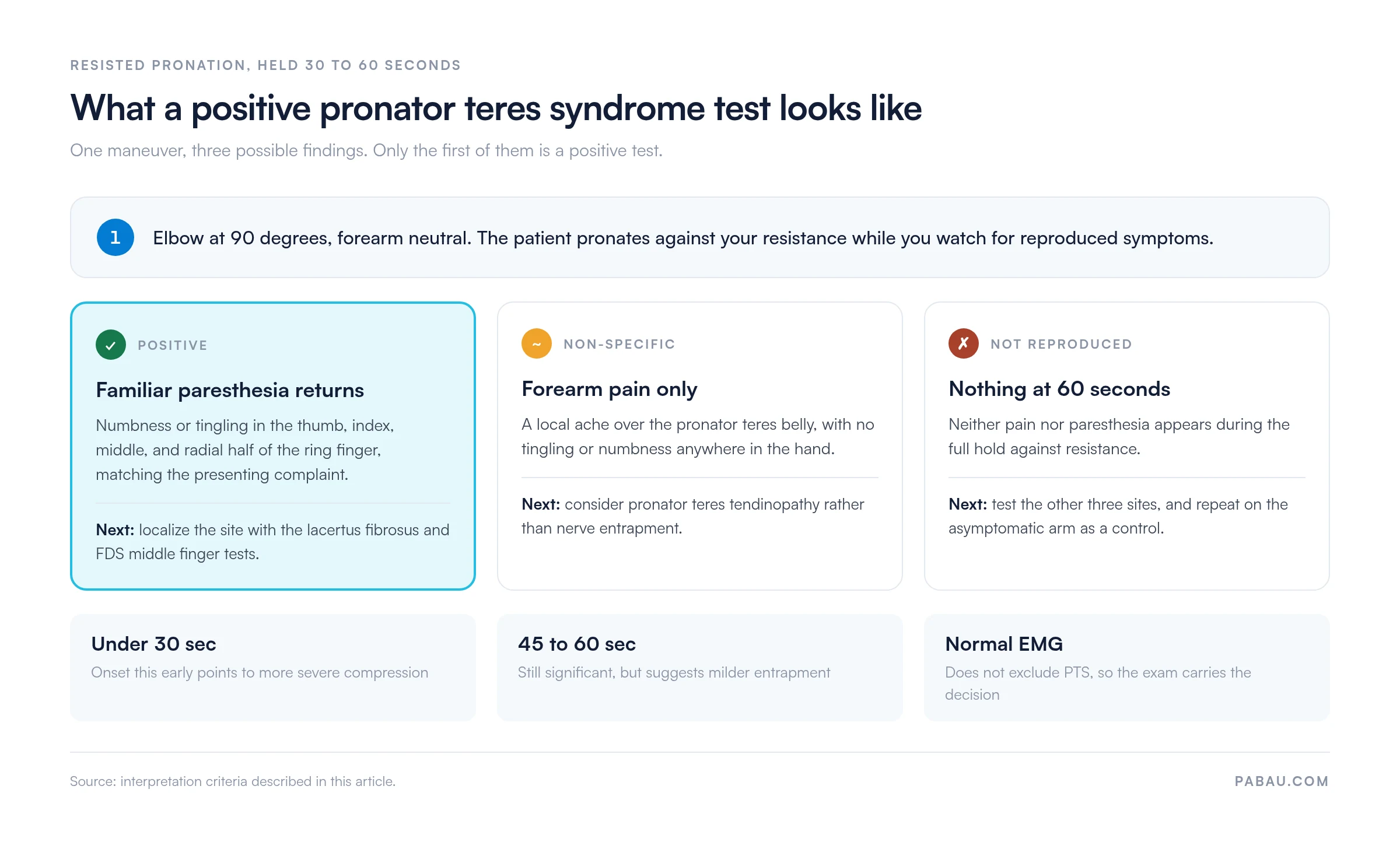
Published definitions of a positive result are not identical, which is worth knowing before you compare notes with a referrer. The Physiopedia clinical reference on the pronator teres syndrome test is one example. It scores the test positive when resisted supination, followed by elbow extension, reproduces the patient’s pain or discomfort. Requiring paresthesia, and not pain alone, sets a stricter threshold and screens out tenderness arising from the muscle belly.
The most important differential in clinical practice is carpal tunnel syndrome, because both conditions produce median nerve sensory disturbance in the same finger distribution. The table below captures the most actionable distinctions:
The thenar sensation row deserves particular attention. The palmar cutaneous branch of the median nerve exits the main trunk approximately 5 cm proximal to the wrist. It therefore bypasses the carpal tunnel entirely. In CTS the branch is therefore spared, while in PTS the compression sits proximal to the branch point and thenar skin sensation is affected. Testing sensation over the thenar eminence takes 30 seconds and adds meaningful diagnostic specificity.
Several conditions can mimic the presentation of PTS and must be considered before committing to a diagnosis:
Where a second, more proximal site is suspected, a thoracic outlet screen is worth adding before you settle on a forearm diagnosis. The patient braces both shoulders back and down while you monitor the radial pulse.
Sports medicine clinicians managing throwing athletes should be particularly alert to combined proximal neuropathies. Episode tracking and outcome measure templates help identify patients who are not progressing as expected, which prompts earlier re-evaluation for the differentials above.
Formal sensitivity and specificity data for this test battery are limited. The published literature leans on case series, case reports, and anatomical studies rather than large diagnostic accuracy trials. The PMC case report on diagnostic pitfalls in pronator teres syndrome is one such account. It describes a proximal median neuropathy initially attributed to carpal tunnel syndrome.
What the evidence does support:
Because no single maneuver settles the question, the documentation has to carry the full battery result rather than one positive or negative line. Anyone treating the patient next, or receiving the referral, then sees what was actually tested. The documentation compliance requirements that physical therapy practices work under point the same way, toward notes a second clinician can read without interpretation.
Clinical examination is the primary diagnostic tool. Electrodiagnostic and imaging investigations play a supporting role and carry specific limitations in this condition.
Nerve conduction studies measure distal motor latency and sensory conduction velocity across the wrist. Because PTS involves the proximal forearm, those distal measurements may appear normal. EMG can sometimes detect denervation in pronator teres-innervated muscles, such as flexor carpi radialis, flexor digitorum superficialis, and flexor pollicis longus. Early or mild compression often produces no detectable change. A normal electrodiagnostic result therefore does not exclude the diagnosis, and the clinical findings should carry the decision when the two disagree.
High-resolution musculoskeletal ultrasound can identify nerve swelling, hypoechogenicity, or perineural fibrosis at the pronator arch in some patients. It also allows dynamic assessment with real-time forearm pronation. MRI of the forearm can demonstrate muscle denervation changes or a compressing structural lesion, such as a fibrous band or an anomalous muscle belly. Both modalities are most useful when a space-occupying lesion is suspected, or when the clinical picture stays complex after a failed conservative trial. Neither replaces the clinical provocative test battery as the initial diagnostic step.
Document the exact compression site for each positive maneuver and the time to symptom onset during the provocative test. A patient whose symptoms appear within 15 seconds of resisted pronation is clinically different from one who takes 55 seconds. That difference guides whether to prioritize proximal nerve mobilization, activity modification, or early surgical referral.
A positive test combined with a consistent clinical picture points toward conservative management as the first-line approach. That care is usually built around activity modification, neural mobilization, and progressive strengthening. It runs as a 6 to 12 week trial before surgical referral is considered.
Core conservative interventions:
Exercise rehabilitation follows two phases: An early phase that reduces neural sensitization, then a progressive loading phase. Mixing them too early prolongs recovery.
Early phase (weeks 1-4): Focus on reducing neural irritability before loading the muscle.
Progressive loading phase (weeks 5-12): Once neural symptoms are diminishing and provocative test severity is reducing on re-examination, introduce forearm loading.
A 12-week program only works if someone checks in between appointments. Automated recall and follow-up messages send structured progress check-ins at the 4-week and 8-week marks, without anyone managing a diary of reminders by hand.
PTS is a multi-appointment problem. One episode holds the test result at assessment, symptom scores at weeks 4 and 8, the re-test after conservative treatment, and the referral decision. Paper notes make that continuity hard to hold together, especially where several clinicians cover the same patient.
Practice management software like Pabau keeps the episode in one place. Its medical records software lets musculoskeletal practices build custom note templates for assessments such as the pronator teres test battery. Laterality, test-specific findings, symptom quality, and outcome measure scores are captured in the same shape every time. A re-test at week 8 is then directly comparable with the baseline.
Some practices run musculoskeletal caseloads across several sites. There, physiotherapy clinic management software centralizes patient records, schedules, and follow-up automation, which cuts the admin behind a structured rehabilitation program. It also makes the 8 to 12 week decision point something the system prompts, rather than something a clinician has to remember.

Pabau’s structured medical records and digital assessment forms let physical therapists and chiropractors capture provocative test findings. Track symptom change and manage follow-up across a whole treatment episode. See how it fits a musculoskeletal caseload.

The decision that matters happens in the first minute of the examination, not in the electrodiagnostic report. Hold the resisted pronation for the full 30 to 60 seconds. Ask what the patient feels rather than whether it hurts. Treat forearm ache without paresthesia as a different problem. Getting that distinction right decides who starts nerve-directed rehabilitation. The alternative is a carpal tunnel release that will not help.
The trade-off worth remembering is that a clean examination buys you a 3-month conservative trial, and a vague one buys nothing. Write down which maneuver was positive, in which fingers, and at what second, so the re-test at week 8 means something. Book a demo to see how Pabau keeps that record consistent across every clinician who treats the patient.

Need a complete physical therapy documentation framework? Physical therapy EMR software by Pabau supports structured clinical note templates, outcome measure tracking, and multi-clinician record continuity for musculoskeletal practices.
Managing a physical therapy practice and need guidance on documentation compliance? Opening a physiotherapy clinic covers the regulatory and operational steps, including the record-keeping rules that apply to clinical assessment notes.
Working up upper limb nerve symptoms that do not fit carpal tunnel? Wright test covers the provocative maneuver for thoracic outlet compression, a differential worth ruling out alongside proximal median nerve entrapment.
The pronator teres syndrome test is a provocative examination that reproduces median nerve symptoms. It stresses the compression sites in the proximal forearm, primarily through resisted forearm pronation. A positive result requires reproduction of the patient’s familiar paresthesia in the median nerve distribution. That covers the thumb, index, middle, and radial half of the ring finger. Local forearm discomfort on its own does not qualify.
Seat the patient with the elbow at 90 degrees of flexion and forearm in neutral rotation. Stabilize the upper arm proximally and apply resistance distally while asking the patient to pronate the forearm for 30-60 seconds. Monitor for reproduction of hand paresthesia. Supplement with the lacertus fibrosus test, which is resisted elbow flexion in supination. Add the FDS middle finger test, which resists proximal interphalangeal flexion of the middle finger. Together they identify the compression level.
Two differences are the most useful clinically. PTS lacks the nocturnal symptoms that are characteristic of CTS. It also affects thenar eminence sensation, because the palmar cutaneous branch exits proximal to the carpal tunnel. That branch is involved in PTS but spared in CTS. Electrodiagnostics are often normal in PTS but typically show slowed distal latency in established CTS.
No. EMG and nerve conduction studies are frequently normal in pronator teres syndrome, particularly in mild to moderate cases. Clinical diagnosis through the provocative test battery, combined with the symptom history, remains the primary diagnostic method. Normal electrodiagnostics should not be used to exclude the diagnosis.
Conservative management is the first-line approach. It starts with activity modification to reduce repetitive pronation loads. Add a 6-12 week physical therapy program covering median nerve mobilization and progressive forearm strengthening. A short course of NSAIDs helps in the acute phase. Wrist splints provide little benefit in PTS, unlike in CTS. Surgical decompression is considered when conservative management fails after 3-4 months.
Yes. Double crush syndrome, where the median nerve is compressed at two levels simultaneously (e.g. the pronator arch and the carpal tunnel), is recognized clinically. Evaluate for dual-level compression where a patient responds incompletely to carpal tunnel treatment. Do the same where proximal forearm pain and nocturnal hand tingling appear together. Both conditions require separate treatment targeting each compression site.