



GLP-1 diet plan
A GLP-1 diet plan is a structured eating framework for patients taking semagluti...

2
Last Updated: Oct 13, 2026
The costoclavicular test is a provocation test for thoracic outlet syndrome (TOS). It narrows the space between the first rib and the clavicle so that neurovascular compression symptoms reappear. The patient braces both shoulders back and down for 30 to 60 seconds while you monitor the radial pulse. The result is positive only when the pulse diminishes and the patient’s familiar symptoms return.
TOS is one of the hardest musculoskeletal conditions to diagnose. Physical examination tests still carry the first clinical decision, according to the Journal of Orthopaedic and Sports Physical Therapy. The test goes by three names: The costoclavicular test, the military brace test, and Eden’s test. All three describe the identical maneuver.
This guide covers the anatomy, the step-by-step protocol, how to read the result, and how the test compares with the rest of the TOS battery. It closes with the five elements your assessment note needs.

The costoclavicular test compresses the space between the first rib and clavicle to provoke TOS symptoms. It is also called the military brace test or Eden’s test.
A positive result requires both radial pulse reduction and reproduction of the patient’s symptoms. Pulse obliteration alone appears in 16% of healthy subjects, so it is not diagnostic.
Published sensitivity ranges from 50% to 94% and specificity from 53% to 100%, depending on TOS subtype and study design.
Run the test alongside the Adson, Roos, and Wright tests rather than on its own, because no single maneuver confirms or excludes TOS.
Pabau’s structured clinical records and digital forms help physical therapists and osteopaths document special test findings, laterality, and grading in a single patient encounter.
The costoclavicular test is a physical examination maneuver that places the shoulder girdle in an exaggerated military brace posture. That posture compresses the costoclavicular space and reproduces the symptoms of thoracic outlet syndrome. Clinicians reach for it when a patient reports unilateral upper limb paresthesia, arm heaviness, or vascular changes that worsen in particular arm positions. It is particularly indicated when:
Musculoskeletal physical therapists, osteopaths, and sports medicine clinicians all use the maneuver as part of a standard TOS provocation battery. Understanding the anatomy it targets comes before administering it.
The costoclavicular space is the narrow passage between the inferior surface of the clavicle and the superior surface of the first rib. It serves as a conduit for the brachial plexus and the subclavian artery and vein. When this space narrows, the neurovascular bundle is compressed, producing the characteristic symptoms of TOS.
The military brace position draws the shoulders sharply back and down, which approximates the clavicle toward the first rib. The costoclavicular space reaches its smallest functional dimension in that position. That is the mechanical rationale for the test.
Precise technique determines whether the costoclavicular test produces a reliable result. Sloppy positioning is the most common reason for false negatives and inconsistent findings between clinicians.
Seat the patient upright on a treatment plinth or chair with no back support, so the shoulder girdle is free. The clinician stands or sits at the patient’s side or slightly behind.
Track three signals at once: Radial pulse amplitude, symptom reproduction by location and quality, and the patient’s ability to sustain the position. A patient who cannot hold the military brace without compensating at the neck or ribcage is not performing the test correctly.
Note the side tested, whether bilateral testing was performed, and the hold duration. This matters for documentation, particularly in medicolegal contexts or when tracking change over serial assessments.
A positive costoclavicular test requires two findings together: Radial pulse diminution and reproduction of the patient’s familiar symptoms. Either finding in isolation is insufficient for a positive interpretation.
One caveat governs the whole interpretation. Pulse obliteration during shoulder retraction and depression happens in plenty of healthy people. Nord and colleagues put five TOS maneuvers to blinded testing on normal volunteers and on patients with carpal tunnel syndrome. The costoclavicular maneuver returned a false positive in 16% of the normal subjects.
The same study shows how widely that error rate varies between maneuvers, which is the argument for choosing a battery rather than a favorite test. The figures below are the share of healthy volunteers who tested positive on each one.
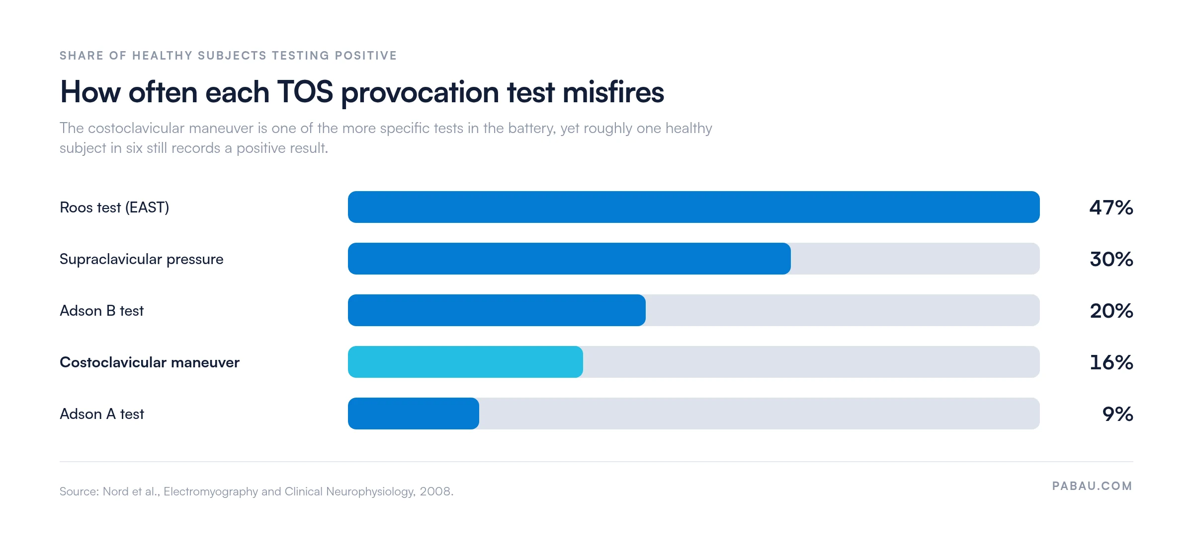
Those rates climbed steeply in the carpal tunnel group, where the costoclavicular maneuver produced a false positive in 48% of patients. A patient with an established distal entrapment will therefore light up this test for reasons that have nothing to do with the thoracic outlet. Always cross-reference the pulse finding against the symptom report.
The diagnostic accuracy of the costoclavicular test varies considerably across published studies, largely because TOS itself has no gold-standard diagnostic criterion. Figures should be interpreted with that limitation in mind.
The American Physical Therapy Association (APTA) clinical practice guidance on TOS assessment consistently recommends using multiple provocation tests. No single test carries sufficient diagnostic accuracy to confirm or rule out TOS on its own.
Run the costoclavicular test bilaterally even when symptoms are unilateral. Asymmetric findings (positive on the symptomatic side, negative on the asymptomatic side) significantly increase diagnostic confidence compared to a unilateral positive alone.
The costoclavicular test is one of four main TOS provocation tests used in clinical practice. Each targets a different anatomical compression site and patient position, which is why combining them improves diagnostic yield.
Reach for the Roos stress test first when a patient reports fatigue-type symptoms that build gradually during overhead activity. Its 3-minute dynamic load replicates occupational strain more closely than a static military brace hold. The trade-off is specificity, since almost half of healthy subjects test positive on it.
The costoclavicular test suits symptoms provoked by shoulder depression loads instead, such as carrying bags or sitting in a slumped desk posture. Using both in the same session maximizes diagnostic coverage across the three TOS compression zones.
The costoclavicular test has meaningful limitations that every clinician should understand before acting on its findings. Recognizing what can mimic a positive result prevents unnecessary onward referral.
When TOS and cervical radiculopathy cannot be separated clinically, thoracic outlet syndrome MRI and nerve conduction studies give the clearest answer. A clean cervical screen strengthens the case for the outlet, so run Spurling’s test before committing the patient to that pathway.
A positive costoclavicular test finding opens a clinical decision pathway, not a diagnosis. The next step depends on whether the presentation suggests neurogenic, arterial, or venous TOS.
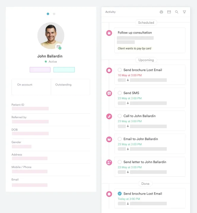
Tracking referral status and follow-up outcomes from the patient record saves a great deal of administrative time across a musculoskeletal caseload. The physiotherapy clinic management software a practice runs on decides how smoothly these steps connect from assessment to onward referral.
Accurate documentation of provocation test findings protects the clinician, supports continuity of care, and ensures onward referrers have a clear clinical picture. The costoclavicular test should be recorded with five elements in the assessment note.
A well-written SOAP note entry might read: “Objective: Costoclavicular test (military brace/Eden’s test) positive bilaterally, right greater than left. Radial pulse obliterated at 20 seconds on the right. Patient reported reproduction of familiar right arm heaviness and fourth/fifth finger paresthesia. Left side: Pulse diminished at 40 seconds, no symptom reproduction. Assessment: Findings consistent with right-sided TOS at the costoclavicular space; left inconclusive.”
Practices running paper-based or generic digital records tend to lose laterality and timing data between encounters. That detail is exactly what a vascular surgeon reads first. For practices with clinical governance obligations, physiotherapy clinic compliance requirements sets out the documentation standards that apply to special test records.
A referral letter that auto-populates from the assessment record cuts duplication and transcription errors across the care pathway. That is one of the things worth testing when you compare an EMR for physical therapists.
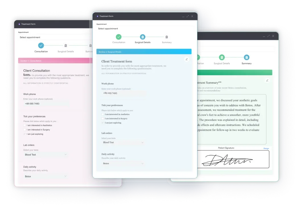
In most musculoskeletal practices the TOS battery gets written up as free text. Laterality, hold duration, and the second of pulse change go into a paragraph that nobody can search later. When the patient returns in six weeks, comparing today’s findings with the last visit means rereading two sets of notes.
Practice management software like Pabau replaces that paragraph with a structured field set. You build the TOS battery once as a digital assessment form. It carries a slot for each test, each side, the pulse grade, and the symptom description. Every clinician in the practice then records the same five elements in the same places.
The form lives on the patient record rather than in a separate document. Serial assessments line up side by side, and a referral letter pulls the findings straight through. Nothing gets rekeyed, so nothing gets dropped between the assessment and the vascular surgeon.

Pabau’s structured clinical records let physical therapists and osteopaths capture TOS test results, laterality, grading, and follow-up actions in a single patient encounter. No paper, no rekeying, no lost findings.

The costoclavicular test is a high-value, low-cost screen for TOS. It earns its diagnostic weight only when pulse change and symptom reproduction appear together. Read the result against the rest of the provocation battery. Treat a lone pulse finding as noise, because 16% of healthy people produce one.
Used that way, the test points you efficiently toward the right confirmatory investigation and the right management pathway. Used on its own, it will send patients for imaging they do not need.
Whatever you find, the note has to carry it. Record laterality, pulse grading, symptom distribution, and the onward referral action in one place. Book a demo to see how Pabau structures that note for a physical therapy or sports medicine caseload.

Need the hyperabduction half of the battery? Wright test for TOS covers the subcoracoid compression site the costoclavicular test cannot reach.
Want a ready-made form for the scalene screen? Adson’s test template gives you the procedure and a recording sheet you can hand straight to a clinician.
Patient confirmed for conservative management? Thoracic outlet syndrome exercises sets out a phased rehab program you can prescribe from the first visit.
Running a musculoskeletal practice and want to stay compliant? Physiotherapy clinic compliance requirements outlines the documentation and governance obligations that apply to special test records.
Managing a physical therapy or sports medicine practice? EMR for physical therapists covers what to look for in purpose-built clinical software for musculoskeletal practices.
The costoclavicular test assesses for thoracic outlet syndrome (TOS). It provokes compression of the brachial plexus and subclavian vessels in the costoclavicular space. It is most useful for identifying the costoclavicular space as the primary compression site. Reach for it when patients report symptoms with shoulder depression loads, such as carrying bags or wearing heavy backpacks.
A positive costoclavicular test indicates probable neurovascular compression in the costoclavicular space consistent with thoracic outlet syndrome. The result counts as positive only when radial pulse diminution and reproduction of the patient’s familiar symptoms both occur during the military brace position. Pulse obliteration without symptom reproduction is not a positive finding.
The costoclavicular test targets the rib-clavicle interval by bracing the shoulders back and down. The Adson test targets the scalene triangle by extending and rotating the neck with a held breath. Use the Adson test when symptoms appear with neck rotation, and the costoclavicular test when symptoms worsen with shoulder depression loads or slumped posture.
Published sensitivity ranges from approximately 50% to 94% and specificity from 53% to 100%, varying by TOS subtype and outcome measure. Those wide ranges reflect the absence of a gold-standard diagnostic criterion for TOS and the subjective element of symptom reporting. No single TOS provocation test is accurate enough to confirm or exclude TOS in isolation.
Nord and colleagues recorded a false positive in 16% of healthy volunteers and in 48% of patients with carpal tunnel syndrome. That places it among the more specific tests in the battery, well ahead of the Roos test at 47% in healthy subjects. A positive result still needs symptom reproduction before it means anything.
No. The hyperabduction test (Wright test) targets the subcoracoid space under the pectoralis minor by taking the arm into full overhead abduction. The costoclavicular test targets the rib-clavicle interval through shoulder retraction and depression. Both are TOS provocation tests but they assess different compression sites.
Order thoracic outlet syndrome MRI when surgical planning needs imaging of the brachial plexus, a suspected cervical rib, or anomalous first rib anatomy. It is also indicated when conservative physical therapy fails after 6 to 12 weeks. Initial vascular workup is typically done with Doppler ultrasound, which is more accessible than MRI.






