CRM for chiropractors: how to manage patients, bookings and marketing
A CRM for chiropractors is a patient relationship management system built around...

Last Updated: Oct 13, 2026

The patellar grind test, also called Clarke’s sign, compresses the patella against the femoral trochlea to provoke patellofemoral pain.
Most clinical references define a positive result as pain on quadriceps contraction, so crepitus on its own is not enough.
Cleveland Clinic uses broader criteria, counting a grinding noise or a failed contraction as positive, so record which definition you applied.
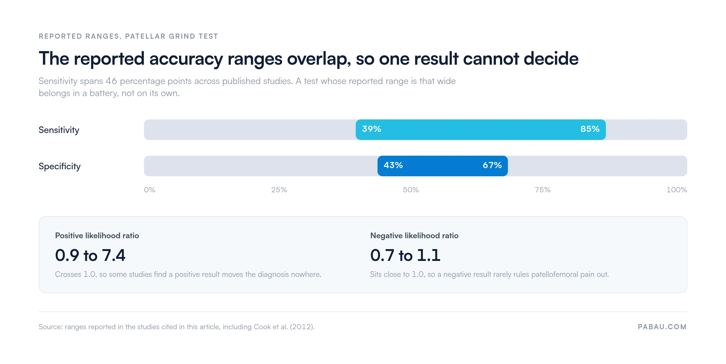
Reported sensitivity runs from 39% to 85% and specificity from 43% to 67%, so pair the test with others.
Practice management software like Pabau captures special-test findings in structured fields, so results stay comparable from visit to visit.
Patellofemoral pain syndrome (PFPS) accounts for up to 25% of sports-related knee injuries, according to a peer-reviewed review published in PMC. Runners, adolescents, and desk-bound patients all arrive with the same complaint of diffuse anterior knee pain.
The patellar grind test is usually the first special test a clinician reaches for. A positive result means pain under the patella while the quadriceps contracts against your compressive hand. What that pain rules in is narrower than most references suggest, and the accuracy evidence below explains why.
The patellar grind test is an orthopedic special test for patellofemoral pain syndrome (PFPS). Clinicians also call it Clarke’s sign or the patellar compression test, and the literature uses all three names interchangeably. You compress the patella into the femoral trochlear groove while the patient actively contracts the quadriceps. Pain during that contraction is the finding you are looking for.
PFPS produces diffuse anterior knee pain that worsens with running, squatting, stair climbing, and long spells of sitting. The test reproduces that pain under controlled conditions. It never diagnoses PFPS on its own. What it does give you is a fast, low-tech read on whether the patellofemoral joint is worth pursuing.
Understanding why the test provokes pain starts with the joint surfaces it loads. Recording that reasoning alongside the finding is what turns a one-word result into a defensible note. Most physical therapy practices now keep those notes in physiotherapy practice software rather than on paper.
The patella is a sesamoid bone embedded in the quadriceps tendon. It articulates with the femoral trochlear groove, the V-shaped channel on the anterior distal femur. Through flexion and extension the patella tracks within that groove. Its path is set by the quadriceps, the patellar tendon, the medial and lateral retinacula, and the shape of the groove itself.
Hyaline cartilage lines the articular surfaces of both the patella and the trochlea, and that cartilage is the structure most often implicated in PFPS. When tracking is disrupted by muscle imbalance, bony malalignment, or overuse, abnormal compressive and shear forces cross the chondral surfaces. The test reproduces that compression mechanically, which is why it hurts in symptomatic patients.
Use the patellar grind test when a patient reports activity-related anterior knee pain that is diffuse rather than focal. These features point toward it:
Skip the test, or apply it cautiously, after an acute patellar fracture, recent knee surgery, or in severe patellar instability. Compression is the wrong load in those knees. Where a patient is apprehensive even at rest, the patellar apprehension test tells you more before you add compressive force.
Consistent technique is what makes the result reproducible. The procedure below reflects the consensus method across the primary clinical references and the Physiotutors diagnostic accuracy resource on Clarke’s sign.
Position the patient supine on the plinth with the knee in full extension at zero degrees. The quadriceps must be fully relaxed before the contraction phase.
Some examiners use a slightly flexed variant of roughly 20 to 30 degrees over a bolster, which can reduce apprehension and allow more patellar mobility. Both variants appear in the literature, though full extension is described most often.
Place the web space of your dominant hand, the skin between thumb and index finger, just proximal to the superior pole of the patella. Apply firm, controlled pressure distally and posteriorly to compress the patella into the trochlear groove.
Enough force to engage the articular surface is the target. Any more risks provoking pain through force alone in an asymptomatic knee.
Hold the compression and ask the patient to contract the quadriceps slowly. “Tighten your thigh muscle” or “push the back of your knee into the table” both work as cues. The contraction drives the patella proximally against your hand and further into the groove. Watch and palpate for pain, resistance to the contraction, crepitus, or the patient pulling away.
A positive patellar grind test is pain or discomfort reproduced during quadriceps contraction while you compress the patella. That is the definition most clinical references use, and it is the one this guide follows.
The criteria are not uniform, though. The Cleveland Clinic treats a grinding noise, an inability to contract the quadriceps, or pain as each sufficient for a positive result. Under those broader criteria, an asymptomatic knee with audible crepitus would score positive.
So write down which definition you applied. A note reading “positive Clarke’s sign” means different things to the next clinician depending on the criterion behind it.
A positive result tells you the patellofemoral joint is likely a pain source. It cannot name the pathology behind it. Treat it as a prompt for further reasoning, additional special tests, and imaging where the presentation justifies it.
The accuracy of the patellar grind test is modest and inconsistent, even by the standards of orthopedic special tests. Read the reported figures before you let a single result carry any weight.
These figures come from several published studies, and the spread reflects differences in populations and methods. Studies drawn from clinical populations with confirmed PFPS report the higher sensitivity. Studies that include asymptomatic controls report the low end. Plotted on one scale, the ranges overlap heavily.
A positive result points at the patellofemoral joint without naming the pathology behind it. Work through these differentials before you settle on PFPS. Practices with a heavy athletic caseload usually track how each one resolves in sports medicine software, so patterns across patients stay visible.
The patellar grind test is rarely used alone. Most clinicians run it alongside other patellofemoral tests to build a fuller picture. The table below sets out the key tests, their procedures, and what each one detects.
Running the grind test with the tilt and apprehension tests tells you whether the dominant problem is articular pain, lateral tightness, or instability. Those three make a reasonable minimum battery for a focused patellofemoral assessment.
A positive finding opens a clinical pathway rather than closing a diagnosis. The sequence below is what usually follows:
Record the criterion you applied, not just the verdict. A note reading “positive: pain on contraction, no crepitus” can be compared against next month’s reassessment. A note reading “positive Clarke’s sign” cannot.
Four weaknesses explain why a single grind test result should never carry a diagnosis on its own. Knowing where the test fails matters as much as knowing how to execute it.
Special-test findings usually end up buried in free text, if they get recorded at all. Three months later nobody can say whether the grind test was positive at the first visit. That turns outcome tracking into guesswork and leaves the record thin if it is ever questioned.
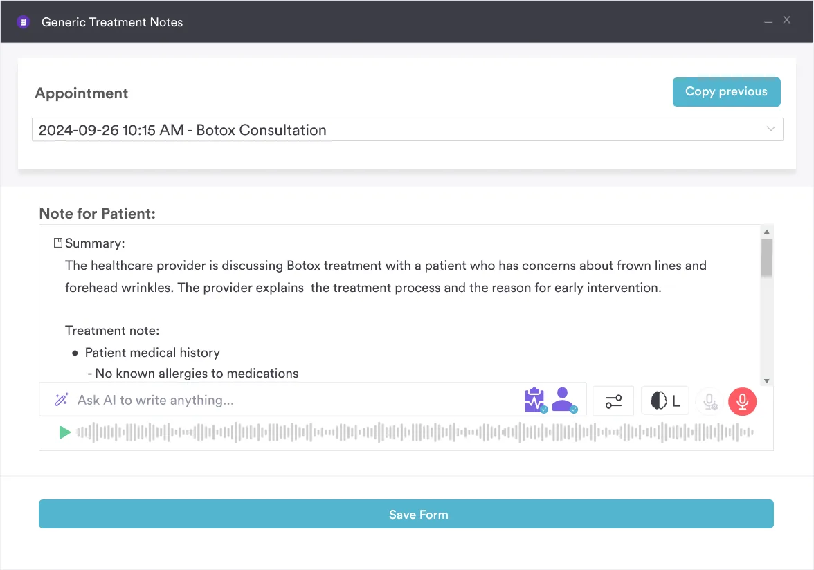
Practice management software like Pabau handles this with structured forms instead. You build a knee assessment template once, with a field for each special test and its result. The finding gets captured at the plinth rather than reconstructed from memory afterwards. It then sits in the client record beside consultation history, imaging notes, and the treatment plan.

For a heavy musculoskeletal caseload, Pabau Scribe, our AI scribe, drafts the note from the spoken consultation. A full knee assessment stops costing you a lunch break in typing. Pabau is software for physical therapists and allied practices, so the record follows the patient from first assessment through to discharge.
Every subscription includes every feature, so structured assessment forms and the audit trail behind them are not something you upgrade into. Book a demo with Pabau to see a knee assessment template built and filled during a live consultation.

Pabau gives physical therapy and sports medicine practices structured assessment forms, client records that hold the whole episode, and an AI scribe for the note. Your special-test results stay comparable from first visit to discharge.

Treat the patellar grind test as a screen and one input among several. Its value is speed. Around 30 seconds of compression tells you whether the patellofemoral joint deserves the rest of your examination time.
The trade-off worth remembering is the definition. Decide whether you count pain only, or pain plus crepitus and a failed contraction, then write down which one you used. Two clinicians applying different criteria to the same knee will disagree, and the note is the only place that disagreement becomes visible.
Do that consistently across a caseload and your reassessments start to mean something. Book a demo to see how Pabau turns knee special tests into structured fields you can compare visit to visit.

Assessing ankle dorsiflexion alongside the knee? Knee to wall test covers how to measure and interpret dorsiflexion range, a frequent contributor to anterior knee pain.
Deciding whether an acute knee needs an X-ray? Pittsburgh Knee Rules sets out the criteria and the walk test, with a template you can use in the room.
Need to exclude a hip or femoral fracture first? Patellar pubic percussion test explains a fast bedside screen for occult proximal femoral fractures.
Unsure what your assessment records have to hold? Mandatory compliance for physiotherapy clinics covers the requirements that shape how you document and store patient assessments.
Building structured intake for a musculoskeletal caseload? Chiropractic intake form template shows the fields worth collecting before a first musculoskeletal assessment.
The patellar grind test is used to detect patellofemoral pain syndrome (PFPS), a condition causing diffuse anterior knee pain. It works by compressing the patella against the femoral trochlear groove while the patient contracts their quadriceps, reproducing the pain characteristic of PFPS. It is also known as Clarke’s sign or the patellar compression test.
A positive patellar grind test means the patient reports pain or discomfort under the patella during active quadriceps contraction. Under the criteria most references use, crepitus alone without pain is not positive, because patellar crepitus is common in asymptomatic knees. Cleveland Clinic applies broader criteria that also count a grinding noise or a failed contraction, so record which definition you applied.
Clarke’s sign is another name for the patellar grind test. Both terms describe the same procedure, compressing the patella into the femoral trochlear groove while the patient actively contracts the quadriceps. The terms Clarke’s sign, patellar grind test, and patellar compression test are used interchangeably in clinical practice and the literature.
The patellar grind test detects patellofemoral pain by compressing the patella while the patient contracts the quadriceps. The patellar apprehension test instead displaces the patella laterally with the knee flexed to about 30 degrees, assessing for subluxation or instability. They target different diagnoses, so most patellofemoral assessments include both.