Nitrates medication list
A nitrates medication list is a point-of-care reference that sets out the main c...

Last Updated: Oct 6, 2026

The olfactory nerve test assesses CN I by presenting non-irritating odors to each nostril separately and recording what the patient reports.
Record detection and identification separately, per nostril, because a patient can detect an odor without being able to name it.
Avoid trigeminal irritants such as ammonia, isopropyl alcohol and peppermint oil, which stimulate CN V rather than CN I.
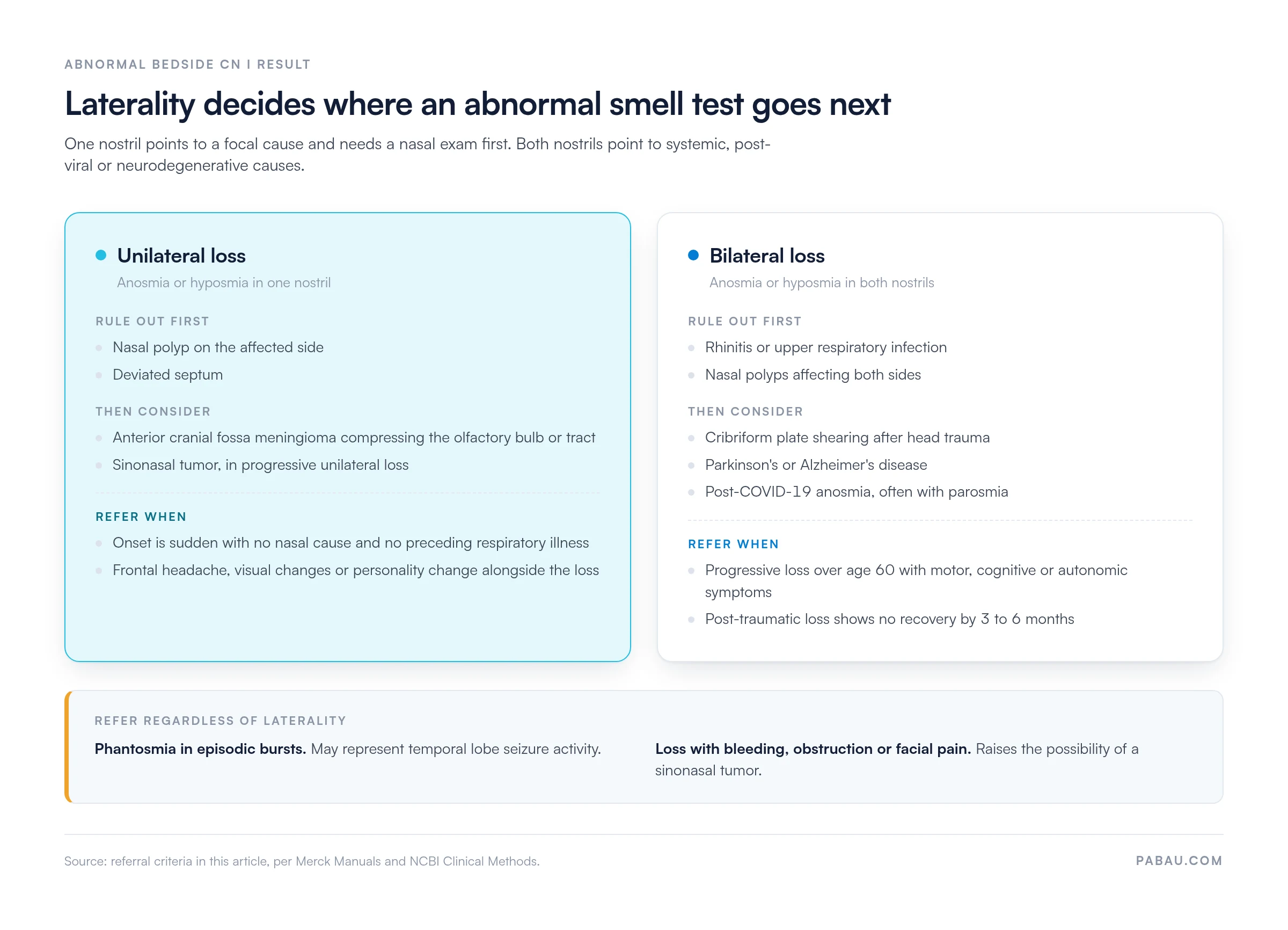
Unilateral loss points toward intracranial pathology or local nasal disease. Bilateral loss more often suggests systemic, post-viral or neurodegenerative causes.
Practice management software like Pabau records the odorant, the side tested and the response as separate fields, so results stay comparable between visits.
The olfactory nerve is one of the most commonly skipped steps in a routine cranial nerve examination. Clinicians cite time pressure, and many assume the patient will volunteer a loss of smell without being asked.
The test itself is short. Occlude one nostril and present a familiar, non-irritating odorant to the open side. Record whether the patient detects it, and whether they can name it. Repeat on the other side with the same odorant.
Two minutes of examination can change a clinical picture. Any of the following can turn up in a patient who otherwise looks neurologically intact:
This guide covers the anatomy, the bedside procedure, and how to read each result. It also sets out what the bedside examination cannot tell you, and when a standardized smell test earns its place.
The olfactory nerve test evaluates cranial nerve I (CN I), the only purely sensory cranial nerve and the one responsible for smell. CN I bypasses the thalamic relay and projects directly to the primary olfactory cortex. Olfactory dysfunction can therefore reflect pathology at four levels: the nasal mucosa, the cribriform plate, the olfactory bulb, or the cortex itself.
According to Merck Manuals, anosmia after head trauma or alongside neurodegenerative symptoms should be evaluated formally rather than assumed. The test earns its place in three situations.
In each case a structured examination gives you documented evidence rather than a self-report. CN I also sits first in the twelve-nerve sequence. The recording habits you set here carry into the rest of the examination, including the hypoglossal nerve test for CN XII.
Each step of the test makes more sense once you know where CN I starts and where it goes. The olfactory receptor neurons sit in the olfactory epithelium, high in the nasal cavity. Their axons bundle into olfactory filaments, pass through the cribriform plate of the ethmoid bone, and synapse in the olfactory bulb. From there the olfactory tract carries signals to the piriform cortex and other limbic structures, with no thalamic relay.
The cribriform plate is the anatomical weak point. It is a thin, perforated bony structure, and the filaments passing through it shear easily under the forces of a head injury. That is why post-traumatic anosmia can follow a fall with no other neurological sign. NCBI Clinical Methods treats this mechanism as well established.
CN I is also unusual among the cranial nerves for its partial regenerative capacity. Olfactory receptor neurons can be replaced from basal cells in the epithelium. That is why some patients recover part of their sense of smell after an infection. Recovery is variable and never guaranteed, particularly where the damage involves the olfactory bulb rather than the peripheral neurons. Keeping structured clinical records lets you compare olfactory function across successive consultations instead of relying on recall.
The bedside test needs almost no equipment. What matters is choosing odorants that stimulate CN I and leave the trigeminal nerve (CN V) alone. Ammonia and isopropyl alcohol are the two items clinicians most often reach for, and both are trigeminal irritants.
A patient who reacts to ammonia has not shown you intact CN I function. They have shown you a CN V response, which a completely anosmic patient can produce just as readily.
Menthol and camphor belong in the same category, which rules out peppermint oil at any concentration. Use familiar, non-irritating, volatile odorants from a kitchen or household supply instead. The table below sets out suitable and unsuitable choices.
The bedside olfactory nerve test follows a consistent sequence. Testing both nostrils at once, or skipping occlusion altogether, strips most of the clinical value out of the result. A practical guide to CN I examination techniques published in PMC makes this point. The quality of bedside testing varies with whether a standardized approach is followed.
Where CN I is tested as part of the full twelve-nerve sequence, work from one form rather than free text. The cranial nerve examination checklist lists each nerve in order, with space for the side tested and the response you recorded.
A normal result is bilateral detection plus correct identification of at least one familiar odorant in each nostril. Anything short of that falls into one of four dysfunction types, and each one carries different clinical implications.
Phantosmia is the one finding here that can reflect central pathology rather than damage to CN I itself. Record whether the patient reports phantom smells alongside other sensory or perceptual symptoms, because that combination shifts the differential.
Laterality is the most diagnostically useful feature of the olfactory nerve test, and it is also the one most often left out of the note. The interpretation changes depending on whether one nostril or both are affected.
Post-traumatic anosmia tends to be bilateral where the mechanism is cribriform plate shearing. It may be the only cranial nerve finding in a patient who otherwise appears neurologically intact. That is the argument for putting CN I into the standard post-head-injury assessment.
Olfactory dysfunction has a broad differential, and the management pathway changes substantially with the cause. Work through an abnormal result against a structured list rather than treating it as a single finding.
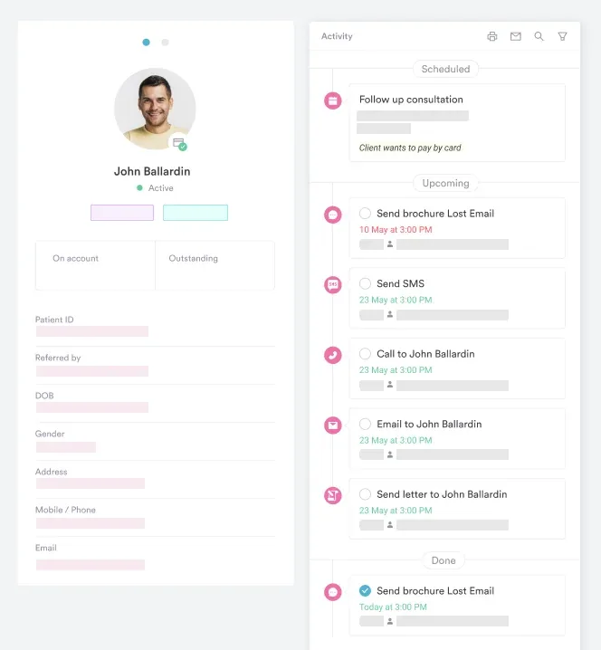
Patients with progressive neurological conditions need CN I re-tested at set intervals, not whenever they happen to mention it. Booking the next review during the same visit, with the odorant and the result already in the record, is what makes the later comparison possible.
The bedside test has clinical utility, but it is not a validated psychometric instrument. Where you need quantitative, reproducible data, a standardized smell identification test is the more reliable option.
The UPSIT is a 40-item scratch-and-sniff test with normative data by age and sex, developed by Sensonics International. Sniffin’ Sticks, produced by Burghart Messtechnik GmbH, generate a composite TDI score covering threshold, discrimination and identification. Both appear in the peer-reviewed literature, including a practical guide to CN I examination techniques in PMC. The bedside test remains the right tool for routine screening, and the standardized instruments earn their cost when you need comparable numbers across visits.
No bedside test is without limits. Five of them matter when you interpret a CN I result.
A normal bedside result does not rule out mild hyposmia, and an abnormal one needs clinical context before you draw a conclusion. Where standardized follow-up is hard to arrange, tie the examination result to a scheduled review in the patient record so the question comes back around.
Most abnormal results are managed conservatively or watched over time. A smaller group needs ENT or neurology input, and laterality is the first thing that separates them.
The referral rationale belongs in the record, not only in a covering letter the receiving clinician may never open. Neurology and rehabilitation teams share many of these patients, and software for rehab practices keeps the CN I finding visible to whoever sees them next.
When documenting a normal olfactory nerve test, record the specific odorant used, the nostril tested first, and the exact patient response. ‘CN I intact bilaterally’ is insufficient for clinical audit or medicolegal review. ‘Patient correctly identified coffee grounds and vanilla extract in both nostrils with eyes closed’ gives a reproducible, defensible record.
Free text is where CN I detail disappears. One clinician writes a full description, the next writes a two-word summary, and the two visits cannot be compared. The fields you actually need are always the same: odorant, side, detection, identification.
Practice management software like Pabau lets you build the cranial nerve examination into a form instead. Structured intake forms capture the odorant, the side tested and the response as separate fields. Every clinician in the practice then records the finding the same way.
Because the fields stay identical, you can put two examinations side by side and see whether smell is recovering or declining. Referral letters and audit reviews then draw on a record that already holds the detail, rather than on a clinician’s memory of the consultation.

Build the cranial nerve examination into a structured form. Pabau captures CN I findings, laterality and the odorant used in the same fields at every visit.

Two minutes of CN I testing buys a finding you cannot get any other way. Skip it and you can miss an early neurodegenerative marker, a post-traumatic lesion, or a unilateral loss pointing at an intracranial cause.
The trade-off is precision. The bedside test tells you that smell is reduced, never by how much. Any patient you plan to follow over months also needs a standardized instrument. Use the bedside test to decide who that is.
What survives the handover is the note, not the examination. Book a demo to see how Pabau keeps CN I findings, laterality and odorants in one comparable record.

Need the rest of the twelve-nerve sequence? Cranial nerve examination checklist lists every nerve in order, with space for laterality and the response you recorded.
Testing CN XII at the same visit? Hypoglossal nerve test covers tongue deviation, atrophy and fasciculation, and how to record each finding.
Assessing balance alongside the cranial nerves? Modified Romberg test explains the stance progression and the timing that keeps results comparable.
Screening peripheral nerve function in the hand? Hand nerve tests works through median, ulnar and radial testing and what each result rules out.
Recording reflexes in the same examination? Deep tendon reflex exam gives a grading scale and a form for logging each reflex by side.
The olfactory nerve test is a bedside examination that assesses cranial nerve I (CN I). You present familiar, non-irritating odorants to each nostril separately and record whether the patient can detect and identify them. It forms part of the routine cranial nerve examination and identifies anosmia, hyposmia, parosmia or phantosmia.
Ask the patient to close their eyes and occlude one nostril. Present a non-irritating odorant such as coffee or vanilla at 2 to 3 cm. Ask them first whether they detect any smell, then ask them to name it. Record a result for each nostril: intact, reduced, absent or distorted. Repeat on the other side with the same odorant, leaving 30 seconds between presentations to avoid adaptation.
Use familiar, volatile, non-irritating odorants: coffee grounds, vanilla extract, cloves and cinnamon are the most commonly recommended. Avoid ammonia, isopropyl alcohol and peppermint oil. All three stimulate the trigeminal nerve (CN V) rather than CN I, so the response carries no diagnostic value.
CN I is tested first in the standard twelve-nerve sequence. It takes under two minutes and needs no specialist equipment. Because it is so often omitted, including it in a structured protocol works as a quality indicator. That matters most in post-head-injury assessments, neurodegenerative screening and post-COVID follow-up.