



GLP-1 diet plan
A GLP-1 diet plan is a structured eating framework for patients taking semagluti...

2
Last Updated: Oct 13, 2026
A neck exam is a systematic clinical assessment of the cervical structures, soft tissues, and neurological function of the neck.
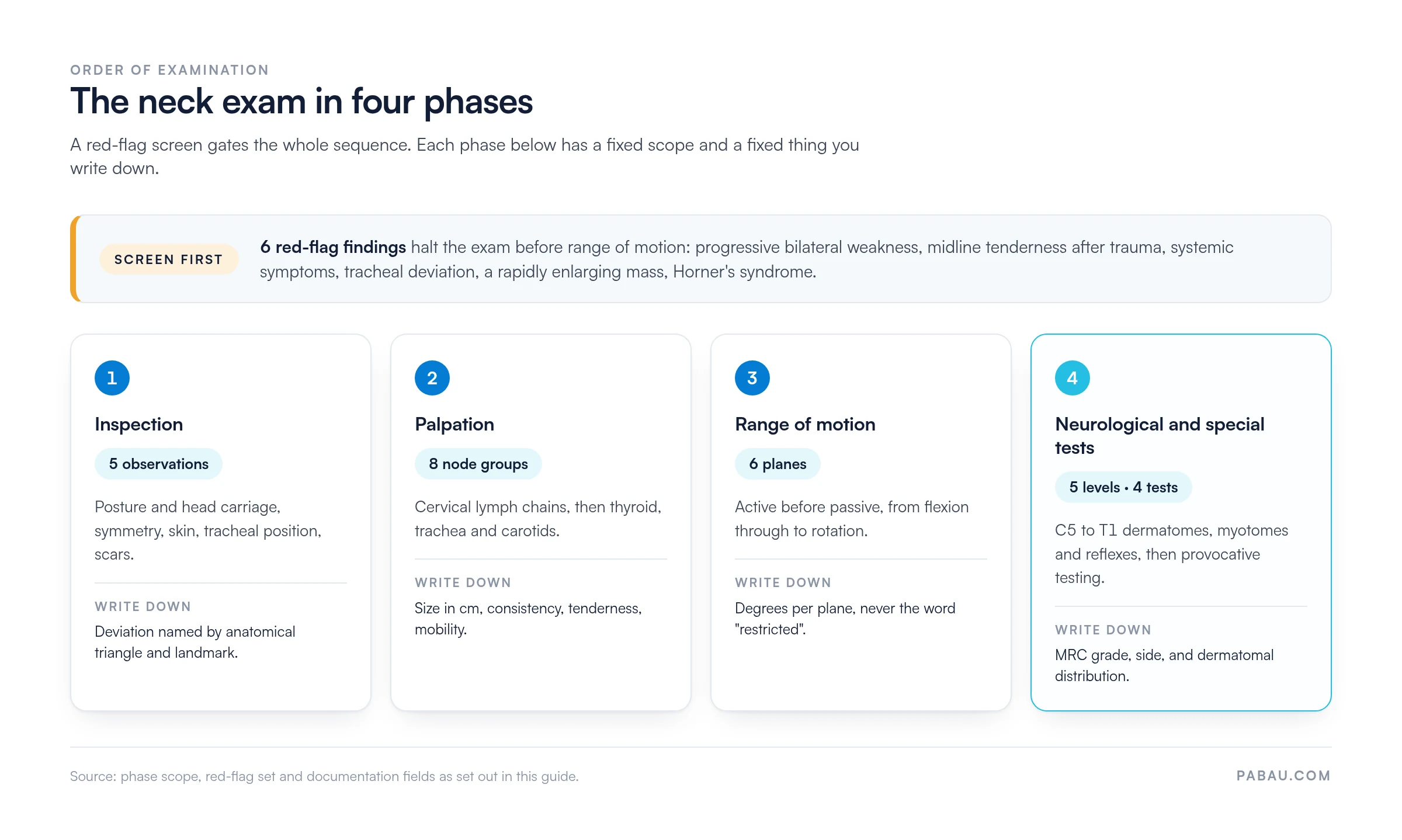
It runs in four phases: inspection, palpation, range of motion, then neurological and special testing. A red-flag screen comes first, because a handful of findings stop the exam before it starts.
The same sequence serves primary care, ENT, orthopedics, sports medicine, and musculoskeletal practice. According to the American Academy of Family Physicians (AAFP), neck pain is among the most common musculoskeletal presentations in primary care. This guide covers each phase in order, then closes on how to write the findings up for coding and medico-legal review.

A neck exam assesses cervical soft tissues, lymph nodes, thyroid, range of motion, and neurological function in a set sequence.
Red flags such as progressive neurological deficit, trauma, or signs of malignancy require urgent escalation before the standard exam continues.
The Spurling test has about 30% sensitivity and 93% specificity for cervical radiculopathy, so it confirms a suspicion rather than screens for one.
Record degrees, MRC grades, and node measurements rather than words like restricted, so a colleague can judge interval change.
Pabau’s customizable clinical note templates and digital forms help practitioners capture structured neck exam findings across multi-practitioner settings.
A neck exam is a structured physical assessment of the cervical spine, surrounding soft tissues, vasculature, glands, and lymphatic system. It identifies pathology, guides diagnosis, and directs management. The same assessment serves a range of encounters. A primary care practitioner uses it for new-onset neck pain, and an ENT clinician for a neck lump. An orthopedic surgeon reaches for it in suspected radiculopathy, and a sports medicine physician when clearing an athlete after a collision.
The exam follows a four-step framework shared across specialties.
Each phase has a fixed scope and a fixed set of findings you write down, and the red-flag screen gates the whole sequence. The diagram below puts those four phases side by side.
The precise emphasis varies by presentation. A soft tissue neck lump warrants deeper palpation of lymph node chains and thyroid. Acute cervicalgia after a motor vehicle collision requires clearing for instability before any range-of-motion testing. Which elements to prioritize is decided before you touch the patient, during history-taking.
History shapes every subsequent examination decision. Five clinical dimensions should be established before the physical neck exam begins.
Document the history findings before examination, so that clinical note templates can pre-populate the correct examination sections. Practices using digital intake questionnaires can capture these history points once and feed them straight into the consultation record.

Certain findings require urgent escalation before a standard neck physical exam is completed. Missing a red flag during a routine assessment carries serious medico-legal and patient safety consequences.
Several of these findings are neurological. Their referral thresholds follow NICE guideline NG127 on suspected neurological conditions, alongside AAFP neck pain evaluation protocols. Documenting the systematic red flag screen is essential for both patient safety and medico-legal defensibility. Practices managing musculoskeletal and ENT caseloads benefit from a consistent red flag prompt built into the note template itself.
Inspection begins the moment the patient enters the room. Observe posture and head position before any formal assessment starts.
Document inspection findings using precise anatomical descriptors. “Right anterior triangle fullness at the level of the angle of mandible” conveys far more than “neck swelling on the right.” Practitioners weighing up chiropractic practice management software or a sports medicine system should check whether its templates enforce that specificity by design.
Systematic palpation maps the neck’s key structures in a logical sequence. It moves from the submental region posteriorly and inferiorly to cover the major lymph node chains, then assesses the thyroid, trachea, and carotid arteries.
Cervical lymph node examination requires palpating eight anatomical groups in sequence. Use the pads of the index and middle fingers with gentle circular movements, comparing left and right simultaneously where possible.
A clinically significant lymph node is firm, non-tender, greater than 1 cm, and non-mobile. Soft, tender, mobile nodes typically represent reactive lymphadenopathy secondary to local infection. Hard, fixed, non-tender nodes raise concern for malignancy. Document size in centimeters, consistency, tenderness, and mobility for each significant finding.
Knowing what each group drains narrows the search for a source. A lymphatic massage chart maps the cervical nodes against the facial, scalp and sinus areas they drain.
Thyroid gland palpation is best performed with the patient seated and the neck slightly flexed, which relaxes the overlying strap muscles. Two techniques are used.
Document the overall gland size, its consistency (soft, firm, nodular), the presence and number of discrete nodules, tenderness, and whether a bruit is present. A normal gland is roughly the size of the patient’s thumb. A solitary hard nodule in a patient over 50 warrants urgent referral even without systemic symptoms. The American Thyroid Association’s guidance on thyroid nodules sets out the referral thresholds.
Cervical spine range of motion is assessed in six planes. Always assess active ROM before passive, to avoid exacerbating pain or causing neurovascular compromise. The normal reference values below are drawn from the AMA Guides to the Evaluation of Permanent Impairment and standard orthopedic references. The same principles apply to any joint, so a practitioner already comfortable with range of motion assessment elsewhere will recognize the method.
Document ROM as actual degrees, or as a fraction of normal such as “50% of expected flexion.” Avoid “restricted” or “reduced” on their own. Precise figures support serial comparison at follow-up and are required for medico-legal reporting. If nerve root symptoms are reproduced on any movement, note the direction and the distribution of those symptoms.
Neurological assessment determines whether symptoms arise from nerve root compression (radiculopathy) or spinal cord compromise (myelopathy). The examination covers three domains at each cervical level: dermatomes, myotomes, and reflexes.
Cervical radiculopathy most commonly affects the C6 and C7 nerve roots, which matches the epidemiology reported in AAFP and UpToDate clinical references. C6 compression typically presents with pain radiating to the lateral forearm and thumb, weakness of elbow flexors, and a diminished brachioradialis reflex. C7 compression causes posterior arm and middle finger pain, triceps weakness, and a reduced triceps reflex.
Myelopathy screening adds Hoffmann’s sign and clonus testing at the wrist or ankle. For Hoffmann’s, flick the middle finger; the sign is abnormal if the thumb and index finger flex. Bilateral corticospinal tract signs alongside neck pain are a surgical emergency until imaging rules out cord compression. Document whether findings are unilateral or bilateral, which levels are involved, and motor weakness on the MRC scale (0-5).
Document neurological findings at the level of precision that lets another clinician assess interval change. ‘Mild weakness of elbow flexors, 4/5 MRC, right C5-C6’ conveys more than ‘some arm weakness.’ Standardized neurology fields in your clinical note templates make serial comparison straightforward.
Special tests confirm or challenge the clinical hypotheses formed during history and inspection. Four tests have the strongest evidence base for the cervical spine exam.
The Spurling test detects cervical nerve root compression by narrowing the neural foramen under load. The examiner laterally flexes and extends the patient’s head toward the symptomatic side, then applies axial compression through the crown. A positive result reproduces radicular arm symptoms in the expected dermatomal distribution.
In the validation study by Tong, Haig and Yamakawa (Spine, 2002), the test showed 30% sensitivity and 93% specificity for cervical radiculopathy against electrodiagnostic findings. Those figures make it a confirmatory test rather than a screening one. A positive finding meaningfully raises the probability of radiculopathy, but a negative result does not exclude it.
The distraction test is the functional inverse of Spurling. The examiner manually distracts the cervical spine by gently lifting the head, which unloads compressed neural structures. Relief of arm symptoms on distraction supports a diagnosis of nerve root compression. It also helps you counsel the patient on the likely benefit of cervical traction within a physical therapy program.
The ULTT assesses neural mechanosensitivity along the brachial plexus. With the patient supine, the examiner depresses the shoulder girdle, then abducts and externally rotates the shoulder. The elbow is extended, the forearm supinated, and the wrist and fingers extended. The cervical spine is then laterally flexed away from the test side. Reproduction of the patient’s arm or hand symptoms is a positive result. The test has higher sensitivity than Spurling but lower specificity, so it works better as an early screening tool.
This is the simplest provocative test in the neck exam. Ask the patient to place a hand on top of their head, which abducts the shoulder and reduces traction on the affected nerve root. Relief of radicular symptoms is a positive sign, and it correlates well with MRI evidence of compression at C5-C6 or C6-C7. The sign suits primary care settings where formal provocative testing is impractical.
Whichever tests you run, record the test name, the technique used, the result, the symptoms reproduced, and the dermatomal distribution. That level of detail supports the medical decision-making element of an E/M code. The CMS guide to evaluation and management services sets out what is expected.
Auscultation is not part of every neck exam, but it is indicated when a vascular cause is suspected. Place the bell of the stethoscope over the carotid arteries bilaterally and ask the patient to hold their breath. A carotid bruit suggests turbulent flow from atherosclerotic stenosis and warrants Doppler ultrasound.
A bruit over a thyroid nodule is more consistent with a hypervascular gland, such as Graves’ disease or a toxic nodule, than with malignancy. Auscultate the heart at the same time, so a transmitted cardiac murmur is not mistaken for a carotid bruit. Document the site, the timing (systolic or diastolic), and the character of any bruit you identify.
Most clinical reference guides stop at the examination itself and say nothing about writing it up. Yet the note determines the legal defensibility of the consultation and the accuracy of E/M coding. It also decides whether a colleague reviewing the record can tell what was found. Structured documentation follows the same sequence as the examination.
A structured template beats free text on three counts. It supports pattern recognition across a patient’s records, cuts documentation time, and stops findings being dropped under time pressure. Practices running musculoskeletal caseloads on an EMR for rehab teams can enforce these fields at the point of entry. The SOAP framework fits neck exam workflows well, because the Objective section maps directly onto the four examination phases.
After completing the neck exam, summarize the findings aloud to the patient and say clearly which investigations or referrals will follow. A well-conducted examination loses much of its value if the patient leaves uncertain about next steps.
For practitioners preparing medico-legal or insurance reports, the structured template described above maps onto the documentation frameworks those commissioning bodies expect. Review your examination templates periodically and update them when guidelines change, so documentation standards stay current across the whole practice.
In most practices the neck exam is written up as free text, minutes or hours after the consultation. Degrees become “restricted,” node measurements go unrecorded, and the next clinician cannot tell whether the patient has improved. That is a documentation problem rather than an examination problem, and it is solved at the template level.
Pabau, practice management software for healthcare practices, includes customizable examination note templates for exactly this job. Instead of free text, you configure field-by-field templates covering inspection, palpation, ROM degrees, neurological levels, and special test results. Those fields sit inside patient record management, so each exam is stored against the patient rather than in a separate document.
Pabau Scribe, our AI scribe, transcribes the consultation and maps what was said into those template fields. That cuts post-consultation writing time without losing precision. A consistent template pays off most in a multi-practitioner setting. Any clinician opening a neck exam record finds each finding in the same place, instead of reading through narrative of varying quality.
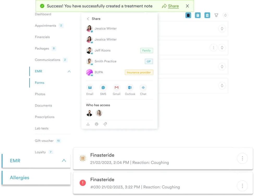

Pabau’s customizable clinical templates and Pabau Scribe let your team capture structured neck exam findings in a fraction of the time. Every field lands where coding, follow-up, and audit need it.

The neck exam itself is rarely where practitioners come unstuck. Run the four phases in order behind a red-flag screen and the findings will be there. What varies between practitioners, and between visits, is how those findings get written down.
So pick the fields before the next consultation, not during it. Degrees instead of “restricted,” centimeters instead of “enlarged,” MRC grades instead of “weak.” The trade-off is a slightly slower first note in exchange for a record any colleague can act on months later.
Standardizing that across every clinician and location is the part software does better than policy. Book a demo to see how structured exam templates cut the documentation burden without thinning the clinical detail.

Want the Spurling test in full detail? Spurling’s test walks through the technique, the grading, and how to record a positive result.
Measuring motion at other joints? Range of motion assessment covers goniometry technique and the normal values worth documenting.
Need to test cervical endurance as well as motion? Neck flexor endurance test gives you the protocol and a recording sheet.
Looking for what comes after the exam? Neck pain exercises sets out the conservative management options you can hand a patient.
Applying the same structure to another joint? Hip examination follows the same inspection, palpation, motion, and special tests sequence.
A neck exam includes four components. Inspection covers posture, symmetry, skin, and tracheal position. Palpation covers lymph nodes, thyroid, trachea, and carotid arteries. Range of motion is assessed in six planes: flexion, extension, bilateral lateral flexion, and bilateral rotation. Neurological testing covers dermatomes, myotomes, and reflexes at C5-T1, with special tests such as the Spurling test when radiculopathy is suspected.
Red flags during a neck exam include progressive bilateral limb weakness and midline cervical tenderness after trauma. Systemic symptoms such as fever, night sweats, and unexplained weight loss count too. Tracheal deviation with respiratory compromise, a rapidly enlarging neck mass, and Horner’s syndrome belong on the same list. Any of these findings requires urgent investigation before a standard examination continues.
The Spurling test is a provocative test for cervical radiculopathy. The examiner laterally flexes and extends the patient’s head toward the symptomatic side, then applies axial compression through the crown. In the 2002 validation study by Tong, Haig and Yamakawa, it showed 30% sensitivity and 93% specificity for nerve root compression. That makes it a confirmatory test: a positive result strongly supports radiculopathy, but a negative result does not exclude it.
Cervical lymph nodes are examined by palpating eight anatomical groups in sequence, using the pads of the index and middle fingers with gentle circular movements. The groups are submental, submandibular, jugulodigastric, the upper, mid and lower deep cervical chain, the posterior triangle, and supraclavicular nodes. For each enlarged node, document size in centimeters, consistency, tenderness, and mobility. Firm, non-tender, fixed nodes over 1 cm are clinically significant and warrant further investigation.
Cervical radiculopathy presents with unilateral arm pain, weakness, or sensory change in a dermatomal distribution. Spurling or the ULTT is typically positive, and the reflex at the affected level is reduced. Myelopathy presents with bilateral limb involvement and upper motor neuron signs. Those include Hoffmann’s sign, clonus, hyperreflexia, an extensor plantar response, and sometimes gait disturbance. Myelopathy is a surgical emergency until cord compression is excluded on MRI.
Neck exam findings belong in the Objective section of a SOAP note, in the order the examination ran. Record inspection findings, then palpation findings for each lymph node group, the thyroid, and the carotid pulses. Record ROM in degrees per plane, then neurological findings with MRC-graded myotome strength and reflex grades. Close with special test results: test name, technique, outcome, and symptoms reproduced. Structured templates that enforce these fields reduce omissions and support E/M coding compliance.






