



GLP-1 diet plan
A GLP-1 diet plan is a structured eating framework for patients taking semagluti...

2
Last Updated: Oct 13, 2026

Homan’s sign test uses passive ankle dorsiflexion to screen for deep vein thrombosis (DVT), but it is no longer a reliable standalone test.
Sensitivity ranges from 10 to 54% and specificity from 39 to 89%, so false positives and false negatives are both common.
Current guidelines favor the Wells score, D-dimer testing, and compression ultrasonography over Homan’s sign for DVT diagnosis.
Pabau’s digital forms and clinical records help practitioners document DVT risk and examination findings the same way at every visit.
Homan’s sign test is a bedside physical examination maneuver with over eight decades of clinical history. John Homans first described it in 1941 and redefined it in 1944. Clinicians used it to screen for deep vein thrombosis (DVT) in patients with calf pain, one-sided leg swelling, or a high clot risk.
DVT occurs when a blood clot forms in a deep vein, usually in the calf or thigh. Left untreated, it can travel to the lungs and cause a fatal pulmonary embolism (PE). Homan’s sign was meant to elicit the pain of venous inflammation. The evidence says it does that job poorly.
So the useful question is no longer whether to perform it. It is what a recorded Homan’s sign should and should not change in your next decision. This guide covers the technique, the accuracy behind the doubt, the conditions that fake a positive, and what to document instead.
The procedure is straightforward. Variations exist in the literature, but the standard technique follows this sequence.
A variant described by some authors adds knee flexion during dorsiflexion, which increases mechanical traction on the posterior tibial vein. Research published in PubMed Central links that traction to the stimulation of pain-sensitive structures. That is the proposed mechanism behind the response the test elicits.
The same movement is measured deliberately elsewhere in rehab work. The ankle dorsiflexion test records range in degrees rather than pain, which gives you a number to track between visits.
A painful lower limb is often several things at once. The Ottawa ankle rules calculator is a useful companion in physical therapy settings, since it tells you when an ankle needs imaging.
A positive Homan’s sign is recorded when the patient reports calf pain during passive dorsiflexion. Some sources widen this to include any posterior calf discomfort, tenderness, or resistance during the movement.
A negative result means no calf pain is produced. A negative Homan’s sign does not exclude DVT. Multiple studies confirm that the test misses a clinically significant share of confirmed clots.
Neither result should be used on its own to confirm or rule out DVT. The test provides a signal, not a diagnosis. Any positive finding warrants further investigation through validated tools.
A positive result tells you the calf hurts on dorsiflexion. It does not confirm that a clot is present. The list of non-DVT conditions that produce the same response is long enough to make the finding ambiguous in most presentations.
Not accurate enough to stand alone, and the evidence has said so for decades. A peer-reviewed analysis in PubMed Central (PMC5485383) reports sensitivity of roughly 10 to 54% and specificity of 39 to 89%. That spread is the problem. When results vary this much across studies, one patient’s finding is hard to read with confidence.
A useful diagnostic test aims to clear 90% on both measures. It should miss few cases, and it should send few healthy patients for scans they do not need. Homan’s sign clears neither bar, even at the top of its reported range.
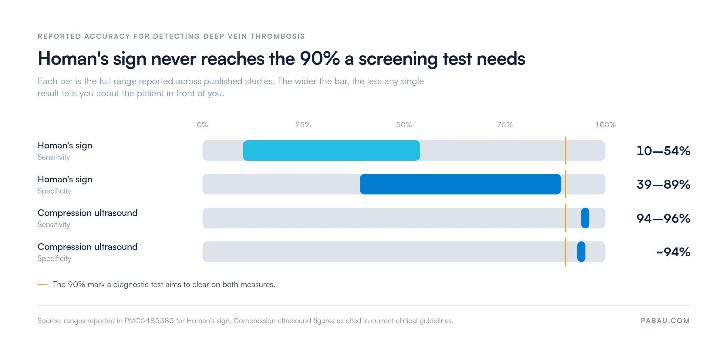
The ultrasound figures reflect data cited across current clinical guidelines, and the Homan’s sign range comes from PMC5485383. None of this dismisses bedside examination. It places one maneuver where it belongs in a modern diagnostic pathway.
This is the core clinical problem. Several common, benign conditions produce calf pain on dorsiflexion, which returns a positive result with no clot present at all.
A patient with any of these conditions can produce a positive Homan’s sign. Without a validated pre-test probability score and objective imaging, that positive finding gives you little to act on.
Lymphedema is also the one condition on that list a therapist may go on to treat. Clear DVT first, because a lymphatic drainage map lists it among the contraindications to screen for before manual drainage starts.
Postpartum women are a high-risk group for DVT. Hormonal shifts, reduced mobility, venous stasis, and increased clotting during and after pregnancy all raise that risk. It matters here because the test’s unreliability gets worse in this population.
Pregnancy itself causes leg edema and calf discomfort. Many pregnant and postpartum patients carry non-DVT calf pain as a baseline. A positive Homan’s sign in this group is therefore even weaker as a guide to the next decision.
DVT symptoms to watch for after birth include one-sided leg swelling, localized warmth, skin discoloration, and new calf tenderness. None of these is specific to DVT on its own. Their presence together, one-sided swelling especially, warrants urgent assessment.
A negative Homan’s sign must never reassure a postpartum patient that DVT has been excluded. Refer anyone with suspected postpartum DVT for compression ultrasonography and Wells score assessment instead of relying on the bedside maneuver.
Document DVT risk factors and physical examination findings in structured clinical notes at every relevant consultation, particularly for postpartum patients. Consistent documentation creates an auditable record if symptoms escalate and the patient requires urgent referral.
Three validated tools sit at the center of evidence-based DVT assessment. Each one covers what Homan’s sign leaves unanswered. Teams in physical therapy practices meet all three in referral pathways and post-operative monitoring protocols.
The Wells DVT score is a validated tool that assigns points for specific risk factors and clinical signs. It sorts patients into low, moderate, or high pre-test probability, which guides how much further testing they need.
A score of 2 or above indicates high probability. Below 2 is low probability, where a negative D-dimer can exclude DVT without imaging in many patients. NICE guidance NG158 builds the Wells score into the recommended UK assessment pathway.
D-dimer is a fibrin degradation product released when a clot breaks down. Raised levels are not specific to DVT, since they also rise with infection, pregnancy, and recent surgery. A low D-dimer in a low-probability patient is highly sensitive for excluding DVT without imaging.
Compression ultrasound is the preferred imaging modality for DVT diagnosis. CDC resources on deep vein thrombosis and international clinical guidelines both describe it as the gold standard. The technique applies pressure with an ultrasound probe over the deep veins of the leg. A normal vein compresses fully, and a vein holding thrombus does not. Sensitivity and specificity both exceed 94% for proximal DVT.
Pratt’s sign involves squeezing the calf with the knee flexed to elicit posterior pain. Moses sign uses front-to-back calf compression rather than side-to-side. Both share Homan’s fundamental limitation, since neither has the sensitivity or specificity to replace a validated tool. They survive in clinical teaching, not in diagnostic algorithms.
Rarely, and never as a primary diagnostic tool. Vascular medicine and emergency medicine protocols have moved away from it. The American College of Chest Physicians (ACCP) and NICE both leave Homan’s sign out of their recommended DVT algorithms.
It survives in clinical teaching as an example of a maneuver that was widely used before imaging became accessible. Some practitioners still perform it inside a broader lower limb examination, as a documentation data point rather than a diagnostic one.
The Physiopedia review of Homan’s sign notes that current evidence does not support it as a reliable standalone maneuver. That reflects the consensus across vascular medicine, emergency medicine, and physical therapy guidelines.
Recording the result is still fair practice, as long as the note says what it did and did not rule out. Written physiotherapy compliance requirements expect examination findings and referral decisions to be documented the same way by every clinician.
A positive Homan’s sign is not grounds to diagnose DVT, and a negative one is not grounds to reassure. Both directions carry risk. The symptoms below warrant urgent medical assessment whatever the bedside test showed.
Pulmonary embolism is a life-threatening complication of untreated DVT. Any suspicion of PE requires immediate emergency referral. Never use a negative Homan’s sign to rule out DVT when other clinical features are present. A negative result carries no safety-net value in a symptomatic patient.
Examination findings only earn their keep when they are recorded the same way every time. Practices managing patients with raised DVT risk usually know the clinical picture well. What slips is the record of who checked, what they found, and what they decided next.
Practice management software like Pabau gives you digital clinical intake forms, so you can build structured assessment templates. They capture Wells score components, examination findings, and referral decisions at every relevant visit. Every clinician then documents DVT risk against the same fields instead of free text.

The patient clinical records module stores examination findings in date order. If a patient’s leg swelling worsens between visits, that trend is visible in the record. Nobody has to hunt through handwritten notes or a separate document to find last month’s measurement.

For multi-practitioner practices, that consistency protects continuity of care. A covering clinician picking up the record can see what was examined and what was decided last time. That matters when the condition being monitored can deteriorate quickly.

Pabau’s digital forms and clinical records let you capture DVT risk factors, examination findings, and referral decisions in structured notes. Every practitioner documents the same way, and every record is ready for audit.

Treat Homan’s sign as a piece of history you may still meet in a chart, not as a decision point. If you perform it, record what it showed and what it could not exclude. Then run the assessment that carries weight.
The trade-off worth remembering is the one that keeps the maneuver alive. It costs nothing and takes seconds. That same low cost tempts clinicians to act on a positive result and to stop looking after a negative one.
The Wells score, D-dimer, and compression ultrasonography are the pathway that holds up. Follow it and you catch more clots early, while sparing healthy patients a scan they never needed.
Consistent records are what make that pathway repeatable across a whole team. Book a demo to see how Pabau keeps DVT risk assessments and referral decisions in one patient record.

Need a validated rule for a painful lower limb? Ottawa ankle rules calculator shows when an ankle injury actually needs an X-ray.
Want another bedside test with a specificity trade-off? Crossed straight leg raise test walks through the steps and how far the result can be trusted.
Screening older or post-surgical patients? Fall risk assessment gives you a scored template you can complete in the same visit.
Managing a patient back to activity after injury? Return to running protocol sets out a staged framework with clear progression checkpoints.
Homan’s sign test is a bedside maneuver once used to screen for deep vein thrombosis (DVT). It applies passive dorsiflexion of the ankle to elicit pain in the back of the calf. Current guidance no longer treats it as a reliable diagnostic tool.
A positive Homan’s sign means the patient reports calf pain during passive ankle dorsiflexion. It does not confirm DVT. Muscle strain, Achilles tendinitis, cellulitis, and a ruptured Baker’s cyst all produce the same response. Treat a positive result as a prompt for the Wells score and D-dimer testing.
No. Published studies report sensitivity of 10 to 54% and specificity of 39 to 89%, which is too variable for clinical diagnosis. The American College of Chest Physicians and NICE both leave it out of their DVT pathways. Both favor the Wells score, D-dimer, and compression ultrasonography.
Three tools carry the work. The Wells DVT score sets pre-test probability, D-dimer testing helps exclude DVT in low-probability patients, and compression duplex ultrasonography confirms it. Ultrasound sensitivity and specificity both exceed 94% for proximal DVT. Most evidence-based pathways use the three together.
Rarely, and not as a primary diagnostic tool. Most current DVT guidelines leave it out of their recommended algorithms. Some practitioners still perform it inside a broader lower limb examination for documentation. It should never be used alone to confirm or exclude DVT.
Yes, often. Muscle strain, Achilles tendinitis, cellulitis, a ruptured Baker’s cyst, superficial thrombophlebitis, and lymphedema all produce a positive result. That false positive rate is one of the main reasons the test is treated as unreliable on its own.






