



GLP-1 diet plan
A GLP-1 diet plan is a structured eating framework for patients taking semagluti...

2
Last Updated: Oct 13, 2026

Confirming a heart attack takes troponin blood work, an ECG, and imaging read together, not one test alone.
High-sensitivity troponin is the primary biomarker, and it can detect cardiac damage within 1-3 hours of symptom onset.
A normal ECG does not rule out a heart attack, because NSTEMI often shows non-specific changes or none at all.
Serial testing beats a single reading, since the rise and fall pattern separates a heart attack from other causes.
Practice management software like Pabau keeps troponin results, ECG findings, and imaging reports in one timestamped patient record.
No single heart attack test confirms or rules out a cardiac event. Instead, a confirmed diagnosis pulls together blood tests, electrical tracings, imaging, and clinical risk scores. In an emergency department, several of those run at the same time.
According to the American Heart Association, diagnosis starts with a history review, a physical exam, and an ECG within 10 minutes of arrival. Blood tests for cardiac biomarkers follow next.
Knowing which test answers which question helps clinicians document findings clearly, and helps patients follow what is happening.
This guide covers each major test used to confirm a myocardial infarction (MI) and how they fit together. It also covers how practices keep records straight when several tests run in parallel. Practices using lab management software can hold those results in one patient record instead of chasing them across systems.
A heart attack is diagnosed by reading an ECG first, then serial troponin blood tests, then imaging where the picture is still unclear. Each step adds evidence, and none of them stands alone.
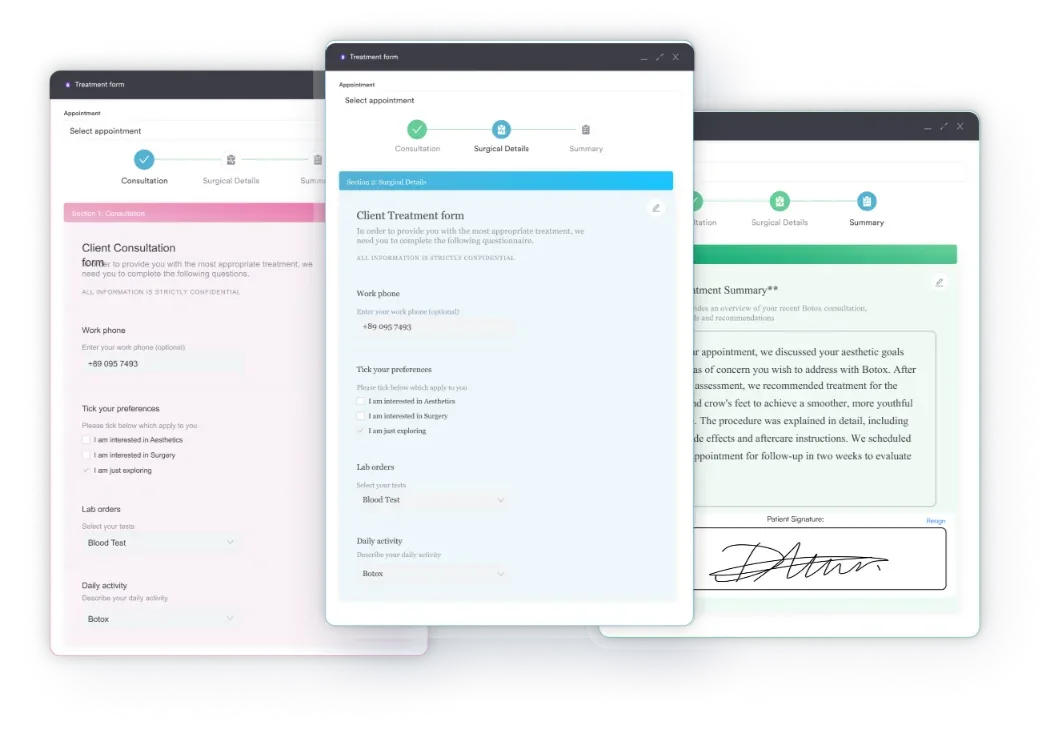
Primary care and urgent care practices lean on digital intake forms at this point. Capturing symptom onset time, risk factors, and current medications gives the assessing clinician a complete starting picture.
Damaged heart muscle releases proteins into the bloodstream. Blood tests measure these cardiac biomarkers to confirm whether damage has happened. That makes the blood test the most direct chemical confirmation of a myocardial infarction.
Heart muscle cells release troponin I and troponin T, together called cardiac troponin, when they are injured. Troponin is now the preferred biomarker for diagnosing a heart attack. The AHA, Mayo Clinic, NHLBI, and the National Library of Medicine all take that position.
Troponin levels start rising within 2-4 hours of cardiac injury. They then peak at 12-24 hours and can stay elevated for 7-14 days, which helps when a patient presents late.
A troponin levels chart gives a visual reference for how those values trend. The guide on interpreting cardiac biomarkers covers how to report findings without overstating certainty.
High-sensitivity troponin (hs-troponin) tests pick up far smaller concentrations of the protein than older assays. According to the US National Library of Medicine, hs-troponin can identify cardiac damage within 1-3 hours of symptom onset.
That speed is what makes rapid rule-out protocols possible, including the ESC 0h/1h and 0h/2h algorithms. Low-risk patients can be discharged safely within a few hours instead of sitting under observation for 6-12. As a result, faster turnaround eases bed pressure without costing diagnostic accuracy.
CK-MB (creatine kinase-MB) was the standard biomarker before high-sensitivity troponin arrived. It rises within 4-6 hours of MI onset and returns to baseline within 48-72 hours. That short window makes it useful for spotting reinfarction while troponin is still elevated from a prior event.
Myoglobin rises earlier than troponin, within 1-3 hours, but it is less specific to cardiac tissue. Skeletal muscle injury raises it too, which limits its value as a standalone heart attack test. As a result, both markers now sit behind troponin in most clinical protocols.
Yes. An elevated troponin does not confirm a heart attack on its own. Other causes include myocarditis, pulmonary embolism, severe sepsis, acute kidney disease, and heart failure.
Clinical context governs the reading. The pattern of rise and fall, the patient’s symptoms, and the ECG findings together determine the diagnosis. Instead, a single high value calls for serial testing and a full clinical assessment before anyone draws a conclusion.
An electrocardiogram (ECG or EKG) records the heart’s electrical activity across 12 leads placed on the chest, arms, and legs. It takes under five minutes. It is usually the first test clinicians perform on a patient presenting with chest pain.
ST-segment elevation indicates a STEMI, the most severe type of heart attack, caused by a completely blocked coronary artery. That finding triggers immediate intervention, typically within 90 minutes of arrival under ACC/AHA guidelines.
A normal ECG does not rule out a heart attack. NSTEMI can show non-specific changes, or a completely normal tracing at first presentation. That is why the ECG always works alongside troponin testing rather than in place of it.
Practices handling cardiac symptoms benefit from safer clinical documentation that flags when ECG and biomarker results disagree.
Blood tests and ECGs confirm biochemical and electrical damage. Imaging shows where the damage sits, how much heart function is left, and whether arteries are blocked.
An echocardiogram uses ultrasound waves to produce live images of the heart’s chambers and valves. After a suspected heart attack, it assesses wall motion abnormalities, the areas where muscle contracts weakly or not at all. It also measures left ventricular ejection fraction, one input to the cardiac output formula.
As a result, an ejection fraction below 40% after MI indicates significant damage. That figure drives decisions about medication, device therapy, and follow-up imaging. It is stroke volume divided by end-diastolic volume, so it starts with the stroke volume equation. Clinicians usually perform the echo within 24 hours of a confirmed event, or sooner if the diagnosis is still uncertain.
Coronary angiography is the definitive imaging test for identifying blocked coronary arteries. The clinician threads a catheter through the wrist or groin to the heart. The team then injects contrast dye, and X-ray imaging maps the coronary circulation, showing blockages as narrowings or complete occlusions.
According to the Mayo Clinic, clinicians perform angiography urgently for STEMI to guide revascularization by balloon angioplasty or stenting. For NSTEMI, timing follows clinical risk. High-risk patients go to the cath lab within 24 hours, while stable patients may be assessed within 72.
Clinicians order a chest X-ray early to check heart size and look for pulmonary edema. That is the fluid that collects in the lungs when the pump fails. It also helps exclude other causes of chest pain, such as pneumothorax or aortic dissection. It does not confirm a heart attack directly.
Clinicians increasingly use cardiac CT angiography in stable chest pain to visualize coronary anatomy without a catheter. Cardiac MRI characterizes tissue in detail, separating viable from non-viable myocardium.
It earns its place when troponin is raised but the clinical picture is unclear, since it can size an infarct and rule out myocarditis. Consequently, both scans often need prior authorization, which a medical necessity letter supports.
A stress test has no role during a suspected acute heart attack. Clinicians order it for stable patients after a cardiac event. It also suits chest pain of unclear origin that needs assessment for underlying ischemia.
During an exercise stress test, the patient walks a treadmill at rising speed and incline while ECG and blood pressure are monitored throughout. ST-segment changes, symptoms, or a drop in blood pressure all point to ischemia. Patients who cannot exercise receive a pharmacological stress agent such as adenosine or dobutamine.
Clinicians record heart rate recovery in the first minute after exercise too, and a heart rate recovery chart supplies age-based reference values. Sports medicine practices similarly run the same protocol when clearing a patient to return to activity.
Nuclear stress tests pair exercise or drug-induced stress with radionuclide imaging to show areas of reduced blood flow. A stress echocardiogram uses ultrasound during stress to catch wall motion abnormalities that only appear under load. Practices offering cardiac follow-up can use AI scribes to record findings and post-test instructions as they go.
Document the exact time of symptom onset in every cardiac presentation record, not just the time the patient arrived. The onset-to-treatment interval drives ACC/AHA performance metrics and determines which rapid rule-out protocol applies. Serial troponin timing is calculated from symptom onset, not from blood draw time.
Risk scores tell clinicians which test to run next and how urgently, without leaning on any single result.
The HEART score (History, ECG, Age, Risk factors, Troponin) assigns 0-2 points across five dimensions. A score of 0-3 marks a low-risk patient suitable for early discharge. A score of 4-6 warrants observation and repeat troponin testing. Meanwhile, anything above 6 signals high risk and usually leads to an early invasive workup.
The TIMI score (Thrombolysis in Myocardial Infarction) is a seven-variable tool for unstable angina and NSTEMI. It estimates the probability of a major cardiac event at 14 days. That estimate guides anticoagulation intensity and the timing of angiography.
Surgical teams reach for a third score to answer a different question. The revised cardiac risk index estimates cardiac risk around non-cardiac surgery, which is a separate judgment from working up chest pain in the emergency department.
Diagnosis runs on a clock. The table below maps each test against the window it belongs in and the decision it feeds.
Timestamped documentation at every stage does double duty. It carries the clinical handover, and it underpins audit trails, payer reporting, and quality metrics. Practices using AI-powered clinical documentation can capture findings as they happen, with notes attached to the patient record rather than dictated afterwards.
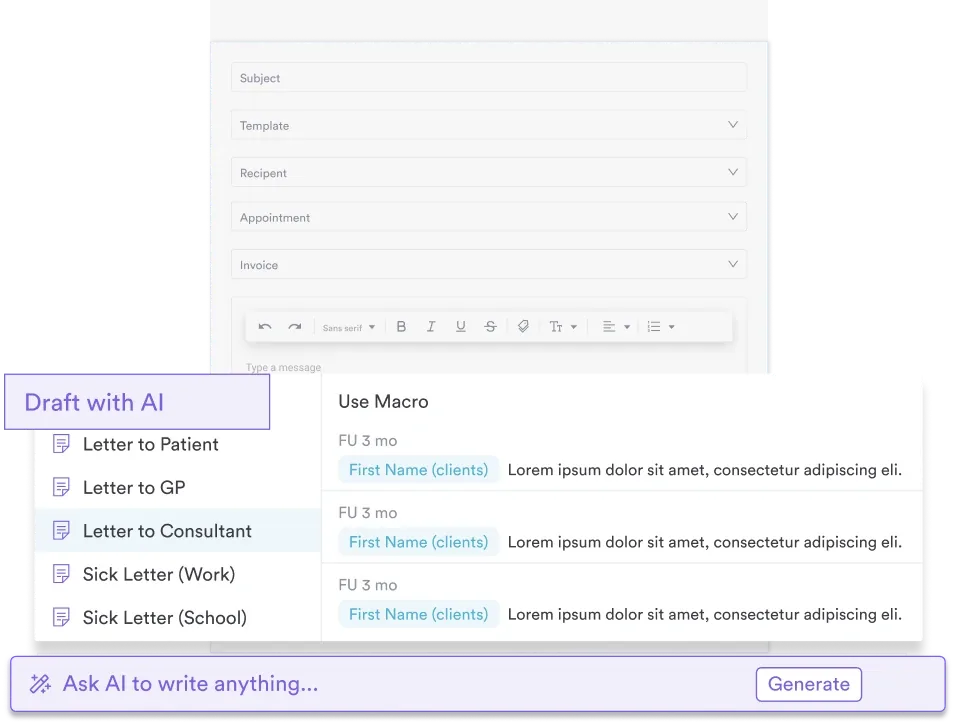
Afterward, the record has to travel with the patient after discharge. A patient discharge form covering medication changes, warning symptoms, and the follow-up date keeps the patient and the next clinician on the same instructions.
A cardiac workup produces results in several places at once. The lab returns serial troponin values, the ECG machine prints a tracing, and radiology sends the echo report separately. Without one place to put them, someone rebuilds the chart by hand from three or four systems.
Practice management software like Pabau holds all of it in one patient record. Intake forms capture symptom onset and risk factors at check-in. Lab results attach to the same chart, so structured patient records show the full sequence rather than the latest value alone.
Pabau Scribe, our AI scribe, writes the consultation note while the clinician talks. The note lands in the record with a timestamp, which matters when the onset-to-treatment interval is audited later. As a result, cardiology and primary care teams finish the day with an accurate chart instead of a dictation backlog.
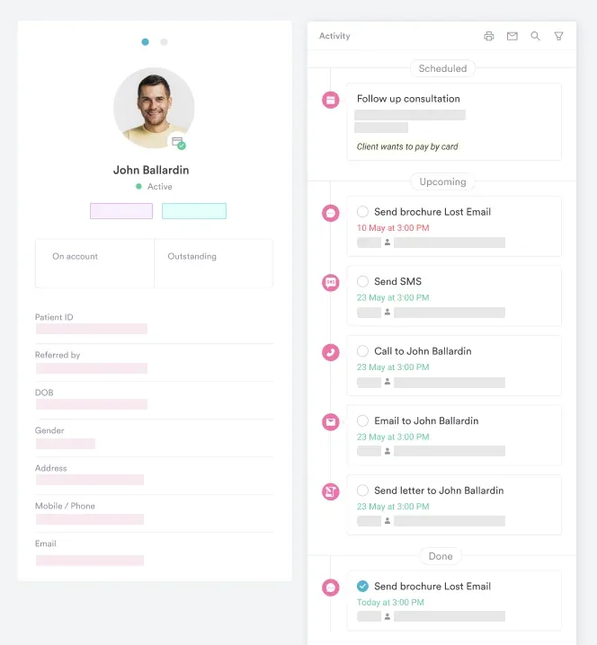

Practice management software like Pabau keeps troponin results, ECG findings, and imaging reports in one timestamped patient record. Digital intake forms and AI-written clinical notes cut the documentation load on your team.

Timing decides a cardiac diagnosis as much as the results do. A troponin drawn too early can read normal in a patient who is having a heart attack. The second sample, an hour or three later, is the one that settles it.
So record the clock, not just the result. Note symptom onset, draw time, ECG time, and the moment each decision was made. That trail is what lets the next clinician read a trend instead of a single number.
Overall, practices that hold the whole workup in one chart spend less time reassembling it later. Primary care practice software keeps intake forms, lab results, and clinical notes together. Book a demo to see how Pabau handles a cardiac workup from arrival to follow-up.

Tracking a patient through cardiac rehab? Sleeping heart rate chart gives age-based overnight ranges to compare against during recovery.
Measuring cardiac output at the bedside? Fick cardiac output calculator walks through the oxygen-consumption method and the values it needs.
Need a patient hygiene checklist? Personal hygiene checklist covers 32 items across eight topics, from oral hygiene to bedding.
A heart attack test is any diagnostic procedure used to confirm or rule out myocardial infarction. The most important are the high-sensitivity troponin blood test and the ECG, typically used together. Supporting tests include echocardiography, coronary angiography, and risk scores such as the HEART score. No single test stands alone.
With high-sensitivity troponin assays and the ESC 0h/1h rapid rule-out protocol, a heart attack can be confirmed or excluded within 1-3 hours of arrival. Clinicians diagnose STEMI within minutes on ECG findings alone, which triggers cath-lab activation before blood results return.
No. A normal ECG does not rule out a heart attack. NSTEMI can present with non-specific changes or a completely normal tracing. Serial troponin blood tests are essential alongside the ECG for any patient with chest pain and cardiac risk factors.
Yes. Conditions that raise troponin without a heart attack include myocarditis, pulmonary embolism, severe sepsis, acute kidney injury, and heart failure. Clinicians must read elevated troponin alongside the clinical picture, the ECG findings, and the pattern of serial results before confirming a diagnosis.
The HEART score assigns 0-2 points across five variables: History, ECG, Age, Risk factors, and Troponin. A score of 0-3 identifies low-risk patients suitable for discharge without further cardiac workup. A score above 6 signals high risk and usually leads to early invasive investigation. Emergency and cardiology teams use it to stratify chest pain patients and guide test ordering.
Clinicians use blood tests for troponin alongside an ECG, echocardiogram, chest X-ray, and, when indicated, coronary angiography. Risk scores such as the HEART score and TIMI score set the timing and urgency of each test. The patient’s overall risk profile drives those decisions.






