



GLP-1 diet plan
A GLP-1 diet plan is a structured eating framework for patients taking semagluti...

2
Last Updated: Oct 13, 2026

The greater trochanter is the bony prominence on the lateral proximal femur, and palpating it is the first step in working up lateral hip pain.
Position the patient side-lying on the unaffected side, hips neutral, then press firmly over each facet to reproduce their familiar lateral hip pain.
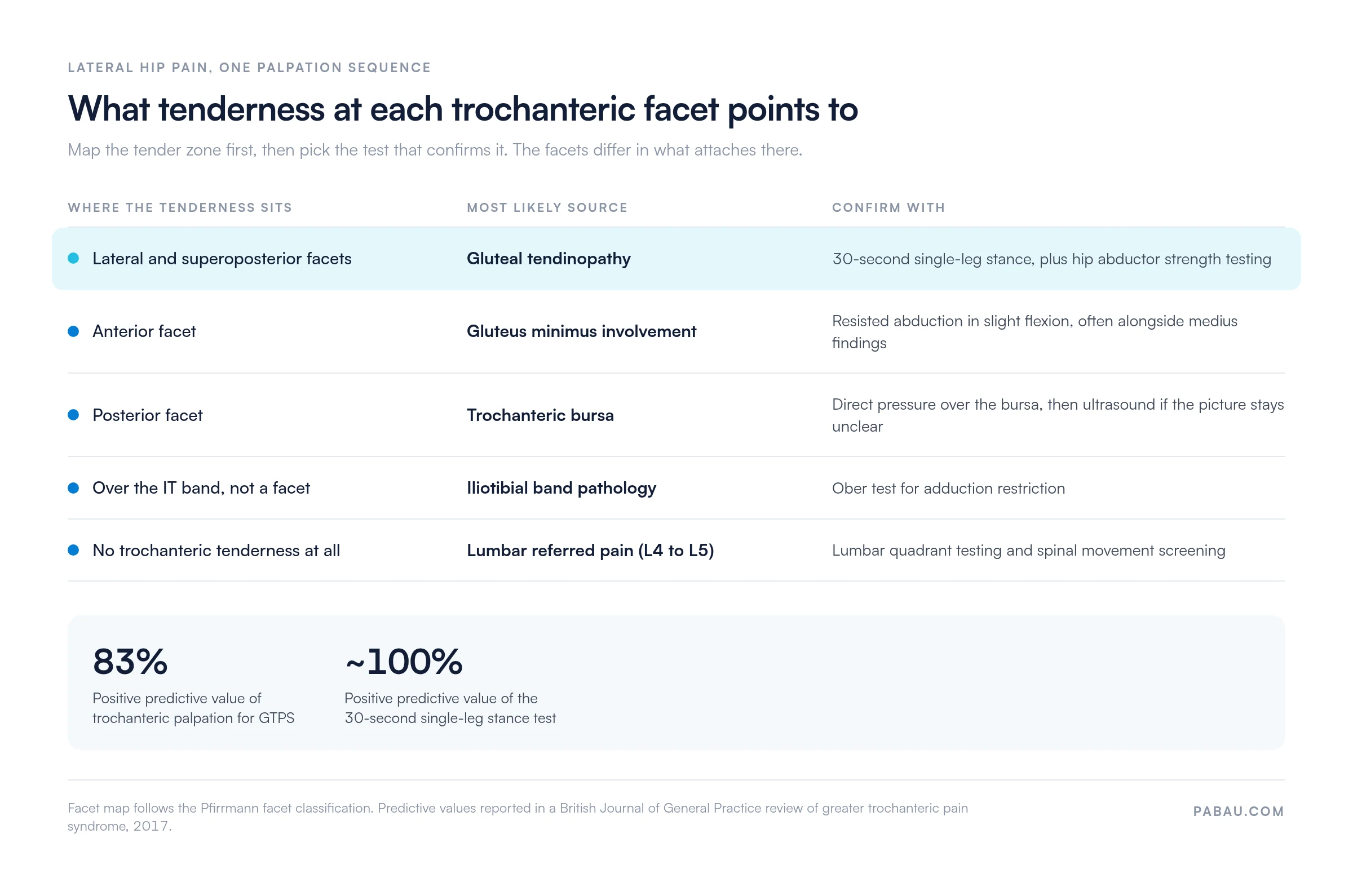
Which facet is tender matters. The lateral and superoposterior facets carry the gluteus medius tendon, while the posterior facet sits under the trochanteric bursa.
A positive finding is not a diagnosis on its own, so pair it with the single-leg stance test before you commit to a management plan.
Practice management software like Pabau lets you capture facet tenderness, pain scores, and the ICD-10 code in one structured note.
Lateral hip pain fills a large share of musculoskeletal (MSK) caseloads in primary care and physical therapy. Most of it traces back to the greater trochanteric region. Misdiagnosis stays common, because trochanteric bursitis, gluteal tendinopathy, and iliotibial band pathology all get filed under one vague label. A precise palpation sequence, run the same way every time, is the fastest way to narrow the field.
This guide covers the surface anatomy, the step-by-step method, and what a positive finding actually means. It also covers how to record the result so your note supports the code. It sits inside a full hip examination rather than replacing one. Whether you are a final-year student or a practicing clinician refreshing your technique, the sequence below gives you a result you can repeat and defend.
The greater trochanter is a large quadrilateral bony eminence at the junction of the femoral neck and shaft. On the body surface it sits roughly 10 cm below the iliac crest on the lateral aspect of the thigh. That makes it one of the most reliably palpable landmarks in the hip and pelvis.
Three reference points help you locate it quickly, before you touch the patient:
Clinicians who skip landmark identification and go straight to pressure tend to miss the lateral and superoposterior facets, where the gluteal tendon fibers concentrate. That produces a falsely negative finding on a hip that is genuinely painful.
The greater trochanter has four facets, and only three of them carry a tendon insertion. Knowing which structure sits on which facet is what gives the location of tenderness its diagnostic weight.
This facet map follows the classification described by Pfirrmann and colleagues, the standard reference for trochanteric anatomy on imaging. Say a patient reports pain radiating down the lateral thigh, and you find tenderness on the lateral facet. The working diagnosis moves toward gluteal tendinopathy rather than bursitis. That changes the management plan you write.
Consistent technique produces consistent findings. The sequence below reflects the approach set out in a British Journal of General Practice review of GTPS and in widely used MSK palpation curricula.
Place the patient in lateral decubitus, side-lying on the unaffected side, with the affected hip facing upward. Ask them to keep both hips in neutral rotation and their knees slightly bent. This relaxes the iliotibial band and the gluteal fascia. The trochanteric region becomes easier to reach, and you get fewer false positives from overlying tissue tension.
Supine with the hip in slight internal rotation is a reasonable alternative when side-lying is not tolerable. It does narrow your access to the posterior facet.
Record the location and severity of tenderness on a standardized scale straight after the assessment. Relying on memory between the examination and the note introduces error, and it does so fastest in a high-volume practice.
Document which facet reproduced concordant pain, not just ‘greater trochanter tenderness positive’. Lateral or superoposterior points to gluteal tendinopathy, posterior to the trochanteric bursa, and pain over the band itself to IT band pathology. That specificity changes both your code and your management pathway.
A positive finding that reproduces concordant lateral hip pain is the cardinal clinical sign of greater trochanteric pain syndrome (GTPS). That comes from a review in the British Journal of General Practice. Tenderness alone does not name one diagnosis, though. Three conditions commonly produce it, and telling them apart is what drives treatment selection.
The same review reports a positive predictive value of 83% for trochanteric palpation, and close to 100% for the 30-second single-leg stance test. So palpation earns its place as a screening step rather than a confirmatory one. Ultrasound or MRI remains the reference standard for separating a tendon tear from bursal irritation. The map below pulls the facet finding, the likely source, and the confirming test into one place.
GTPS covers gluteal tendinopathy, trochanteric bursitis, and IT band pathology as overlapping entities under one clinical umbrella. The same GTPS review reports that the condition is seen more commonly in women between the ages of 40 and 60. Risk factors include obesity, low back pain, ipsilateral knee osteoarthritis, and hip abductor weakness.
Clinical presentation typically includes lateral hip pain that worsens with:
Diagnosis combines palpation with special testing and, where the picture stays ambiguous, imaging. Load management and hip abductor strengthening carry most of the treatment, so a written exercise plan is worth handing over at the first appointment. Our gluteal tendinopathy exercises handout gives you a starting point to adapt to the patient in front of you.
Palpation alone does not separate the subtypes of GTPS precisely enough for treatment planning. The tests below, summarized from Physio-pedia’s GTPS resource, are routinely run alongside the palpation sequence.
When palpation reproduces concordant pain and the single-leg stance test is positive, the likelihood of gluteal tendinopathy rises well above either finding alone. That pairing is the most useful combination in routine MSK assessment.
Not all lateral hip pain starts at the greater trochanteric region, and lumbar referred pain mimics GTPS closely. A framework for ruling the alternatives in and out belongs at the front of the assessment, before you commit to a GTPS management plan.
One clinical rule is worth holding on to. If lumbar provocation reproduces the lateral hip pain before you palpate the trochanter, work the spine first. Palpation is most informative once lumbar screening has been documented as negative or contributory.
Documentation of palpation findings is a compliance and continuity matter, not a paperwork chore. In ICD-10-CM, trochanteric bursitis maps to M70.61 for the right hip and M70.62 for the left. Gluteal tendinopathy sits under gluteal tendinitis, M76.0, coded M76.01 on the right and M76.02 on the left. Picking the wrong code because the note was vague creates audit risk and billing errors.
A structured note for this examination should capture at minimum:
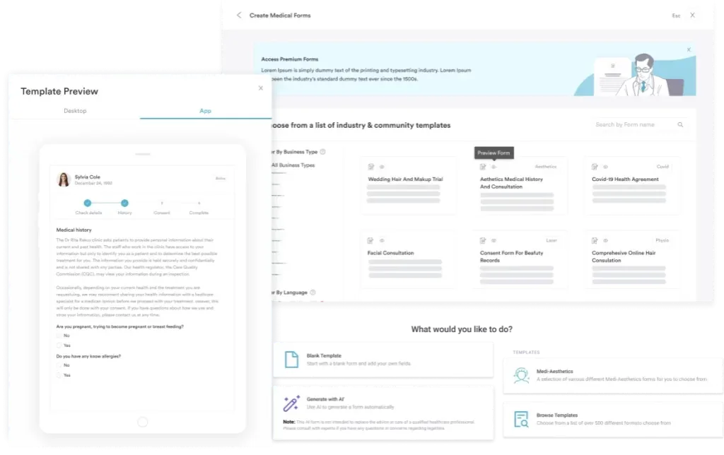
Practices carrying large MSK caseloads get the most from digital assessment forms. Those forms pre-populate the tenderness fields and tag the ICD-10 code from the examination findings. That removes the manual code lookup. It also cuts transcription errors between the assessment and the bill, which is where most coding disputes start.
MSK and physical therapy practices carrying GTPS caseloads face one recurring documentation problem. Palpation findings, special test results, outcome measure scores, and billing codes all have to be captured the same way across practitioners and across appointments. When one clinician fills in a paper form and another types free text, the records stop lining up, which complicates both handover and billing.
Practice management software like Pabau lets you build assessment templates for hip evaluation, with tenderness grids, special test checklists, and outcome measure scoring. Those structured patient records connect to the appointment, the treatment plan, and the coded diagnosis. You get one workflow from assessment to bill instead of three manual steps.
For an ongoing GTPS caseload, a physical therapy EMR tracks rehab progress across sessions and sends follow-up prompts. It also flags patients who have not returned within their planned interval. That is what stops a 12-week loading program from quietly ending at week four.
Want to see how this fits your own documentation workflow? Book a demo and ask about the clinical note builder.

Pabau lets MSK and physical therapy practices build hip assessment templates that capture facet tenderness, special test results, and the ICD-10 code in one note. The diagnosis reaches the bill without a second round of data entry.

The hard part of lateral hip pain is not finding tenderness at the greater trochanter. It is mapping that tenderness to the right facet and pairing it with the test that confirms it. Then it is writing the result down in a way that survives a handover and an audit.
Get the facet into the note and the rest follows. The code stops being a guess, the reassessment has a baseline to compare against, and a colleague picking up the case knows what you found.
Book a demo to see how Pabau turns a hip assessment template into a coded, auditable note in a single pass.

Need an outcome measure to track GTPS rehab? Harris hip score gives you a scored baseline you can repeat at every reassessment.
Want the range-of-motion half of the hip exam? Hip range of motion test sets out the normal values and how to record them.
Ruling out an anterior hip source? Hip flexor strain test covers the tests that separate flexor pain from lateral hip pain.
Building a standard intake for MSK patients? Physical therapy intake form lists the fields worth collecting before the first assessment.
Checking your documentation against the rules? Mandatory compliance for physiotherapy clinics covers the regulatory requirements MSK practices need to meet.
Greater trochanter palpation is a manual clinical technique where the examiner applies firm pressure over the lateral proximal femur. It identifies tenderness associated with greater trochanteric pain syndrome (GTPS), gluteal tendinopathy, or trochanteric bursitis. A positive finding is reproduction of the patient’s familiar lateral hip pain, not simply pressure sensitivity.
Tenderness at the greater trochanter most commonly points to gluteal tendinopathy, trochanteric bursitis, or iliotibial band pathology. The lateral and superoposterior facets carry the gluteus medius tendon, so tenderness there suggests tendinopathy. The posterior facet has no tendon insertion and sits under the trochanteric bursa instead.
Trochanteric bursitis is coded M70.61 for the right hip and M70.62 for the left hip in ICD-10-CM. Gluteal tendinopathy sits under gluteal tendinitis, M76.0, coded M76.01 on the right and M76.02 on the left. Which code applies depends on the structure your examination identifies as the primary pain source.
Yes. Lateral decubitus, side-lying on the unaffected hip, is preferred. It relaxes the overlying fascia and gives the best access to the lateral and posterior facets. Supine with the hip in slight internal rotation works for patients who cannot tolerate side-lying, though access to the posterior facet is reduced.
GTPS is diagnosed clinically. You combine palpation that reproduces concordant lateral hip pain with positive special tests, particularly single-leg stance and the Trendelenburg sign. Ultrasound or MRI is used when the clinical picture stays ambiguous. It also helps separate a tendon tear from bursitis before an interventional procedure.






