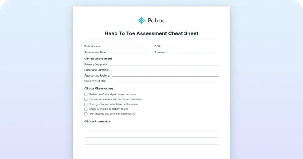
Head to toe assessment cheat sheet: Printable nursing guide
A head to toe assessment cheat sheet gives you one fixed order at the bedside, s...

3
Last Updated: Oct 23, 2026

Patient positions in bed are standardized body arrangements that protect the skin, support breathing, and give safe access for care.
Seven core positions cover most bedside scenarios: supine, prone, lateral, Fowler’s, Sims’, Trendelenburg, and lithotomy.
Turning every two hours is the usual standard, and high-risk patients need shorter intervals with a pressure-redistributing surface.
The free download is a one-page, text-only list of 13 bedside and procedural positions, each with a short description.
Practice management software like Pabau lets nurses log each turn, skin check, and position change in the patient’s record.
A one-page clinical reference listing 13 positions, from supine and Fowler’s to lithotomy, jackknife, and the Wilson frame. Each entry describes how the patient is placed and what the position is used for.
Download templatePatient positions in bed are standardized ways of arranging the body, and each one serves a clinical goal. Leave a patient flat on their back for long enough and pressure damage over the sacrum can start within hours. Turn that same patient onto one side, with a pillow between the knees, and the skin keeps its blood supply. Primary care depends on continuity, and continuity depends on the previous consultation being easy to find.
Below is every standard position, when to use it, when to avoid it, and what to record.
The download is one page of plain text, sized for printing. It names 13 positions and mixes everyday bedside postures with positions used in the operating room:
Each position gets a short written description of how the patient is placed and what it is generally used for. That is the whole document.
It carries no diagrams, no indication and contraindication columns, no pressure point map, and no repositioning checklist. Sims’ position is not on it either. For that detail, use the quick reference table further down this page alongside your own unit policy.
Every position is a body arrangement chosen to serve one clinical goal. That goal might be protecting the skin, easing breathing, or opening access for a procedure. Each choice balances comfort, safety, and what the care team needs to do next.
Seven core positions cover nearly every bedside scenario: supine, prone, lateral, Fowler’s, Sims’, Trendelenburg, and lithotomy.
Each has defined indications, contraindications, and pressure points that need watching. Knowing which to choose, and when to move on from it, is core nursing competency.
Incorrect positioning causes harm quickly. Pressure damage can start within a couple of hours over a bony prominence when nothing moves. Poor alignment compresses nerves, which is how brachial plexus injury and peroneal nerve palsy happen. Lying flat also restricts chest wall expansion and makes breathing harder.
Position drives comfort, dignity, and how well a procedure goes.
A patient in pain from an awkward position will not stay still or cooperate. So it belongs in the care plan alongside the rest of your patient care management, not squeezed between other tasks.
Each position below carries its own indications, its own pressure points, and one thing that catches people out. Read them as a set, because most shifts move a patient through three or four of them.
The supine position lays the patient flat on the back with the legs extended. It is the default for rest, central line placement, and abdominal examination.
Watch the sacrum, heels, and scapulae. Raise the head of the bed, or choose another position, if the patient is breathless or at risk of aspiration.
The prone position turns the patient face down. It improves oxygenation in ARDS and other causes of severe hypoxemia, gives access to the back, and suits some spinal procedures.
Watch the forehead, cheekbones, elbows, knees, and toes. Avoid it after facial trauma, with an unstable spine, or when the patient cannot tell you they are in pain.
The patient lies on one side with the lower leg extended, the upper leg flexed, and a pillow between the knees. Alternating left and right lateral is the workhorse of pressure relief.
Watch the hip, knee, and ankle on the weight-bearing side. Keep a fractured hip off the weight-bearing side.
Fowler’s position raises the head of the bed. Low Fowler’s, at 15 to 30 degrees, suits general rest. Semi-Fowler’s, at 30 to 45 degrees, is standard for eating and talking. High Fowler’s, at 60 to 90 degrees, helps breathlessness, recovery after surgery, and feeding without aspiration.
Watch the sacrum and heels, because shear rises with the angle. Raise the head slowly if blood pressure is low.
Sims’ position mixes supine and lateral. The patient lies on the left side with the left arm behind the body and the right arm forward. The left leg stays extended and the right leg is flexed. It is used for enemas, rectal examination, and as the basis of the recovery position.
Watch the hip, knee, and ankle.
Trendelenburg tilts the whole bed so the head sits lower than the feet, usually by 15 to 30 degrees. It was long used for shock, and that use is now questioned. Reverse Trendelenburg raises the head above the feet, which helps reflux, gastric emptying, and breathing after surgery.
Trendelenburg raises intracranial pressure, and reverse Trendelenburg increases shear at the sacrum, so both need watching.
Lithotomy holds the patient supine with the hips and knees flexed and the legs apart, which is standard for gynecologic, urologic, and rectal work. The orthopneic or tripod position sits the patient upright, leaning forward onto an overbed table.
It is a fast win in acute breathlessness, and patients on a heart failure care plan often find it themselves. The recovery position is a modified Sims’ used to keep an unconscious patient’s airway clear.
Scan the row before you move the patient, then check the care plan for anything specific to them.
A good turn protects two people at once, the patient’s skin and your own back. The sequence below covers a single move from supine to lateral, and it scales with the number of staff you have.
Once all five are settled, the move itself is short.
Every two hours is the working standard, and it is a starting point rather than a fixed rule.
Risk sets the interval. A stable post-operative patient on a pressure-redistributing mattress may be safe at three hours. A patient with a spinal injury, an existing pressure injury, or poor nutrition needs hourly attention.
Write the agreed interval in the care plan and post it where the team can see it, so every shift keeps the same rhythm. Explaining the plan to the patient helps too, because patient compliance is what keeps the turn happening when staff are stretched. Reminders in your EMR stop the cycle drifting overnight.
The right equipment makes every turn safer and more comfortable:
Bring all of it to the bedside before you start. A turn that pauses halfway while somebody hunts for a wedge is exactly where shear happens.
The mechanism is simple. Unrelieved pressure cuts blood flow to skin and soft tissue, and the tissue dies from the inside out. Shear and moisture make it worse, which is why a patient sliding down a raised bed carries more risk than the angle alone suggests.
Picture a 78-year-old admitted with pneumonia, eating little and barely moving. On a standard mattress at four-hour turns, the sacrum is often red before the second day. Two-hourly turns on a pressure-redistributing surface usually hold the same skin intact.
Three habits prevent most of it. Keep to the interval in the care plan, match the support surface to the risk, and inspect the skin at every turn. Look for redness that does not blanch, unusual warmth, and any break in the surface.
Most unit policies are built on the international EPUAP, NPIAP, and PPPIA pressure injury guideline. NPIAP was renamed from NPUAP in 2019, so older documents still cite the old initials.
Weight changes the physics. Contact pressure rises, and manual repositioning risks injury to staff and patient. Use a mechanical lift or a slide sheet with two people every time. Check the safe working load of the bed, and use a wider frame where one is needed.
Post-surgical patients follow the surgeon’s restrictions, which usually means no hip flexion after a hip replacement and no spinal movement after a fusion. Spinal precautions call for log rolling. Align the spine, move the patient as one unit, and use a draw sheet under the trunk and hips.
High-risk patients need the shortest interval and the best surface available. That group includes anyone with an existing pressure injury, limited mobility, poor nutrition, diabetes, or advanced age. A functional status questionnaire at admission tells you how much of the move the patient can take on themselves.
Whatever the risk level, the skin check is the part that gets skipped first when the unit is busy. Digital intake forms tied to each turn keep it on the record instead.
Your chart is the legal record of the care you gave. For each turn, record:
A standard form keeps the wording consistent between staff, which matters more than it sounds when three people chart the same patient. Records held in a secure system also survive the shift, unlike a sheet taped above the bed.
The plan then has to survive handoff. Name the position and the interval when you hand over, the same way you would in an ICU presentation. Keep it on the written sheet the team works from too. A CVICU report sheet has a line for exactly this.
Share the positioning plan with physical therapy, occupational therapy, and the medical team. A plan only nursing knows about breaks the first time the patient leaves the unit for a scan.
Treat the sheet as a memory aid that sits alongside your unit policy.
Read the indication table on this page before you choose a position for a patient with precautions. The sheet names positions, and the decision still belongs to the assessment.
Most units record turns on a paper chart taped above the bed. It works until the sheet gets wet, leaves with the linen, or reaches an audit with three different handwritings on it. Nobody can then say when the last skin check actually happened.
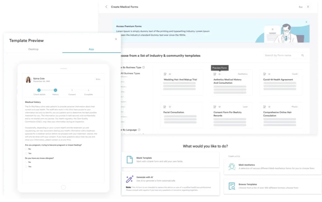
Pabau is an all-in-one practice management system, and its digital forms let you build one repositioning and skin-check form for your unit. Staff complete it on a tablet at the bedside. Each entry lands in the patient’s record with a time and a name against it, so the history is written once and read by everyone.
Treatment notes, photos, and care plans sit in the same client record, which makes a skin change easy to follow across days instead of shifts. Task assignment puts the next check in front of whoever is on duty. Records held in one system give therapy staff and the medical team the same turn history you see.

Pabau’s digital forms and client records give your team one place to log repositioning, skin checks, and the equipment used. The next shift picks up the history without chasing a paper chart.

Positioning is one of the few interventions where the cost of getting it wrong shows on the skin within hours. Learn the seven core positions well enough to choose one without stopping to think. The schedule and the chart then carry the rest of the work.
One trade-off is worth remembering. A two-hour cycle suits the average patient and fails the frail one. Anyone with poor nutrition, limited mobility, or a stage 1 injury already showing needs a shorter interval and a better surface. The record is what proves they got both.
So print the reference sheet for the bedside, and keep the evidence of every turn where the whole team can see it. Is your unit still tracking turns on paper? Book a demo to see how Pabau records repositioning, skin checks, and care plans in one patient record.

Need the assessment that comes before the turn? Health assessment in nursing walks through a head-to-toe assessment you can chart at the bedside.
Confirming allergies before you begin? Food allergy form template documents allergies so the team can respond correctly in an emergency.
Assessing cognition in an older patient? Free memory test for seniors compares SAGE, MoCA and SLUMS score cutoffs and how to administer each.
Admitting a patient into hospice care? Hospice admission note template turns intake into a structured, CMS-aligned conversation that avoids missed directives.
Watching for a hypoglycemic episode too? Hypoglycemia nursing diagnosis template structures rapid assessment for a patient at risk of seizures or altered consciousness.
Heels carry a thin layer of tissue over bone and press through a small contact area. Float them with a pillow placed lengthwise under the calves, keeping the knees slightly bent. Check that the heels hang clear of the surface.
No. A ring cushion concentrates pressure around the edge of the hole and restricts blood flow to the tissue inside it. Use a pressure-redistributing cushion or mattress instead, and keep to the turning schedule.
It is a shallow side-lying position held with pillows or a wedge behind the back. The tilt keeps weight off the hip bone and the sacrum at the same time. Many units prefer it to a full side-lying turn for frail patients.
Support the weak shoulder and arm on a pillow, and keep the shoulder forward rather than pulled back. Avoid long spells lying on the weak shoulder. Ask physical therapy for the patient’s agreed positioning plan.
In severe hypoxemia, prone sessions often run 12 to 16 hours before the patient is turned back. That needs continuous monitoring, planned pressure care, and staff trained for the turn. Follow your unit protocol.