PCS pain catastrophizing scale: Scoring, cutoffs, and free template
The PCS pain catastrophizing scale is a 13-item self-report questionnaire that m...

2
Last Updated: Oct 15, 2026

A leg muscle diagram maps the position, attachments, action, and nerve supply of every major muscle group between hip and ankle.
The leg holds over 50 muscles in seven compartments: three in the thigh and four below the knee.
Knowing a muscle’s action and nerve supply narrows an assessment from a whole limb down to one compartment.
Practice management software like Pabau attaches the printable diagram to consultation notes, so patient education is stored with the record.
A printable anatomical reference covering the thigh and lower leg muscle groups, their attachments, their actions, and their nerve supply. Hand it to a patient during a consultation, or keep a copy at the treatment table.
Download templateA leg muscle diagram is a visual reference that maps the major muscle groups of the lower limb.
It shows how each muscle attaches to bone, the action it produces, and the nerve that controls it.
That map earns its place in a physical therapy, sports medicine, or podiatry practice. It narrows an assessment from a whole limb to one compartment, and it gives you something to point at while you explain the plan. Storing it inside your physical therapy EMR puts the same reference at intake, in the note, and in the take-home summary.
The leg holds over 50 individual muscles, and they are grouped rather than listed. Fascia divides them into compartments, and the muscles inside a compartment share a nerve supply and pull in roughly the same direction. That is why a compartment, not a single muscle, is usually the unit of assessment.
The map below puts all seven compartments in one place. Use it to work from the region a patient points to down to the muscles inside it.
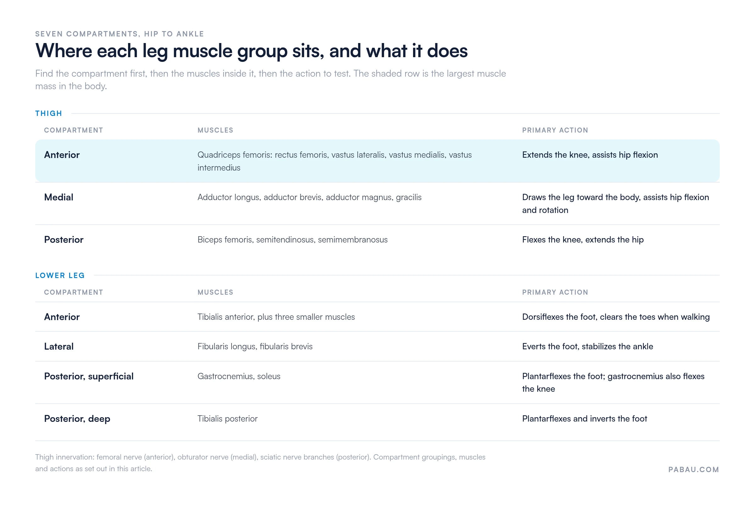
The thigh holds the body’s largest and most powerful muscle groups. Quadriceps, hamstrings, and adductors work together to generate force during walking, running, and every change of position. Both the quadriceps and the hamstrings cross the knee, so the knee muscle diagram is the natural companion sheet for a knee complaint.
The quadriceps comprises four muscles: rectus femoris, vastus lateralis, vastus medialis, and vastus intermedius. Together they form the largest muscle mass in the human body. Rectus femoris crosses both the hip and the knee, so it tightens when a patient sits for long stretches. The vastus muscles cross the knee only, and they build strength through squatting and climbing.
The hamstring group runs down the back of the thigh and contains biceps femoris, semitendinosus, and semimembranosus. These muscles flex the knee and extend the hip, which makes them central to sprinting and jumping. Chronic tightness here often surfaces as lower back pain and limited hip mobility.
The adductors are adductor longus, adductor brevis, adductor magnus, and gracilis. They form the medial thigh and steady the pelvis during standing and walking. Groin strains involve these muscles and are common in athletes. The adductor strain exercises handout gives the patient a loading progression to take home.
Below the knee, fascia divides the muscles into four compartments, each holding muscles with similar actions. That design gives precise control of ankle and foot position. It also means swelling inside one compartment has nowhere to go, which is the mechanism behind compartment syndrome.
The anterior compartment holds four muscles that dorsiflex the foot, meaning they lift the toes upward. Tibialis anterior is the largest of them and the easiest to palpate. It lifts the foot during walking so the toes clear the ground. Shin splints involve inflammation of tibialis anterior or its tendon sheath, and runners account for most of those cases.
The lateral compartment holds fibularis longus and fibularis brevis, also called the peroneal muscles. They evert the foot and hold the ankle steady. Both are commonly injured during an inversion sprain, which is why lateral support matters so much in the first weeks of recovery.
The posterior compartment splits into superficial and deep layers. The superficial group is the calf, made up of gastrocnemius and soleus, and both plantarflex the foot. Gastrocnemius crosses the knee and the ankle, while soleus attaches only at the ankle. A return-to-running protocol after a calf injury therefore has to load the two separately.
The diagram earns its keep when it sits inside the consultation rather than on a wall. Pointing at the muscle a patient just described turns an abstract diagnosis into a location they can see. That lowers anxiety and improves how closely they follow the plan.
Body contouring and aesthetic practitioners use the same sheet differently. They point at calf shape, quadriceps development, or hamstring definition to agree on what a treatment is meant to change.

Leg anatomy drives how you sort one injury from another. Strains, tears, compartment syndrome, and tendon inflammation each produce a distinct pain pattern. Each one also maps to a structure shown on the diagram.
Practices that specialize in these injuries usually run sports medicine software, so assessment findings, exercise prescriptions, and progress notes stay in one record. A record of where an injury sat also pays off years later. When the same patient returns with a similar complaint, you have a baseline to compare against.

The intrinsic foot muscles sit inside the foot itself and support the arch and toe control. They rarely lead a treatment plan. They do become the focus in flat foot development, plantar fasciitis, and toe dysmobility. A reference sheet is worth little if it stops at the ankle.
In most practices the diagram lives on a wall or in a folder of printouts. The practitioner points at it during the consultation, then writes a note that never references it. The patient leaves with a photocopy that never reaches the chart.
Practice management software like Pabau treats the diagram as part of the record instead. You attach it to a digital intake form, so the patient marks the painful area before the appointment. You attach the annotated version to the treatment note, so the assessment and the picture stay together.
Pabau Scribe, our AI scribe, drafts the treatment note from the consultation you just had. The muscle you discussed ends up named in the record rather than in your memory. A follow-up months later starts from your original assessment, not from a blank page.
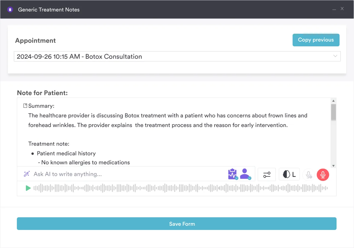

Pabau attaches printable diagrams to intake forms, treatment notes, and take-home summaries. The reference a patient saw in the room stays with their record.

Download the diagram, print it, and put it where the consultation happens. A patient who can see which compartment the pain sits in asks better questions and sticks to the plan.
The trade-off worth remembering is that a printout only helps the visit it is used in. Once the sheet leaves the room, the assessment it captured leaves with it, unless the diagram lives in the record. Book a demo to see how Pabau stores anatomy references, marked-up diagrams, and treatment notes against one patient file.

Treating a knee complaint next? Knee muscle diagram covers the muscles that cross the joint from above and below.
Need to test hamstring length? 90/90 hamstring test sets out the positioning and how to record the result.
Checking quadriceps integrity after a knee injury? Quadriceps active test covers the setup and what a positive finding means.
Assessing the anterior compartment? Ankle dorsiflexion test measures the range tibialis anterior needs to clear the toes.
Grading strength across a whole limb? Manual muscle testing explains the grading scale and how to document each muscle group.
The leg contains thigh muscles (quadriceps, hamstrings, adductors), lower leg compartment muscles (anterior, lateral, posterior), and intrinsic foot muscles. A leg muscle diagram organizes these into anatomical groups, showing how each crosses joints and produces movement.
The gluteus maximus (buttock muscle) is the largest single muscle, but the quadriceps femoris is the largest in the thigh itself. Both are visible on a detailed leg muscle diagram.
The lower leg contains anterior, lateral, and posterior compartments. A fourth (deep posterior) subdivides the posterior group. Each compartment is enclosed by fascia and contains muscles with distinct actions.
During consultations, use the diagram to show patients exactly where their injury sits, which muscles are affected, and what actions will be restricted. This visual explanation increases patient compliance and reduces treatment anxiety.
Both plantarflex the foot, but the gastrocnemius crosses the knee joint while the soleus does not. This means the gastrocnemius also flexes the knee, and the two muscles are active in different movement patterns. A leg muscle diagram shows this anatomical difference clearly.