Vegan based diet plan: Free template and nutrition guide
A vegan based diet plan is a structured dietary plan that builds every meal arou...

3
Last Updated: Oct 14, 2026

A heart diagram labels the four chambers, four valves, great vessels and conduction system on one page. Anatomy questions get answered in seconds, not paragraphs.
The labeled page works for consultations and staff training. The blank page, with numbered callouts, works for self-testing and quizzes.
Pictures closely tied to text raise both attention and recall, and patients with lower literacy skills gain the most.
Note what you showed and what the patient understood, otherwise the education you delivered leaves no trace in the record.
Two pages, print ready. Page one labels 20 structures, from the venae cavae to the Purkinje fibers. Page two repeats the diagram with numbered callouts and blank answer lines for teaching and quizzes.
Download templateHeart disease is the leading cause of death in the United States, and it accounted for 20.1% of all deaths in 2021. So a fair number of your consultations touch the heart. Most of them stall at the same point: the patient cannot picture the anatomy you are describing.
A labeled diagram solves that faster than any explanation. Point at the valve, name it, and the conversation moves on to what you plan to do about it.
The template is a schematic anterior view, drawn the way a patient faces you: their right side sits on your left. Oxygen-poor blood is blue, oxygen-rich blood is red, and the conduction system is amber.
Page one names 20 structures. Page two repeats the same drawing with numbered callouts and blank lines, so it doubles as a quiz sheet.
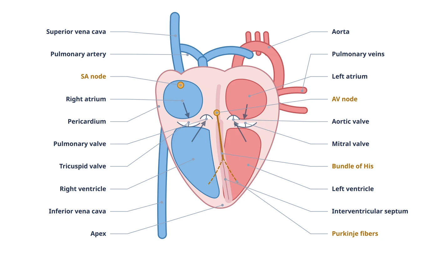
Twenty structures carry labels on page one of the template. These are the ones worth naming out loud in a consultation.
The National Heart, Lung, and Blood Institute describes the same four chambers and three tissue layers. It’s a useful second reference for a patient who wants to read more at home.
Blood flow is the part patients grasp quickest, because it is a sequence rather than a list.
That circuit repeats roughly 60 to 100 times a minute at rest. Tracing it with a finger on the diagram turns a physiology lecture into a 30 second explanation.
Print the labeled page for the consultation room wall and keep a stack of the blank page in the drawer. Handing a patient the blank version and asking them to point to where their valve sits tells you more about their understanding than any yes or no question.
The amber pathway on the diagram is the electrical system. You need it when the conversation turns to palpitations, rate control or a pacemaker.
As StatPearls sets out, the AV node is the only normal electrical connection between the atria and the ventricles. Showing that single crossing point on a diagram explains a block better than describing it does.
A review in Patient Education and Counseling found that pictures closely tied to text raise both attention and recall. The largest gains came among patients with lower literacy skills.
The practical uses fall into a short list.
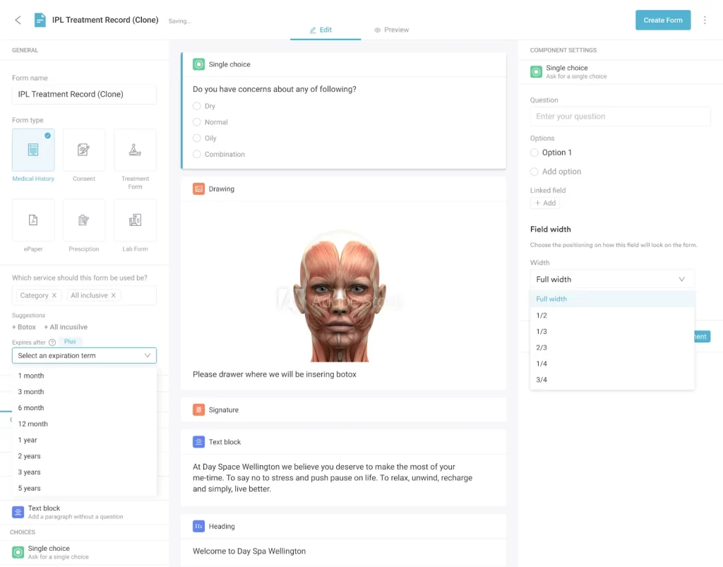
Sending the diagram ahead of the visit also changes the opening of the consultation. Attach it to a universal health form through digital patient forms, and the patient arrives having already seen the anatomy you are about to discuss.
The template was drawn for clinical teams rather than exam illustrators, so it stays readable at arm’s length across a desk.
One drawing serving all of those roles is the point. The poster on the wall, the handout in the folder and the quiz sheet in the drawer all use the same colors and structures. Nobody has to translate between versions.
Page two is the same drawing with 20 numbered callouts and blank answer lines. Labeling it from memory sticks better than rereading a finished sheet, which is why both pages are in the file.
The blank page also works in front of a patient. Ask them to point out where their own valve or stent sits, and any misunderstanding shows up straight away.
Patient education that leaves no trace in the record is a problem at audit. It is also a problem for the next clinician who sees the patient.
Practice management software like Pabau keeps the handout and the note in one place. Pabau Scribe, our AI medical scribe, drafts the consultation note from the conversation. The structures you named end up in the record without a second typing pass.
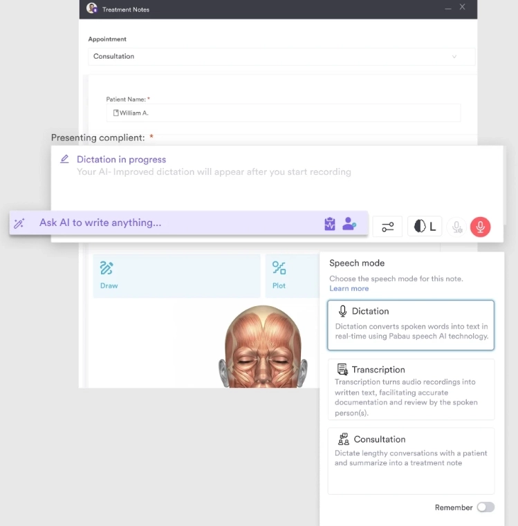
From there the note behaves like any other clinical entry. It is searchable, it carries an audit trail, and it holds up better than a line reading “anatomy discussed”.
If your team writes these up by hand, a structured progress notes template keeps the education you delivered in a consistent place in the record.

Pabau’s digital forms and client portal deliver patient education material ahead of the visit. The answers come back as structured fields in the client record.

Cardiac anatomy is hard to hold in your head from words alone. That is why a single labeled page still earns its place next to imaging and 3D models. Print the labeled version for the consultation room, and keep the blank version for teaching. Pair it with a written plan of care so the explanation carries through to what happens next.
Practices that get the most from a handout like this put it in front of the patient early. Then they write down what was understood, not just what was said.
Book a demo with Pabau to see how digital forms, the client portal and AI-assisted notes keep patient education and documentation in the same record.

Need the numbers behind the pump, not just the parts? The cardiac output equation shows how rate and stroke volume combine, which is the next question once a patient understands the chambers.
Explaining why a weaker ventricle matters? Stroke volume equation covers what each beat actually ejects, and what changes it.
Moving from anatomy to a care plan? The hypertension nursing care plan gives you diagnoses, goals and interventions for the risk factor that comes up most in these conversations.
A heart diagram is a labeled anatomical illustration of the heart. It shows the four chambers, four valves, great vessels and often the electrical conduction system on one page. Clinicians use it to explain cardiac anatomy during consultations, and educators use it for staff training and student teaching.
A full heart diagram labels the four chambers (right atrium, left atrium, right ventricle, left ventricle) and the four valves (tricuspid, pulmonary, mitral, aortic). It also labels the great vessels (superior and inferior vena cava, pulmonary artery, pulmonary veins, aorta), the interventricular septum, the apex and the pericardium. The conduction system rounds it out: the SA node, AV node, bundle of His and Purkinje fibers.
Blood arrives from the body in the right atrium. It passes the tricuspid valve into the right ventricle, then leaves through the pulmonary valve and the pulmonary artery for the lungs. Oxygenated blood returns through the pulmonary veins into the left atrium. It passes the mitral valve into the left ventricle, then leaves through the aortic valve into the aorta for the rest of the body.
The four valves keep blood moving one way. The tricuspid and mitral valves sit between the atria and ventricles and shut during ventricular contraction to stop blood washing back into the atria. The pulmonary and aortic valves sit at the ventricular outflows and shut during relaxation to stop blood draining back out of the arteries.
A labeled diagram names every structure, which suits consultations, quick reference and teaching. A blank diagram shows the same outline with numbered callouts and no names, which suits self-testing and quizzes. This template includes both, so the same illustration serves teaching and assessment.
Use the standard anatomical names and keep them consistent across your practice. Write right atrium rather than upper right chamber, call the bicuspid valve the mitral valve, and spell out SVC and IVC on patient-facing material. Consistent naming avoids confusion when several staff document the same case.
Yes. The PDF is free, prints on standard paper and carries no usage restriction for patient education, staff training or classroom teaching. Print it for the consultation room, attach it to a digital form, or send it through a patient portal before the appointment.