



GLP-1 diet plan
A GLP-1 diet plan is a structured eating framework for patients taking semagluti...

2
Last Updated: Oct 13, 2026

A thallium stress test uses radioactive thallium-201 to map blood flow through the heart muscle, first under stress and then at rest.
The scan separates a reversible defect, which signals ischemia, from a fixed defect, which signals scar from an old heart attack.
Rest images are captured 3 to 4 hours later, because thallium-201 redistributes into viable muscle over that window.
The radiation dose runs roughly 16 to 35 mSv, several times higher than a technetium-based perfusion scan.
Practice management software like Pabau delivers preparation instructions and intake forms before the visit, so fewer scans get canceled on the day.
Chest pain with a normal ECG leaves a cardiologist holding a decision and very little evidence. A thallium stress test fills in what the ECG cannot show. It maps how much blood reaches each part of the heart muscle, once under stress and again at rest.
That difference decides treatment. Muscle starved of blood during exertion can recover once a stent or a bypass restores flow. Scar tissue from an old heart attack will not, no matter how well the artery above it is opened.
Thallium-201 can show both pictures from a single injection. That one property is why the test has outlived four decades of newer imaging technology.
A thallium stress test is a nuclear scan that uses thallium-201, a radioactive tracer, to image blood flow through the heart muscle. It belongs to a family of studies called myocardial perfusion imaging, or MPI. A gamma camera captures the pictures using SPECT, short for single-photon emission computed tomography.
The principle is simple. Thallium-201 behaves much like potassium inside the body. Healthy muscle cells with a good blood supply pull it in. Areas with reduced flow take up less tracer and show up as cold spots on the image.
Redistribution is what sets thallium-201 apart from other tracers. After the stress injection, the tracer slowly moves into muscle that was underperfused at peak exercise. Over 3 to 4 hours, that shift lets one injection produce both a stress picture and a rest picture.
You may see the study called a thallium scan, a nuclear stress test, or a Tl-201 perfusion scan. All three describe the same thing.
The scan sorts each region of heart muscle into one of three states, and those labels drive what happens next.
The test also grades earlier treatment. A patient who has had bypass surgery or angioplasty can be re-imaged to check whether flow into the old ischemic territory improved.
Biomarkers sit alongside the images, and this guide to troponin levels shows how the two lines of evidence fit together.
Cardiologists order the test when a functional look at blood flow adds something an ECG or a plain treadmill test cannot. Most referrals come down to that one question.
Common indications include:
That last point carries weight in scheduling. Severe arthritis, peripheral artery disease, or plain deconditioning can keep a patient from reaching a target heart rate. Drugs then stand in for the treadmill, which changes both the preparation instructions and the room time.
Referral volume is the other pressure. Preventive cardiology and longevity practices now send patients for risk stratification in numbers that fill a nuclear schedule fast.
Careful appointment scheduling keeps wait times honest, because each study holds a camera and a bay for most of a day.
Preparation decides image quality more than any other factor you control. Poor fasting or an unheld medication can produce a false positive or a false negative, which sends the patient down the wrong path.
Standard instructions cover five areas:
Practices that send digital intake forms at booking get these rules in front of the patient early, instead of the night before. A new patient questionnaire collected in advance also flags the asthma history that rules adenosine out.
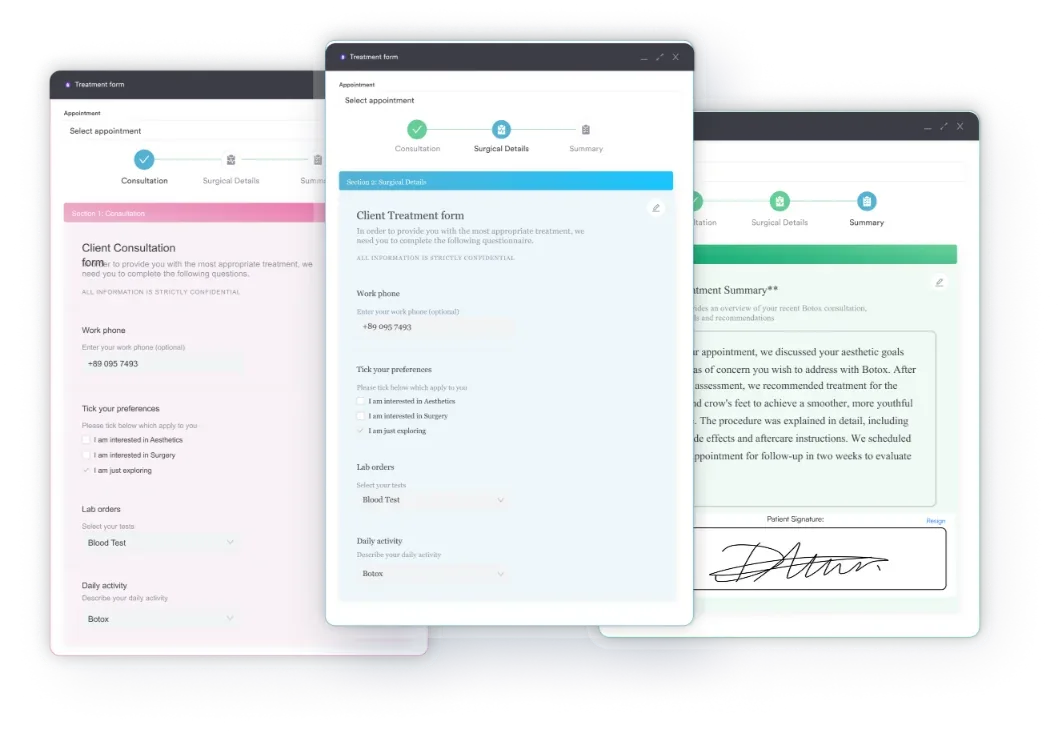
Medication lists matter as much as fasting. Capturing them on a pre-procedure form lets the team spot contraindications while there is still time to change the protocol.
Caseloads heavy with diabetes need extra care here. Practices running metabolic health programs often see patients on insulin, GLP-1 drugs, and beta-blockers at once. Each of those changes the morning of the test.
Most day-of cancellations trace back to a call that never happened. A short checklist the day before catches nearly all of them.
Keeping that script in one place stops it drifting between staff. A shared medical information form gives the front desk the same fields to work through every time.
Build the caffeine reminder into your pre-appointment workflow, not into the morning phone call. A patient who arrives after a coffee cannot have adenosine or regadenoson that day, and the slot is lost. Instructions sent automatically 48 hours ahead remove most of these cancellations.
The study runs in two phases on the same day, though some redistribution protocols push the second image set to the next morning. Here is the sequence.
Exercise wins whenever the patient can reach 85% of maximum predicted heart rate. It also adds prognostic detail a drug cannot, including ECG changes, blood pressure response, exercise capacity, and whether the symptoms reproduce.
When exercise is out, three agents stand in:
A nuclear cardiologist compares the paired image sets and writes the report. Four patterns cover almost everything a report will show.
Size and location then set the plan. A small reversible defect in a low-risk territory may end in medical management. Anything large in the left anterior descending territory usually triggers angiography and a revascularization discussion.
Numbers on a report always need context from the patient in front of you. This piece on interpreting biomarkers makes the same case for laboratory data, and the discipline is identical here.
One caution for patients reading their own report. An imaging result is one input into a clinical judgment, and it needs the symptoms and history beside it. Book the follow-up with the cardiologist who ordered the study.
The test is safe for the large majority of patients. Four risk categories are worth naming before consent.
Radiation exposure: Thallium-201 delivers ionizing radiation, and the dose sits at the high end for cardiac imaging. A stress-rest study runs roughly 16 to 35 mSv, while natural background radiation is about 3 mSv a year. That works out near 6 to 13 years of background exposure.
That is materially more than a technetium-99m scan, which is one reason sestamibi has taken over routine perfusion imaging. The American Society of Nuclear Cardiology, or ASNC, publishes the imaging guidelines most US labs work from. Patients should mention earlier nuclear scans or radiation therapy so cumulative dose can be weighed.
Exercise risk: Treadmill testing carries a small chance of arrhythmia, infarction, or cardiac arrest, estimated at 1 to 2 events per 10,000 tests. Testing happens in a monitored room with resuscitation equipment at hand.
Stress agent side effects: Adenosine and dipyridamole are contraindicated in significant bronchospastic disease, including active asthma, because they can trigger severe bronchospasm. Regadenoson is gentler on the lungs but still needs caution in reactive airways disease. Flushing, chest discomfort, headache, and brief dizziness are common, and they usually settle within minutes. GP clinic software keeps consultations, results and recalls against one patient record rather than three systems.
Allergic reactions: True allergy to thallium-201 or to a stress agent is rare. Labs still keep an anaphylaxis protocol ready.
Documentation carries its own risk. Contraindication screening, consent, and post-procedure monitoring all belong in the record, and nuclear reports travel between referring and specialist teams constantly. That traffic is why patient data security deserves attention alongside the clinical protocol.
Thallium-201 has a physical half-life of about 73 hours, so the activity halves every three days. Urine and sweat clear it faster than decay alone. Most of the dose is delivered in the first 24 to 48 hours.
Practical precautions usually include:
Normal activity can restart straight away, and workplaces or public transport pose no risk. Patients whose work puts them close to pregnant colleagues or young children should ask about timing. The Nuclear Regulatory Commission sets the radiation safety framework for these procedures in the US.
Once the rest images are done, the line comes out and the patient can leave. No recovery period is needed for the imaging itself, though a pharmacologic protocol can leave mild fatigue or flushing for 30 to 60 minutes.
Reports that sit unread are the quiet failure here. Automated follow-up inside a patient care management workflow shortens the distance between a finished read and a phone call to the patient.
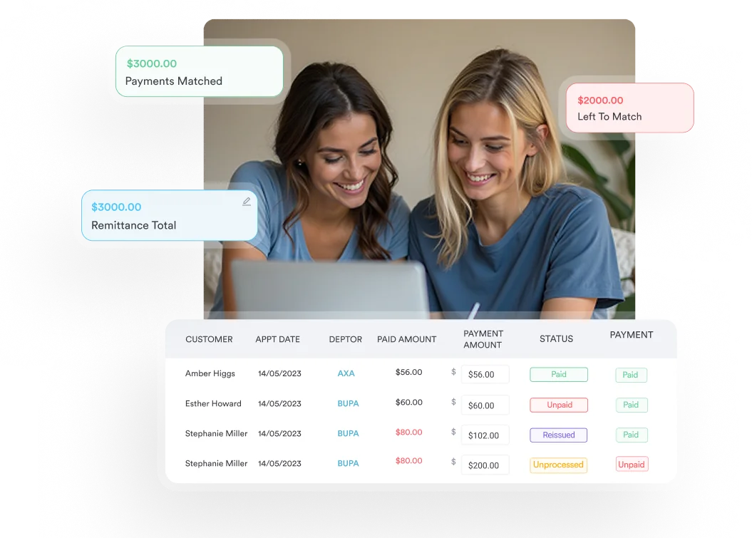
A nuclear stress test rarely travels as one line on a claim. The imaging study, the tracer supply, and any stress drug are documented and billed separately, so one missing detail can hold the whole encounter.
Denials cluster around a few predictable causes. The indication is not documented, the tracer dose never made it into the record, or the report does not say which stress method was used.
Run through this before you submit:
Teams new to nuclear billing usually want a shared reference. This medical coding cheat sheet works as a starting point for the front office.
It is faster to check payer details before submission than to fix a rejection later. Claims management tools validate the insurer fields on an invoice, so an obvious error is caught before the claim leaves the practice.
Thallium-201 was the first nuclear cardiology tracer. Technetium-99m sestamibi, sold as Cardiolite, now handles most routine perfusion imaging. Dose and image quality drove that shift, and thallium kept one clear advantage.
The American Heart Association treats both tracers as validated for perfusion imaging, so the choice comes down to the clinical question and local protocol.
Thallium still wins on viability. When the question is how much muscle can be saved in a patient with poor left ventricular function, redistribution answers it better than sestamibi. For routine screening and risk stratification, the lower dose and sharper images make sestamibi the default.
A thallium stress test asks more of the schedule than a standard consultation. One patient occupies a stress bay, a camera slot, and a second camera slot hours later. The whole day slips if that patient arrives after a coffee.
Practice management software like Pabau handles the work around the scan. Appointment scheduling books the stress and rest slots against one patient record, and automated messages carry the fasting and caffeine rules 48 hours ahead.
Everything the patient signs or completes lands in their patient record, next to the contraindication screening and the report. Invoicing and claims sit in the same place, with insurer details validated before a claim goes out.
The outcome is fewer scans canceled on the morning, fewer reports waiting on a callback, and a schedule your nuclear team can actually hold.

Pabau’s automated pre-appointment messages, digital intake forms, and shared patient records keep multi-phase imaging appointments running to plan. Your team spends less time chasing cancellations and unread reports.

The thallium stress test earns its place by answering a question no ECG can. Reversible ischemia and fixed scar look different on these images. That difference decides whether a patient heads to the cath lab or to medical therapy.
The trade-off is dose and time. Thallium carries more radiation than sestamibi and ties up most of a day, so reserve it for the questions it answers best, chiefly viability.
Operationally, the unglamorous work is what decides the day. Instructions have to reach the patient early, the checklist has to happen the day before, and the claim has to leave with its documentation intact. Book a demo to see how Pabau keeps multi-phase imaging appointments and their paperwork on track.

Handling a different medical emergency on shift? Meningitis nursing care plan gives nurses a framework for fever, altered consciousness, and neck stiffness.
Want the physiology behind the numbers? Cardiac output formula explains how stroke volume and heart rate shape perfusion.
Triaging chest pain at the front desk? Chest pain location charts give your team a visual reference for describing symptoms.
Plan for 4 to 5 hours in total. Setup and the stress phase take about an hour, stress imaging another 20 minutes, then rest images follow the 3 to 4 hour wait.
Studies of appropriately referred patients with chest pain report sensitivity around 70% to 90% for disease that needs revascularization. Accuracy falls when preparation slips, which is why caffeine and held medications matter.
A stress echo uses ultrasound to watch wall motion and adds no radiation. A thallium scan measures perfusion directly and reads through many resting ECG abnormalities. Availability and the clinical question decide.
Most patients can. Anyone given adenosine, dipyridamole, or regadenoson should sit until the flushing and dizziness pass. No sedation is used, so driving is usually fine once symptoms settle.
Pregnancy is a reason to postpone, or to pick a test without ionizing radiation. Nursing mothers should ask the nuclear medicine team whether to interrupt breastfeeding after the injection.
Cost depends on the payer, the facility, and whether a drug replaces exercise. Ask the practice for a written estimate, and confirm prior authorization is on file before the appointment.






