Yeoman’s Test Template
Yeoman’s test is a prone provocation test for sacroiliac joint (SIJ) pain....

1
Last Updated: Sep 28, 2026

A reflex test measures the motor response to a tendon tap, graded on a standardized 0 to 4+ scale.
Grade 2+ is normal, while 3+ to 4+ points toward upper motor neuron pathology and 0 to 1+ toward peripheral nerve involvement.
Patient relaxation and precise hammer technique drive accuracy, and poor positioning accounts for most false-negative findings.
Practice management software like Pabau stores reflex grades, reinforcement status, and examination notes in one structured client record.
A single absent knee jerk can be the first sign of an L4 radiculopathy. A brisk ankle reflex in a patient with neck pain raises the suspicion of cervical myelopathy. The reflex test is one of the fastest, lowest-cost neurological assessments a clinician has. It is also one of the most frequently performed badly.
Reflexes are involuntary, stereotyped motor responses generated by the spinal reflex arc. A sensory neuron detects tendon stretch, synapses directly with a motor neuron in the spinal cord, and drives a muscle contraction. Deep tendon reflexes travel through a monosynaptic arc, so no cortical input is required.
That is what makes the test so useful. It interrogates the peripheral and central nervous system independently of patient cooperation. The Stanford Medicine 25 clinical examination program frames it the same way. Reflexes evaluate nervous system integrity from peripheral nerve to spinal cord to corticospinal tract.
The sections below work through the grading scale, the equipment, and patient positioning. They then cover technique for each major deep tendon reflex, reinforcement, and the errors that skew results. It is written as a bedside reference for clinicians, medical students, nurses, and allied health professionals.
Every reflex test result is recorded on the same standardized 0 to 4+ grading scale. Knowing what each grade means, and where the clinical thresholds sit, is the foundation of interpreting any neurological examination.
Grade 2+ is the expected normal, verified by both Cleveland Clinic’s deep tendon reflex reference and Stanford Medicine 25. Symmetry matters as much as the absolute grade. A bilateral 3+ patellar reflex in an otherwise asymptomatic patient is far less concerning than a unilateral 3+ with ipsilateral spasticity.
Always compare left against right, and upper limb against lower limb, within the same patient. The scale also carries known inter-rater variability, so two clinicians examining one patient may record grades a level apart. Consistent technique, proper positioning, and reinforcement improve reproducibility, but do not overstate precision when you write the finding down.

The most common reason for a false-negative reflex test is patient tension, not pathology. A muscle that is already contracting will not respond to a tendon tap, however well the hammer is swung. Reliable results begin with preparation, before you pick the hammer up.
Where responses are consistently diminished, attempt the Jendrassik maneuver before recording a finding as hyporeflexic or absent. It has its own section below. Documenting that reinforcement was tried, along with the result with and without it, is what makes the grade usable at the next visit.
According to the Merck Manual Professional Edition, technique decides the result. The patient must be relaxed, the limb properly positioned, and the hammer swing smooth. Each of the five standard deep tendon reflexes has its own strike point, position, and expected response.
Support the patient’s elbow in about 90 degrees of flexion with the forearm resting in semi-pronation on your own arm. Place your thumb or index finger firmly over the biceps tendon in the antecubital fossa. Strike your finger, not the tendon directly. The expected response is elbow flexion and visible biceps contraction. A brisk response may spread into forearm pronation. Absent or diminished biceps reflex warrants consideration of C5 or C6 radiculopathy.
The forearm rests in semi-pronation on your arm or a surface. Strike the brachioradialis tendon at the distal radius, approximately 5 cm proximal to the wrist. The expected response is forearm flexion and slight supination. This is sometimes called the supinator reflex. A paradoxical inversion, meaning finger flexion rather than forearm flexion, may indicate a C5/C6 cord lesion and warrants urgent neurological review.
Support the arm in about 90 degrees of elbow flexion, with the forearm hanging freely. Strike the triceps tendon just above the olecranon. The expected response is elbow extension. The triceps reflex confirms C7 nerve root integrity. That matters most in patients with posterior shoulder or elbow pain, and in those presenting with cervical disc symptoms at the C6/C7 level.
The patient sits with the knee at 90 degrees, leg freely hanging. Strike the patellar tendon briskly, just below the patella. The expected response is quadriceps contraction and knee extension. In supine patients, support the knee in slight flexion (about 30 degrees) by placing your hand under the popliteal fossa. The patellar reflex is the most commonly assessed lower-limb reflex and the most sensitive indicator of L3/L4 radiculopathy. If the response is absent, attempt the Jendrassik maneuver before you record the finding.
With the patient seated, dorsiflex the foot gently to a neutral position and hold it there with light pressure. Strike the Achilles tendon directly. The expected response is plantarflexion. The Achilles is the most commonly absent reflex at the bedside. S1 radiculopathy, diabetic peripheral neuropathy, and healthy aging all reduce it. On a painful ankle, pair the finding with validated ankle assessment decision rules so you separate a neurological loss from a bony injury.
The Jendrassik maneuver is the standard reinforcement technique for a deep tendon reflex that appears absent or diminished. The patient interlocks their fingers and tries to pull their hands apart as you strike the tendon. The sustained isometric effort distracts them and activates descending motor pathways. Those pathways lower the reflex threshold and let a genuine response emerge.
Run the same short sequence every time a reflex looks absent. It takes under a minute, and it decides what you are entitled to write in the note.
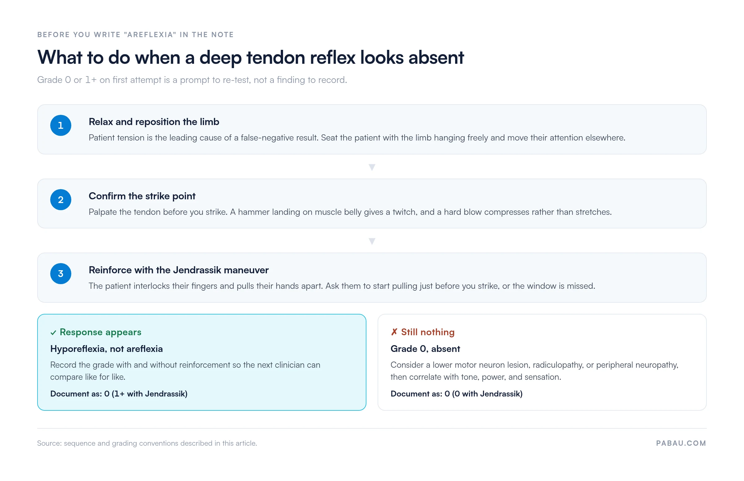
Reinforcement matters most on the patellar reflex, where suspected lumbar radiculopathy and peripheral neuropathy both sit in the differential. A reinforcement field on the examination form is the simplest way to make sure the step is recorded rather than assumed.
Deep tendon reflexes form the backbone of reflex testing, but a complete neurological assessment includes superficial and pathological reflexes. These tests evaluate different components of the nervous system. They pick up upper motor neuron pathology that deep tendon reflexes alone may not localize.
Run a blunt instrument along the lateral border of the sole from heel to ball of the foot, then curve medially across the metatarsal heads. The handle of the reflex hammer, an orange stick, or a key all work. The normal flexor response in adults is downward curling of all toes.
A positive Babinski sign is dorsiflexion of the great toe, often with fanning of the other toes. This extensor plantar response is pathological in adults and indicates corticospinal tract disruption, a hallmark of an upper motor neuron lesion. In infants under roughly 2 years it is normal, because those tracts are not yet fully myelinated.
Hold the patient’s middle finger loosely, flick the fingernail downward, and release. A positive Hoffman sign is reflex flexion of the thumb and index finger. Like the Babinski sign in the lower limbs, it is a pathological upper-limb reflex that may suggest cervical cord compression.
It is not diagnostic in isolation. A bilateral Hoffman sign without other upper motor neuron features may occur in anxious or hypermobile individuals, and asymmetry carries more weight than bilateral positivity. Pair it with a root-level provocation test such as Spurling’s test before you commit to a level.
Clonus is rhythmic, involuntary muscle contraction triggered by sudden sustained stretch. To test for ankle clonus, hold the patient’s knee in slight flexion, rapidly dorsiflex the foot, and hold that position. Sustained clonus indicates significant upper motor neuron involvement. Thresholds vary across sources, but beyond 3 to 5 beats is generally considered pathological.
A few beats of non-sustained clonus may occur in anxious patients and is less specific. Wrist clonus is tested the same way, by rapidly extending the patient’s wrist and holding the position.
Abnormal reflex findings are signs, not diagnoses. They narrow the differential and direct further investigation, without confirming anything on their own. The two primary abnormal patterns are hyperreflexia and hyporeflexia, and each points toward a different level of nervous system involvement.
Hyperreflexia points to upper motor neuron lesions and hyporeflexia to lower motor neuron or peripheral nerve pathology, as set out in Cleveland Clinic’s DTR reference. Always read a reflex finding alongside tone, power, coordination, and sensory testing.
Rehab teams repeat this examination across a course of care, so the grades have to sit somewhere comparable. That is a job for purpose-built physical therapy software rather than a free-text note that nobody can search six visits later.
One of the most practical uses of the reflex test is localizing a lesion to a specific spinal level. The table below maps each standard deep tendon reflex to its nerve root and myotome. It also names the radiculopathy that most often explains a loss.
A lost patellar or Achilles reflex rarely travels alone. Pair the finding with a nerve tension test such as the crossed straight leg raise before you commit to a level in the note.
The errors below account for most inaccurate reflex findings, and every one of them is avoidable at the bedside. Ruling them out first is what makes a grade worth recording.
In a busy practice or ward, ask the patient ‘did you feel anything?’ after a zero-grade response. Some patients notice a subtle contraction before the examiner can see it. Use that cue to re-examine closely, or attempt reinforcement, before you record areflexia.
Reflex testing is routine in chiropractic and physical therapy examinations. The difficulty is not performing it. It is capturing the finding consistently enough that another clinician can compare it six weeks later.
A grade written as “2+” by one practitioner and “normal” by another at the next visit cannot be compared at all. Free-text notes make that mismatch close to inevitable, because nothing prompts for the scale or for whether reinforcement was used.
Practice management software like Pabau closes it at the form level. A digital form carries a fixed grading field for each tendon, a reinforcement checkbox, and a nerve root note. Every entry is timestamped against the client record, so comparing visit one with visit six takes seconds instead of a chart review.
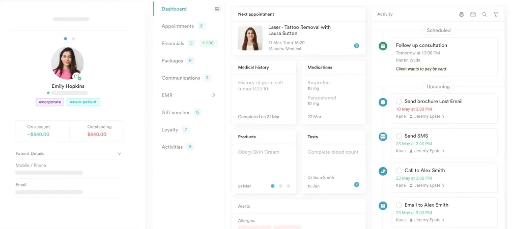
If a visiting clinician still works on paper, the deep tendon reflex exam template mirrors the same fields. Findings then transfer into the record without a second interpretation.

Pabau lets physical therapy, chiropractic, and neurology practices capture reflex grades, reinforcement status, and nerve root notes on a structured form. Findings stay attached to the client record, so comparing visits takes seconds.

The reflex test holds its place at the bedside because it needs no imaging, no laboratory, and no effort from the patient beyond relaxation. What it does need is discipline. Use controlled technique, one consistent scale, and reinforcement before anyone writes “absent” in the note.
The grade on its own is worth less than the comparison. A 1+ Achilles reflex means little in isolation and a great deal when the same practice recorded 2+ four weeks earlier. That only holds if the grade lives in a fixed field rather than a paragraph of prose. Book a demo to see how Pabau structures neurological examination records for a whole team.

Working through a full neurological exam? Cranial nerve examination checklist covers the twelve cranial nerves in the same structured order you would grade reflexes.
Chasing a cervical radiculopathy? Upper limb tension tests add the neural mobility findings that sit alongside a diminished biceps or brachioradialis reflex.
Need a sciatic nerve root test? Bragard’s test explains the dorsiflexion step that separates nerve root pain from hamstring tightness.
Assessing balance as well as reflexes? Modified Romberg test walks through the proprioceptive screen that pairs with an absent Achilles reflex.
Suspecting lumbar instability? Passive lumbar extension test gives you the segmental check to run after the neurological screen.
A reflex test is a neurological examination in which a clinician strikes a tendon with a reflex hammer. It checks the reflex arc, the sensory-motor loop connecting peripheral nerve, spinal cord, and muscle. Results are graded from 0 to 4+, with 2+ considered normal. The test identifies upper and lower motor neuron lesions without imaging or patient effort.
Hyperreflexia (grade 3+ to 4+) may suggest an upper motor neuron lesion disrupting the corticospinal tract. Common causes include stroke, multiple sclerosis, cervical myelopathy, and traumatic brain injury. It is a sign that warrants correlation with tone, power, and sensory testing, not a standalone diagnosis.
The Jendrassik maneuver is a reinforcement technique used when a reflex appears absent or diminished. The patient interlocks their fingers and tries to pull them apart while the tendon is struck. The sustained effort lowers the reflex threshold and amplifies the response. That separates genuine hyporeflexia from a false negative caused by patient tension. Attempt it before recording a reflex as absent.
Absent reflexes that persist through reinforcement may indicate a lower motor neuron lesion, nerve root compression, or peripheral neuropathy. Acute spinal shock also suppresses reflex activity temporarily. In older adults, an absent Achilles reflex can be a normal age-related finding. Attempt the Jendrassik maneuver and correlate with other neurological signs before drawing conclusions.