Best chiropractic billing software in 2026: Top 5 compared
Chiropractic billing software automates insurance claims, CPT coding, eligibilit...

Last Updated: Oct 8, 2026
The Gillet test is a standing clinical examination of sacroiliac joint (SIJ) motion, also known as the stork test.
The examiner rests one thumb on the posterior superior iliac spine (PSIS) and another on the sacrum. The patient then lifts the knee on that side. A positive result is a PSIS that fails to drop relative to the sacrum, which suggests restricted motion on the side tested.
Sacroiliac joint dysfunction accounts for roughly 25% of chronic low back pain in adults, according to the American Academy of Family Physicians. That makes a quick pelvic screen worth running. The Gillet test gives physiotherapists, chiropractors, and allied health clinicians one, without imaging or equipment.
What it cannot do is confirm the diagnosis on its own. Reported agreement between examiners is fair at best, and one study group argues the test reads stance strategy rather than joint movement. This article covers the procedure, how to read the result, what the evidence supports, and which SIJ tests belong alongside it.

The Gillet test, also called the stork test or marching test, assesses sacroiliac joint motion by monitoring PSIS movement during hip flexion.
A positive result means the PSIS fails to move inferiorly relative to the sacrum, suggesting hypomobility on the tested side.
Published kappa values run from 0.22 to 0.68, which is fair-to-moderate reliability. Agreement between two examiners is consistently lower than one examiner’s agreement with themselves.
Reported sensitivity spans roughly 0.08 to 0.80 across studies, so treat any single published accuracy figure with caution.
No single SIJ test is diagnostic in isolation. Confidence rises once three or more provocation tests come back positive.
The Gillet test is a clinical motion test for the sacroiliac joint. The examiner monitors PSIS movement while the patient performs single-leg standing hip flexion on the same side. It is one of the most commonly taught SIJ special tests in physiotherapy and chiropractic curricula.
The test goes by several names, which creates confusion in the clinical literature. All three terms refer to the same examination:
In practice, stork test and Gillet test are used interchangeably. Older references cite it as the Gillet marching test. Some systematic reviews fold it into a group of sacroiliac provocation tests without naming it at all.
The sacroiliac joint connects the sacrum to the ilium on each side of the pelvis. It transmits load between the spine and the lower limb and contributes to lumbopelvic stability. Movement at the joint is small, around 1 to 4 degrees of rotation and 1 to 2 mm of translation.
Stability depends on both form closure, meaning bony geometry, and force closure, meaning muscular and ligamentous tension. That combination is why a motion test at this joint detects so little travel.
The posterior superior iliac spine is the key palpation landmark. It is the bony prominence at the dimples of the lower back, just medial to the gluteal cleft on each side.
During normal SIJ motion, the PSIS on the flexing side should move inferiorly relative to the sacrum as nutation occurs. Movement that is absent or reversed raises the suspicion of restricted sacroiliac motion.
SIJ hypomobility, the condition the Gillet test screens for, is abnormally restricted movement at the joint. It turns up alongside low back pain, posterior pelvic girdle pain, and referred gluteal or posterior thigh pain. Practices running physical therapy EMR software see this presentation across musculoskeletal and post-partum caseloads.
Performing the test correctly depends on precise palpation and consistent patient instruction. Small variations in hand placement or movement speed change what you feel, which partly explains the variable agreement between examiners.
The patient stands barefoot on a flat, non-slip surface. They should face away from the clinician, feet about hip-width apart, with weight evenly distributed.
Ask the patient to relax the gluteal and abdominal muscles before you begin. Patients who habitually brace the pelvis will move the PSIS independently of any joint motion.
Stand directly behind the patient. Place one thumb on the ipsilateral PSIS, the side being tested, and the other on the sacrum at the second sacral segment (S2). S2 sits roughly level with the opposite PSIS dimple, or about 2 cm below the lumbosacral junction in most adults.
Both thumbs rest with light, even pressure. Pressing hard restricts the subtle movement you are trying to detect. The palpation point is the uppermost bony prominence of the posterior ilium, just lateral to the sacrum.
Instruct the patient to flex the ipsilateral hip and knee to roughly 90 degrees, as if taking a slow marching step. The movement should be smooth and deliberate rather than a rapid kick.
Feel what happens to the ipsilateral PSIS against your sacral thumb throughout the lift. Then repeat on the other side and compare.
Interpretation means knowing what normal and abnormal PSIS movement look like, and what conclusion the finding will and will not support.
A positive Gillet test is present when the ipsilateral PSIS fails to move inferiorly relative to the sacrum during hip flexion. A PSIS that travels superiorly counts as positive too.
That pattern suggests restricted SIJ motion, or hypomobility, on the tested side. A negative result is a PSIS that moves inferiorly as expected, suggesting adequate mobility on that side.
A positive Gillet test alone does not confirm SIJ dysfunction. Interpretation requires correlation with patient history, symptom pattern, and results from additional SIJ provocation tests. Never document a positive result as a definitive diagnosis.
The evidence is not settled, and one 2018 study makes a case for stance strategy over joint motion. That question decides how much weight the finding deserves in your assessment.
A 2018 peer-reviewed study published on PubMed Central challenged the traditional interpretation. Researchers found the test may primarily reflect asymmetric single-leg stance strategies, involving gluteus medius and biceps femoris activation. On that reading, a positive finding records a neuromuscular compensation pattern rather than a restricted joint.
That does not strip the test of clinical value. It does mean the finding belongs in the screening column rather than the confirmation column.
Published reliability data shows a consistent pattern. A clinician who tests the same patient twice agrees with themselves more often than two clinicians agree with each other.
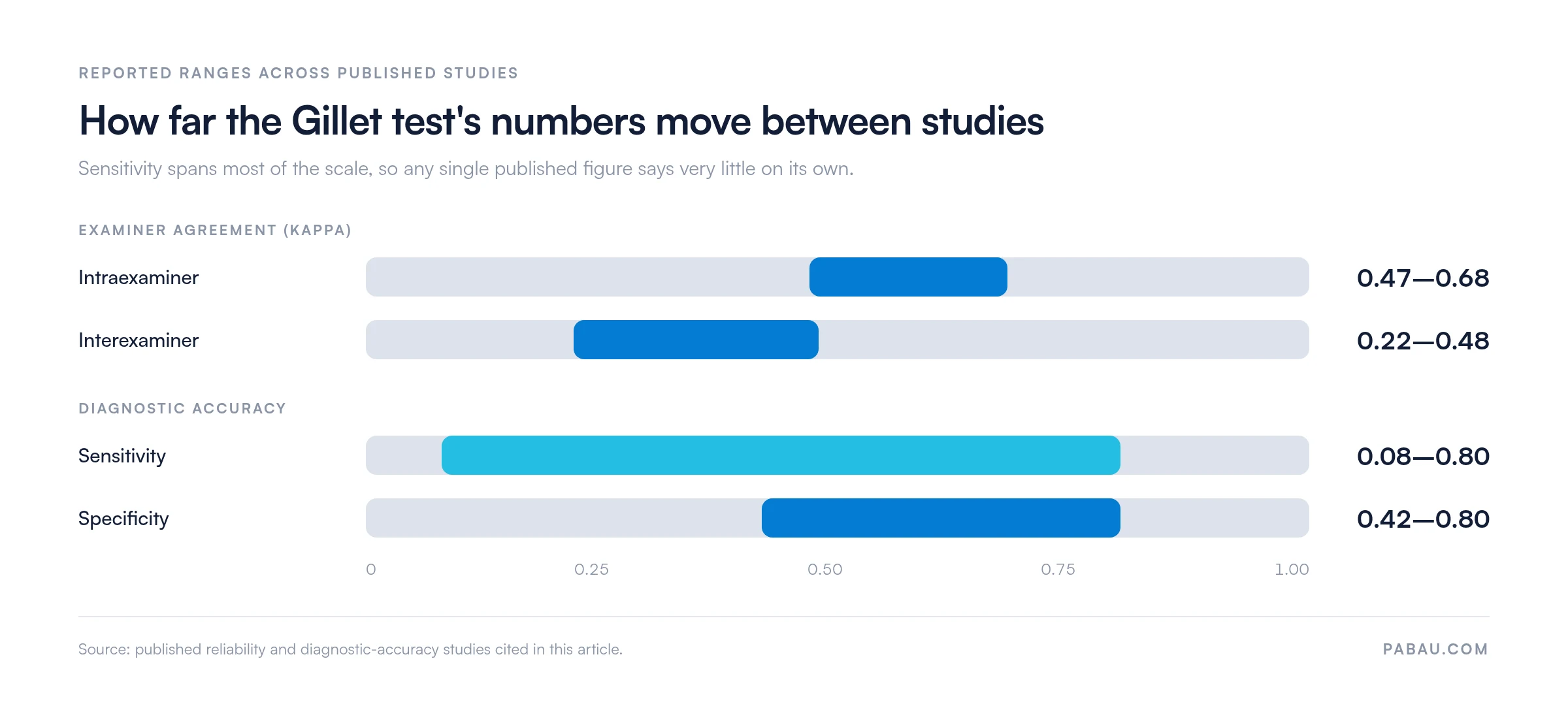
Intraexaminer kappa values typically run from 0.47 to 0.68, which counts as moderate agreement. Interexaminer kappa values run from 0.22 to 0.48, which is fair to moderate.
The practical consequence is that one practitioner’s result should not be read straight across against another’s. That matters most in physiotherapy practice management software settings where several clinicians share a caseload and review each other’s assessments.
Published accuracy figures for the Gillet test vary widely between studies. Reported sensitivity runs from roughly 0.08 at the low end to about 0.80 at the high end. Reported specificity sits between about 0.42 and 0.80.
The spread comes from small samples, differing patient populations, and the absence of an agreed gold standard for SIJ dysfunction. Systematic reviews of SIJ motion tests report the same heterogeneity, which is why a single quoted sensitivity figure should not drive a clinical decision.
Set the four published ranges side by side and the difference in spread becomes obvious.
Track Gillet test results across appointments rather than reading each one alone. A patient whose PSIS movement returns between the first and fourth visit gives you meaningful data on SIJ mobility recovery. That progression carries weight even though the initial positive was not diagnostic on its own.
No single special test diagnoses sacroiliac joint dysfunction with acceptable accuracy on its own. Laslett and colleagues showed that three or more positive provocation tests raise diagnostic confidence meaningfully.
The Gillet test fits at the start of an SIJ battery. It provides a motion-based screen before the provocation tests add stress loading.
A minimum three-test screen usually pairs the Gillet test with the thigh thrust and the Gaenslen test. Three or more positive findings from that battery carry far more weight than any single positive result.
Clinicians managing return-to-running protocols after pelvic injury often run the same three tests at every reassessment point. Comparing the pattern over time tells you more than the first result did.
The Gillet test earns its place in a systematic SIJ screen for patients with unilateral low back pain, posterior gluteal pain, or pelvic girdle pain. It adds most where the pain distribution, history, and functional limits all point the same way. That means an SIJ source rather than a lumbar disc, facet, or hip one.
Chiropractors and physiotherapists should consider applying the test in these scenarios:
The test is not appropriate as a standalone screen for acute radicular pain, or for hip joint pathology without posterior pelvic pain. Skip it where the PSIS cannot be palpated reliably because of adipose tissue depth or patient discomfort.
A positive Gillet test at the initial assessment that turns negative by session four is clinically meaningful. Without structured documentation, that progression never surfaces in the patient record.
Free-text notes make that comparison slow. Reading visit one against visit four means rereading four blocks of prose. Practice management software like Pabau replaces them with digital assessment forms you build yourself.
You can build a custom SIJ examination template that records each special test finding, grades a positive result, and attaches it to the appointment. Results link straight to the SOAP note, so the clinician writing a follow-up sees whether the Gillet test was positive last visit.

Across a multi-practitioner team, any clinician can open the prior assessment before the patient walks in. That cuts redundant re-testing and keeps the clinical picture continuous, which matters given how much Gillet test results vary between examiners.


Pabau gives physiotherapists and chiropractors customizable SOAP note templates and digital assessment forms. Record special test findings, track SIJ progress over time, and link results to the treatment plan.

The Gillet test earns a place at the front of an SIJ assessment rather than at the end of one. Its fair-to-moderate reliability and the open question about what it measures both land in the same place.
Run it, then let the provocation tests decide. Pair it with the thigh thrust and Gaenslen tests, correlate against symptom history, and record the finding somewhere you can compare it next visit.
That comparison is only as good as the record it sits in. Book a demo to see how Pabau captures SIJ assessment findings your whole team can read back.

Need a framework for clinical documentation compliance? Mandatory compliance for physiotherapy clinics covers the documentation standards UK physiotherapy practices must meet.
Assessing pelvic alignment alongside the SIJ? Anterior pelvic tilt explains how to spot it, what drives it, and how it changes a treatment plan.
Need to rule out lumbar instability? Passive lumbar extension test covers the procedure and how its accuracy compares with other lumbar screens.
Looking for a documentation template? SOAP notes for chiropractic gives you a structured format for recording special test findings at every visit.
The Gillet test is a clinical motion assessment of sacroiliac joint (SIJ) mobility. Clinicians use it to screen for SIJ hypomobility or dysfunction. It suits patients presenting with unilateral low back pain, posterior pelvic girdle pain, or suspected SIJ involvement. It is usually one part of a wider provocation battery.
The patient stands barefoot with feet hip-width apart, facing away from the clinician. The clinician places one thumb on the ipsilateral PSIS and the other on the sacrum at S2 level. The patient then flexes the ipsilateral hip and knee to roughly 90 degrees. The clinician monitors whether the PSIS moves inferiorly relative to the sacrum, then repeats on the opposite side for comparison.
A positive Gillet test means the ipsilateral PSIS failed to move inferiorly relative to the sacrum during hip flexion. That suggests restricted SIJ motion, or hypomobility, on the tested side. It does not confirm SIJ dysfunction in isolation, so correlate it with other provocation tests and the clinical history.
Yes, the Gillet test and the stork test are the same clinical examination. Both names describe the standing single-leg hip flexion assessment of PSIS movement relative to the sacrum. The test is also called the marching test. All three terms appear in the clinical literature and are used interchangeably.
The Gillet test has fair-to-moderate reliability. Intraexaminer kappa values range from 0.47 to 0.68, so a single clinician testing the same patient twice will usually agree with themselves. Interexaminer kappa runs from 0.22 to 0.48, so two clinicians examining the same patient may reach different conclusions. Reported sensitivity varies even more widely, from roughly 0.08 to 0.80 across published studies.
A 2018 peer-reviewed study suggests the Gillet test may reflect asymmetric gluteus medius and biceps femoris activation during single-leg stance. On that reading it records a stance strategy rather than movement at the sacroiliac joint. The test still has clinical value, but the finding should be treated as screening information rather than direct evidence of restricted SIJ movement.
The Gillet test works best as part of a multi-test battery. Clinicians typically pair it with the thigh thrust test and the Gaenslen test as a minimum. When three or more SIJ provocation tests come back positive, diagnostic confidence rises well above what any single positive finding supports.