



GLP-1 diet plan
A GLP-1 diet plan is a structured eating framework for patients taking semagluti...

2
Last Updated: Oct 13, 2026
The prone instability test is a hands-on clinical assessment for lumbar segmental instability.
It compares a patient’s pain response to posteroanterior pressure in two positions. The feet rest on the floor for the first phase, then the legs are actively raised for the second. Pain that eases in the second phase is a positive result.
A systematic review in the Journal of Orthopaedic and Sports Physical Therapy reports moderate sensitivity and moderate-to-high specificity for radiographic lumbar instability. That holds when the test is read alongside a clinical cluster. Physical therapists, chiropractors, and sports medicine clinicians use the result to decide whether a stabilization program is warranted.
This guide covers the procedure step by step, and how to read positive and negative findings. It also sets out what the evidence says about diagnostic accuracy, and how to record the result.

The prone instability test detects lumbar segmental instability by comparing pain during passive versus active leg positioning under posteroanterior pressure.
A positive result occurs when PA-provoked pain reduces during the active leg-raise phase, suggesting the active muscular subsystem can offset segmental instability.
Reported sensitivity is roughly 0.61 and specificity roughly 0.57, per Fritz, Piva and Childs (2005), so pair it with other lumbar tests.
A positive finding gives a clinical basis for referral to a lumbar stabilization program and structured follow-up documentation.
The prone instability test is a clinical special test for lumbar segmental instability. It separates pain produced by passive mechanical load on the lumbar spine from pain that eases once the active muscular subsystem is engaged. It was first described as a way to identify radiographic lumbar instability. Few clinical tests target the active-passive stabilizing relationship this directly.
Use it when a patient reports non-specific low back pain that worsens with sustained postures, a catch or apprehension on movement, or repeated episodes. Patients who say that bracing the abdomen eases their pain are strong candidates. Screen for contraindications against your professional body’s guidance before you load the spine.
Chiropractors working from chiropractic software and physical therapists across musculoskeletal caseloads both run this test inside a lumbar instability screening cluster. On its own it settles very little. Its value comes from how it combines with the patient history, imaging findings, and the other special tests.
The procedure takes roughly three minutes from setup to assessment. Accuracy depends on correct patient positioning, consistent examiner hand placement, and a methodical comparison between the two phases.
Position the patient prone with the hips at the edge of the treatment table and both feet flat on the floor. The hips sit at roughly 90 degrees of flexion with the knees straight, so the legs hang but the feet stay grounded. This loads the lumbar spine in neutral to slight extension and keeps the erector spinae and multifidus quiet at rest.
Check that the arms are relaxed alongside the body or hanging off the table edge. Any trunk bracing before pressure is applied will confound the first phase of the test.
Stand at the patient’s side. Place both thumbs over the spinous process of the lumbar vertebra being assessed, or use the pisiform of one hand reinforced by the other. Begin at L4 or L5 and work systematically through each lumbar level.
Apply a graded posteroanterior (PA) pressure directed anteriorly through the spinous process. The force should be enough to provoke pain or discomfort where instability is present. Note the pain response at each level and record which levels are symptomatic. That is phase one.
Ask the patient to lift both legs off the floor and hold them in extension. The feet should clear the floor by about 4 to 6 inches. This recruits the lumbar erector spinae, multifidus, and hip extensors, loading the posterior stabilizing subsystem.
With the legs held up, re-apply PA pressure to the same lumbar levels that were symptomatic in phase one. Ask the patient to compare the pain against phase one. Record whether it is reduced, unchanged, or increased.
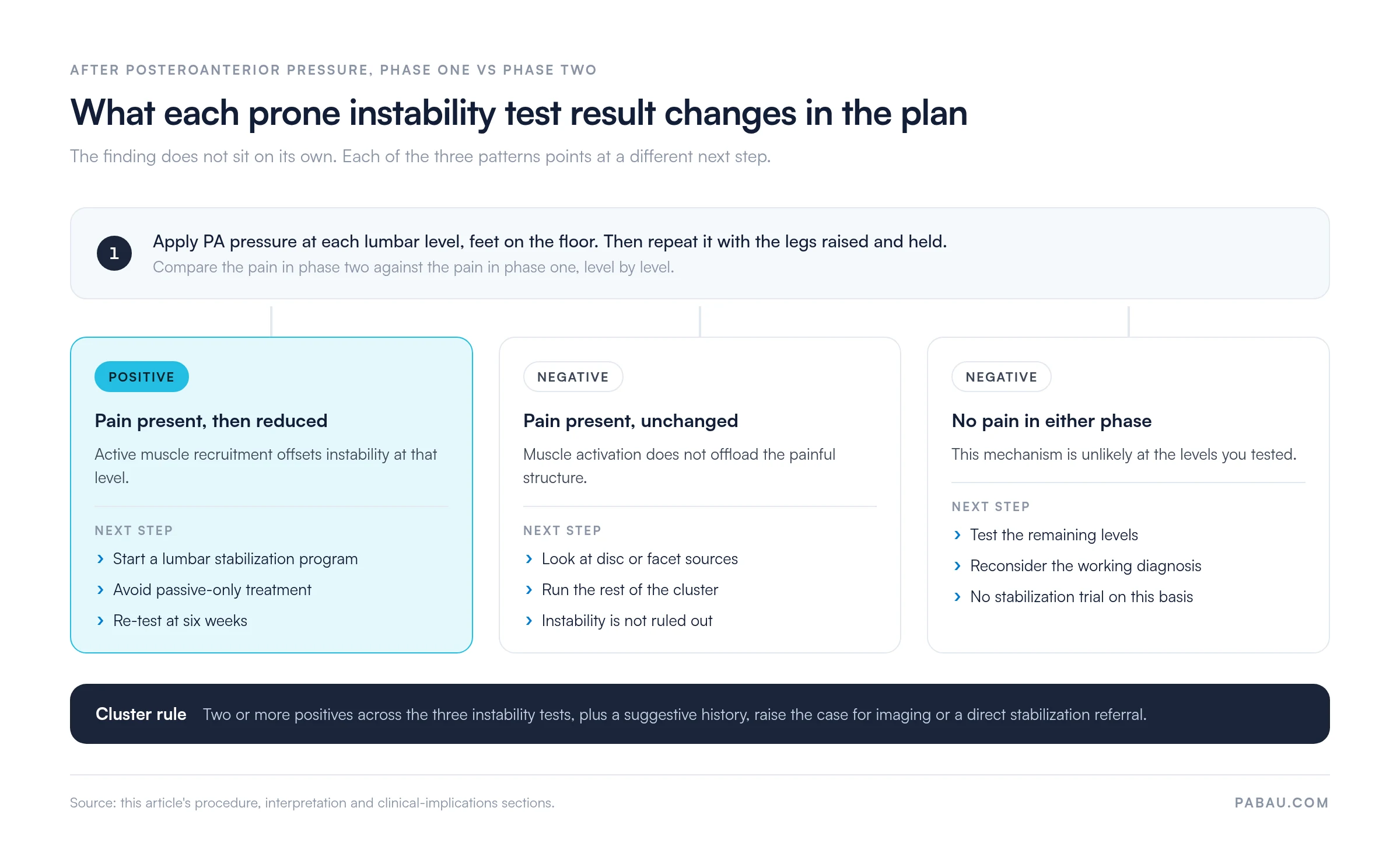
A positive prone instability test is recorded when PA-provoked pain from phase one is reduced or eliminated in phase two. That pattern shows active muscle engagement compensating for segmental instability at the level you tested.
A negative prone instability test does not rule out lumbar segmental instability. It means active muscle recruitment during this specific test did not reduce the pain.
Instability may still be present if the patient cannot engage the stabilizing muscles under load, or if the pattern is flexion-based rather than extension-based. Treat a negative as a prompt to run the rest of the cluster, not as a reason to drop the diagnosis.
The test rests on a three-subsystem model of spinal stability. The passive subsystem is the ligaments and disc. The active subsystem is the paraspinal musculature, and neural control coordinates the two. Lumbar segmental instability occurs when the passive subsystem cannot hold a segment within its neutral zone under load.
When the patient raises the legs, the erector spinae and multifidus contract bilaterally to hold the extended position. That recruitment compresses the lumbar segments and increases stiffness across the motion segment, substituting for lost passive restraint. Fritz, Piva and Childs (2005) describe this compensatory mechanism in the European Spine Journal. It is the clinical basis for how the test is read.
The practical consequence shapes the treatment plan. If muscle activation resolves the pain during the test, a structured stabilization program targeting those same muscles has a reasonable chance of producing functional benefit. That is why a positive finding points toward motor control and stabilization training rather than passive modalities.
Diagnostic accuracy for the prone instability test has been evaluated in several studies, and the values vary by population and reference standard. The figures below are the ones most often cited, from Fritz, Piva and Childs (2005) and the JOSPT systematic review. Read them as indicators rather than benchmarks, against the pre-test probability for your patient.
These modest likelihood ratios reinforce a clinical principle. No single lumbar instability test is diagnostically sufficient. The APTA’s low back pain guideline advocates test clustering and multi-factor clinical reasoning over reliance on individual special tests.
Inter-rater reliability data for the prone instability test is limited. Kappa values are not consistently reported across studies, and the evidence base is smaller than for other lumbar special tests. Training examiners on consistent hand placement, force magnitude, and the criteria for recording a pain response reduces variability between clinicians.
Intra-rater reliability is generally considered acceptable in practice, though formal studies are sparse. Practices running the test at volume should standardize the protocol, recording the spinal levels tested and the force applied, so results stay comparable across sessions.
The prone instability test is one of three widely studied clinical tests for lumbar segmental instability. The passive lumbar extension test and the posterior shear test target similar pathology. They differ in position, technique, and the type of instability each preferentially loads. Running the three as a cluster rather than in isolation improves diagnostic confidence substantially.
Values above come from the JOSPT systematic review and are approximate, given variation in study populations and reference standards. The passive lumbar extension test has the strongest individual accuracy figures, which makes it a useful first-line test when clinical suspicion is high.
The prone instability test contributes most clearly when its positive finding matches what the patient describes, namely pain that eases when they brace actively.
Research on lumbar instability testing consistently shows that combining two or more tests from the same diagnostic cluster outperforms any single test. When all three are positive, post-test probability for radiographic lumbar instability rises substantially. That holds even with moderate individual likelihood ratios.
A pragmatic approach for a busy musculoskeletal practice is to run all three tests in the same session and record the number of positives. One positive warrants monitoring and an exercise trial.
Two or more, alongside a suggestive history, support either an imaging pathway or a direct move to a lumbar stabilization program. Hicks, Fritz, Delitto and McGill (2005) set out the clinical prediction rule framework behind that lumbar stabilization decision.
A positive prone instability test points to a specific management pathway. It is not a reason to discharge without intervention, and on its own it does not trigger an imaging referral. The chart below sets out what each result pattern changes in the plan.
Practices comparing physiotherapy practice management software should check that a treatment plan can be tied to the assessment that triggered it. That link is what keeps a stabilization program scheduled, tracked, and adjusted on reassessment data rather than on the calendar.
When a prone instability test is positive at multiple lumbar levels, start stabilization loading at the level of least provocation. Overloading the most unstable segment first can flare symptoms and reduce patient compliance. Build from adjacent stable segments inward.
Accurate documentation of the prone instability test supports continuity of care and informs handover between clinicians. It also creates the clinical record that a later review of the patient’s management would rely on. Inconsistent documentation is the most common failure point in musculoskeletal assessment records.
A well-structured note records the levels tested individually, from L1 to L5. It then records the pain response in each phase and the interpretation, whether positive, negative, or equivocal. Asymmetric findings belong at level, so L4-5 positive with L5-S1 negative is written that way.
Anchor every special test result to the clinical question it answers. A usable entry reads: “Prone instability test positive at L4-5. PA pain reduced from 7/10 to 2/10 with active leg raise. Supports segmental instability at L4-5.”
Most musculoskeletal practices record special test results in free-text notes, which makes them hard to find at reassessment. Practice management software like Pabau stores them as structured fields instead. The phase one and phase two responses then sit in the patient record in the same shape every time.
Practices running a physical therapy EMR can build the prone instability test into their assessment and reassessment templates. Every clinician then captures the same fields, and the six-week comparison becomes a lookup rather than a hunt through old notes.

Pabau Scribe, our AI medical scribe, drafts a structured note from what you dictate during the assessment. You review and sign it, which cuts the admin time that usually lands after a full lumbar screen.

Pabau captures clinical findings, special test results, and treatment plans in structured patient records for physical therapy and musculoskeletal practices. No paper, and no re-entry between sessions.

The prone instability test gives you a low-equipment way to separate pain driven by segmental instability from pain with another mechanical basis. Its value rises inside a cluster with the passive lumbar extension test and the posterior shear test. Two or more positives carry a much stronger rationale for a stabilization-focused plan.
The trade-off worth remembering is that moderate accuracy cuts both ways. A negative result buys you very little, so treat it as a reason to keep testing rather than a reason to stop.
Consistent recording is what turns a single finding into a trend you can act on at six weeks. Book a demo to see how Pabau structures assessment notes for physical therapy and musculoskeletal practices.

Testing the same muscle group another way? Multifidus lift test covers the procedure, the scoring, and how to read a segmental motor control deficit.
Want the strongest single instability test? Passive lumbar extension test sets out the procedure and the accuracy figures that make it a first-line choice.
Screening for a radicular cause instead? Crossed straight leg raise test walks through the steps and what a positive side-to-side response means.
Assessing femoral nerve tension in prone? Prone knee bend test explains how to perform, grade, and document the result.
The prone instability test detects lumbar segmental instability. It identifies whether active muscle contraction during a leg raise reduces the pain that posteroanterior pressure provokes. A positive finding guides clinicians toward a stabilization exercise-focused treatment approach.
Position the patient prone with hips at the table edge and feet on the floor. Apply posteroanterior pressure to each lumbar spinous process and record the pain. Then ask the patient to raise both legs off the floor and re-apply the same pressure. A positive result is recorded when pain in phase one is reduced in phase two.
A positive result indicates that active paraspinal and extensor muscle recruitment can offset lumbar segmental instability, reducing pain during PA pressure loading. It supports classifying the back pain as stabilization-relevant, and indicates that a motor control exercise program is likely to help.
Reported sensitivity is approximately 0.61 and specificity approximately 0.57, based on Fritz, Piva and Childs (2005) in the European Spine Journal. The reference standard was radiographic lumbar instability. These moderate values mean the test works best inside a clinical cluster rather than alone.
The prone instability test uses an active patient response, the leg raise, to see whether muscle activation reduces instability-related pain. In the passive lumbar extension test the examiner lifts the patient’s legs to load the lumbar spine. The passive test has higher reported sensitivity and specificity, so it is often run first in the cluster.
Inter-rater reliability data for the prone instability test is limited and formal kappa values are not consistently published. Reliability is acceptable for clinical use when examiners standardize hand placement and force application. It should not be your sole clinical measure. Pairing it with a structured assessment protocol and clear documentation improves consistency across sessions.






