



GLP-1 diet plan
A GLP-1 diet plan is a structured eating framework for patients taking semagluti...

2
Last Updated: Oct 13, 2026

The pronator drift test detects upper motor neuron (UMN) lesions by observing whether an outstretched, supinated arm drifts downward and pronates with eyes closed.
A positive result indicates a contralateral corticospinal tract lesion, commonly seen in stroke, TIA, multiple sclerosis, and brain tumors.
The test is quick, equipment-free, and sensitive enough to catch subtle hemiparesis that standard strength grading may miss.
Pabau’s digital clinical documentation tools help neurology and physical therapy teams record, grade, and track pronator drift findings consistently across visits.
Missed hemiparesis is one of the costliest diagnostic errors in acute neurology. A patient presents with vague arm heaviness, normal grip strength on direct testing, and no obvious facial droop. Standard motor grading returns a 5/5. Yet the pronator drift test catches the lesion in under 30 seconds. That difference between manual testing and the pronator drift test is why the assessment has endured. Nurses, physicians, and allied health clinicians have relied on it for decades.
This guide covers:
The pronator drift test is a bedside neurological assessment that detects upper motor neuron, or UMN, dysfunction. It works by exploiting a specific biomechanical signature of corticospinal tract lesions. When the descending motor pathways are interrupted, the arm’s flexor muscles receive relatively stronger signals than the extensors. The forearm supinators also lose tone relative to the pronators. Holding the arms outstretched with palms up and eyes closed exposes this imbalance. The affected arm drifts downward and rotates palm-down, or pronates, because the weaker anti-gravity muscles can no longer hold position against gravity without visual feedback.
The test is also called the Barré test or the upper extremity drift test. Jean Alexandre Barré described the arm-drifting phenomenon in the early 20th century as a reliable indicator of pyramidal tract pathology. Today it is embedded in acute stroke protocols, routine neurological examinations, and nursing assessments for patients on neurology, medical, and post-surgical wards. It requires no equipment and takes less than 30 seconds to perform. That makes it practical for frequent serial monitoring, which matters in conditions like patient care management protocols for acute neurological deterioration.
Understanding why the test works prevents misinterpretation. The corticospinal, or pyramidal, tract originates in the motor cortex and crosses in the medulla. It then descends contralaterally to synapse on lower motor neurons in the spinal cord’s anterior horn. A lesion anywhere along this pathway, from the cortex to the level above the anterior horn, constitutes an upper motor neuron lesion.
UMN lesions produce a characteristic imbalance: extensor and abductor muscle groups lose more tone than flexors. In the upper limb, this means forearm pronation and elbow flexion overcome supination and extension. With eyes closed, removing visual compensation, the proprioceptive and cortical motor signals become insufficient to maintain the supinated position against gravity. This happens when the pyramidal tract is compromised. The result is pronation and downward drift. The lateralization is predictable: a lesion in the left hemisphere produces pronator drift in the right arm, and vice versa. A posterior fossa or brainstem lesion above the decussation may also produce contralateral findings.
This neuroanatomical specificity is what separates pronator drift from tests that assess cerebellar function or proprioception. The mechanism targets the pyramidal motor system exclusively, which is why it is categorized alongside other UMN signs such as the Babinski reflex and clonus. Healthcare teams working with neurological physical therapy workflows encounter these distinctions regularly when differentiating UMN from lower motor neuron (LMN) presentations.
Precision in technique prevents false positives and false negatives. The following steps reflect the procedure used consistently across clinical literature and nursing education resources.
A common variation adds a light downward tap on both arms simultaneously after the patient is positioned. The clinician gently pushes each arm down by 5 to 10 cm, then releases. A healthy arm springs back to position; an arm with subtle UMN dysfunction returns slowly or not at all, and may pronate during the rebound. This maneuver increases sensitivity for very mild lesions that do not produce drift under static conditions alone.
Several clinically useful enhancements are underused. The Mingazzini sign is the lower limb equivalent. The patient lies supine and raises both thighs and knees to 90 degrees, the “bent-knee” position, then closes their eyes. The examiner watches for the affected leg to drop slowly. This can be performed sequentially with the arm test to screen both levels of the neuraxis in one clinical encounter.
For patients who cannot cooperate fully, a modified version using a 5-second observation window after arm positioning still captures significant drift. Always document which variation was used, as this matters for serial comparison. Using standardized assessment tools within electronic records removes ambiguity from these distinctions across different clinicians performing the same assessment on different shifts.
The result is not simply positive or negative. Three distinct findings carry different clinical meanings, and conflating them leads to documentation errors.
Lateralization is crucial. The affected arm is on the opposite side from the brain lesion. If the right arm drifts and pronates, the lesion is in the left hemisphere or the left corticospinal pathway above the decussation. Recording “left arm drift, pronation at 8 seconds” is more useful than “positive pronator drift” alone. Using structured neurological assessment frameworks keeps this level of detail consistent.
A positive pronator drift test signals UMN dysfunction, but the clinical context determines what you do next. The test is most powerful as a screening and monitoring tool, not as a standalone diagnostic instrument. Its value lies in detecting findings that manual strength grading would score as 5/5.
In the acute setting, a new positive pronator drift in a patient without a prior neurological diagnosis warrants urgent escalation. The differential includes ischemic stroke, hemorrhagic stroke, and transient ischemic attack (TIA). All three require time-sensitive imaging. A positive pronator drift in a patient with sudden-onset neurological symptoms should trigger the same response as an abnormal NIHSS arm motor score. Both assessments identify the same pathophysiology through different methods.
In the subacute or outpatient setting, a positive result in a patient with known multiple sclerosis may indicate a new or expanding demyelinating lesion. In a patient recovering from neurosurgery, it may represent post-operative edema affecting the motor cortex. In both scenarios, serial testing, meaning repeated assessments at defined intervals, converts the binary finding into a trend. That trend reflects disease activity or recovery. Documenting these serial results clearly means any clinician picking up the case can interpret the trend without ambiguity.
Any pathology that interrupts the corticospinal tract above the anterior horn can produce a positive result. The most common conditions encountered in practice are listed below.
The test does not distinguish between these conditions. That is not its purpose. Its purpose is to flag corticospinal tract involvement so that the appropriate next steps, whether urgent imaging, neurology review, or enhanced monitoring, can begin. For practices managing neurological patient populations, documentation workflows that flag positive findings automatically reduce the risk of escalation delays on busy wards.
Document the time to onset of drift in seconds, not just whether drift occurred. A patient who drifts at 4 seconds has more significant UMN dysfunction than one who drifts at 18 seconds. That difference guides urgency of escalation and provides a baseline for serial comparison.
The pronator drift test’s diagnostic performance has been studied in the context of hemiparesis detection and stroke triage. Peer-reviewed research published in the National Library of Medicine (PMC) evaluated the iPronator smartphone app, an inertial-sensor tool that measures arm drift and pronation objectively. That study confirmed the movement itself is measurable and reproducible with sensors. It enrolled only patients who already showed visible drift, so it could not establish sensitivity or specificity for detecting hemiparesis.
The most cited reference for sensitivity and specificity is Teitelbaum et al. (2002), a study of 170 patients using CT-confirmed lesions as the reference standard. The figures below come from that study and vary by population and threshold used.
These limitations matter. Always interpret the test alongside the full neurological examination, history, and imaging findings. Never use it as the sole basis for ruling out a significant intracranial event. Consistent technique and threshold definitions improve the reliability of any bedside test.
The pronator drift test integrates directly into acute stroke pathways. In the NIHSS (National Institutes of Health Stroke Scale), item 5 (arm motor) assesses whether the arm holds position for 10 seconds. That window is functionally equivalent to the pronator drift observation period. A score of 1 (drift without limb falling) or higher on the NIHSS arm motor item corresponds closely to a positive pronator drift finding.
In pre-hospital and emergency department settings, the FAST mnemonic (Face, Arm, Speech, Time) uses “arm drift” as the “A” component. The pronator drift test formalizes this screening step and adds the pronation component that increases specificity beyond simple arm drop. For nurse-led neurological assessments on acute medical wards, adding the pronation criterion to arm-drop observation reduces false positives from generalized weakness or pain-related guarding.
Serial monitoring is particularly valuable in TIA patients during the observation window. A patient who initially tests negative may develop a positive pronator drift as ischemia evolves before imaging captures the lesion. Documenting the result with a timestamp and the affected arm lets the team track this fluctuation over time.
Choosing the right bedside test requires understanding what each one actually measures. Clinicians frequently conflate pronator drift with the Romberg test because both involve eyes-closed positioning, but they assess entirely different systems.
The Romberg test and pronator drift both use eye closure. The Romberg assesses whether removing visual input destabilizes standing balance, which implicates dorsal column proprioception or cerebellar function rather than the pyramidal tract. A patient can have a strongly positive pronator drift with a completely normal Romberg, and vice versa. Using both in sequence gives a clinician a rapid three-system screen: corticospinal, proprioceptive, and cerebellar. The whole screen, including pronator drift, Romberg, and finger-nose-finger, takes two to three minutes at the bedside. For teams building acute clinical escalation strategies, this three-test sequence provides a structured rapid neurological screen that any trained clinician can perform without specialist equipment.
Vague documentation undermines the clinical value of the assessment. “Pronator drift checked, negative” tells the next clinician very little. Structured entries enable meaningful serial comparison and support safe handover.
The following elements should appear in every pronator drift entry in the clinical record. The resources on writing effective clinical notes reinforce this principle across assessment types, and the same logic applies in neurology.
Using templated fields within a patient record management system removes the variability that free-text entries introduce. When a neurological observation chart includes a pronator drift field with laterality and severity drop-downs, every clinician on every shift captures the same data points. Reviewing safer clinical notes standards helps clinical teams see what level of detail protects both patient and clinician during an outcome review.
Many practices still log pronator drift findings as free text on paper charts or scattered EHR notes. That format makes it hard to compare results across visits or spot a trend before it becomes urgent.
Practice management software like Pabau replaces that with structured fields for laterality, severity, and time to onset. Every clinician then records the finding the same way.
Pabau Scribe, our AI scribe, turns what the clinician says during the exam into structured notes. The pronator drift result lands in the record without extra typing, and the next clinician sees the same structured fields at the following visit.
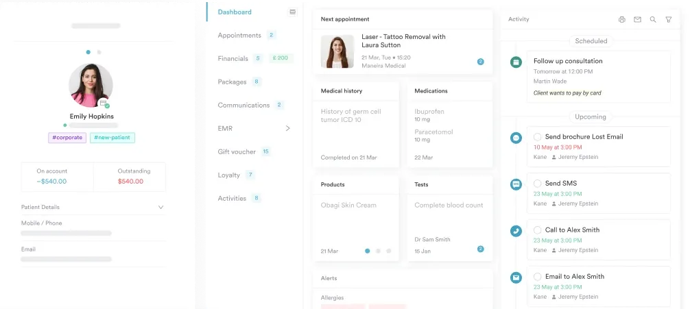

Pabau helps neurology, physical therapy, and acute care teams build structured assessment workflows, capture serial findings, and keep clinical records current across every patient visit.

The pronator drift test remains one of the most cost-effective neurological screening tools available at the bedside. It takes 30 seconds, requires no equipment, and detects corticospinal tract dysfunction that standard motor grading misses. Performed correctly, with supinated arms, closed eyes, and an observation window long enough to detect slow drift, the test gives reliable information. It supports acute triage, serial monitoring, and safe clinical handover.
The weakness is not in the test itself but in inconsistent technique and documentation. Standardizing both, through structured clinical note templates and digital record systems, converts a subjective bedside observation into a reproducible, longitudinally trackable data point. Pabau’s digital clinical documentation tools help neurology and physical therapy teams build exactly that structure into every patient encounter. To see how Pabau supports neurological assessment workflows from first contact to discharge, book a demo with the team.

Need a structured neurological assessment framework? Psychiatric evaluation template covers how to structure comprehensive clinical assessment documentation for complex neurological and mental health presentations.
Looking to improve how your team writes clinical notes? Writing effective clinical notes provides a step-by-step framework applicable across neurology, therapy, and acute care settings.
Managing neurological patients across a physical therapy caseload? Neurological physical therapy workflows shows how specialist EMR tools support motor assessment documentation and serial tracking at scale.
The pronator drift test is a bedside neurological assessment where a patient holds both arms outstretched with palms facing up and eyes closed. A positive result occurs when one arm drifts downward and rotates palm-down (pronates), indicating an upper motor neuron lesion in the contralateral corticospinal tract. The test is also called the Barré test or upper extremity drift test.
Ask the patient to extend both arms forward at shoulder height with palms fully supinated, then close their eyes. Observe for 10 to 20 seconds. A positive result is downward arm drift combined with forearm pronation. Eye closure is essential; testing with eyes open will miss subtle findings because visual compensation masks the motor deficit.
A positive pronator drift indicates dysfunction in the contralateral upper motor neuron pathway, specifically the corticospinal (pyramidal) tract. Common causes include ischemic stroke, hemorrhagic stroke, TIA, multiple sclerosis, and brain tumors. In any patient without a prior neurological diagnosis, a new positive result warrants urgent escalation and imaging.
The pronator drift test assesses the upper motor neuron (corticospinal tract), while the Romberg test assesses proprioception and dorsal column integrity. Both use eye closure, but they target different neurological systems. A patient can have a positive pronator drift with a normal Romberg, and vice versa. The two tests are complementary, not interchangeable.
The Barré test is an alternative name for the pronator drift test, referring to the arm-drifting component described by French neurologist Jean Alexandre Barré. The terms are used interchangeably in most clinical literature. Some sources draw a distinction between the Barré sign, or pure arm drift, and pronator drift, which adds pronation. In everyday clinical use, the terms refer to the same bedside assessment.
Sensitivity and specificity come from Teitelbaum et al. (2002), a study of 170 patients using CT-confirmed lesions as the reference standard. It found sensitivity of approximately 92% and specificity of approximately 90%, rising to about 97% sensitivity when combined with finger-tap and reflex testing. The test is most valuable as a complement to clinical history and imaging, not as a standalone diagnostic tool.
Effective nursing documentation names the test performed and the patient’s cooperation status. It records the affected arm, whether drift occurred with or without pronation, and the approximate time to onset. It also compares the finding to the prior assessment and states the clinical response taken. An entry like “positive right arm, drift at 8 seconds, senior clinician notified at 14:32” beats “arm drift noted” for follow-up care.






