CRM for chiropractors: how to manage patients, bookings and marketing
A CRM for chiropractors is a patient relationship management system built around...

Last Updated: Oct 13, 2026

A hip mobility test is a structured self-assessment that measures how freely your hip joint moves through flexion, extension, internal rotation, and external rotation.
The Thomas test and the seated rotation test are the most clinically referenced at-home assessments. A side-to-side difference above 10 to 15 degrees matters more than any single number.
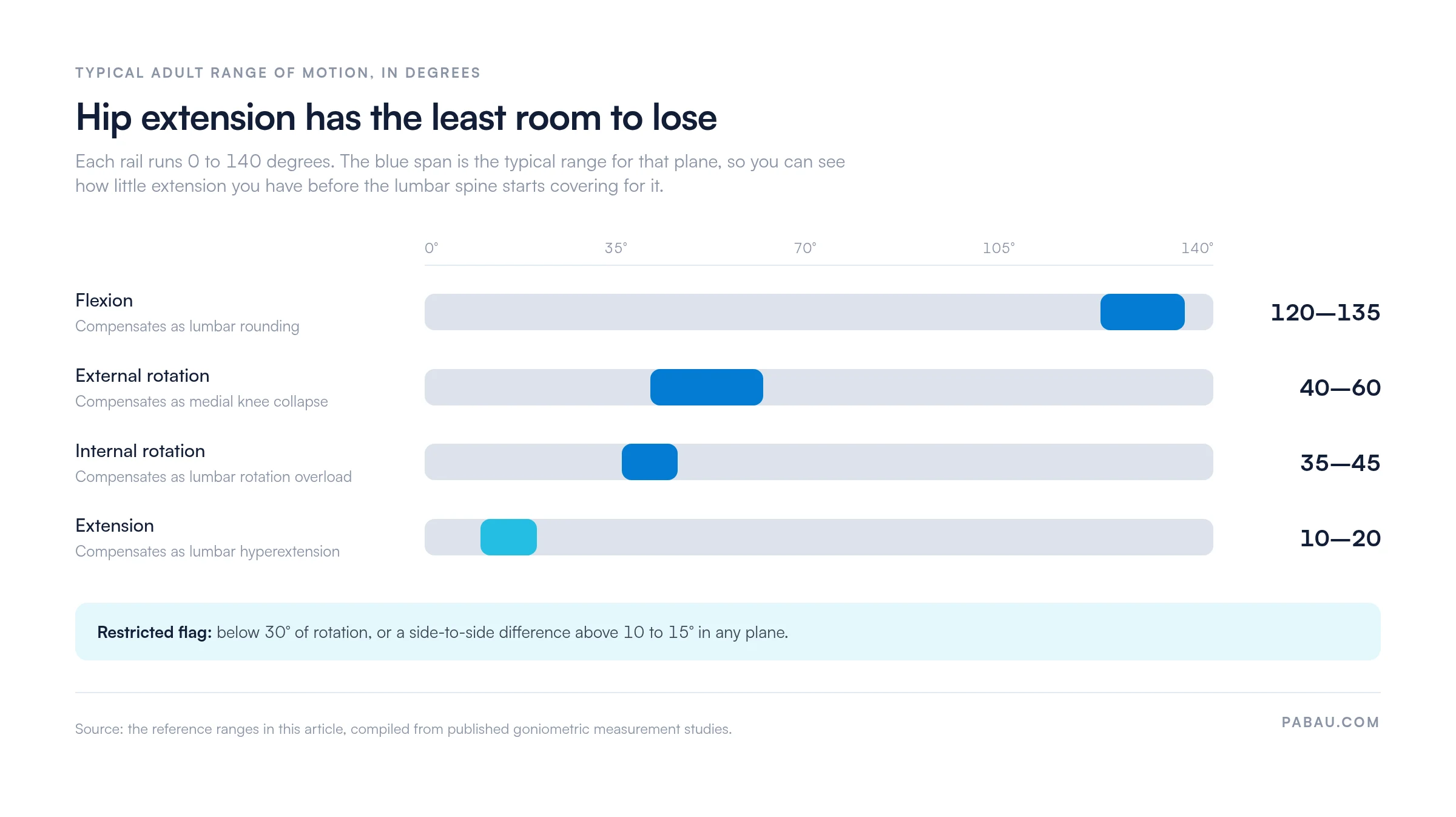
Hip extension has the narrowest normal range, roughly 10 to 20 degrees, which is why it is the first plane to force lumbar compensation.
Restricted hip mobility is a well-established contributing factor to lower back stiffness. Testing the hips before treating the back often points to where the restriction started.
Practice management software like Pabau records mobility assessments at intake and tracks range of motion progress between appointments.
A hip mobility test is a structured self-assessment that checks how freely your hip joint moves through its full range of motion. It is not a clinical diagnosis. It is a screening tool that tells you where restrictions sit, so you can address them before they turn into injuries or chronic pain patterns.
The hip is a ball-and-socket joint that moves in several planes: flexion, extension, internal rotation, external rotation, and abduction. When one of those planes becomes restricted, the body compensates. That compensation usually shows up first in the lower back, the knees, or the ankles.
Testing the hips tells you which plane is short, which makes the fix far more specific. Practices running physical therapy EMR software often build a structured hip screen into their intake paperwork for the same reason.
What you need: a flat surface (floor or firm table), a partner is helpful but not required, and about 15 minutes. No equipment is needed for the tests covered here.
Lower back stiffness is one of the most commonly misattributed complaints in musculoskeletal health. Before treating the back directly, it is worth running a hip mobility test first. Restricted hip mobility is a well-established contributing factor to lumbar compensation patterns.
When the hips cannot move freely, the lumbar spine takes on movement it was not built to repeat. Over time that drives stiffness, disc loading, and the kind of ache that feels like a back problem but starts elsewhere.
That is why a hip screen belongs before any back-focused program. Treating the lumbar spine while the hips stay short usually buys short-lived relief.
Preparation changes the result. Asymmetry between sides is the most useful finding self-testing gives you, and cold muscles hide it. Five minutes of setup separates a number you can act on from a number you cannot.
The Thomas test is the most widely used clinical screen for hip flexor tightness. A positive result suggests the iliopsoas or rectus femoris is restricting hip extension range. It gives you a strong directional signal, but a specific diagnosis still needs professional confirmation.
Interpreting your result: if the thigh floats instead of resting flat, hip flexor tightness is likely restricting hip extension. If the thigh is flat but the knee does not bend to 90 degrees, the rectus femoris is the likely culprit. The modified Thomas test adds a goniometer measurement of the thigh angle, which separates iliopsoas restriction from rectus femoris restriction more precisely.
Test both sides before you interpret either result. A thigh that floats 10 degrees on one side and 25 degrees on the other tells you far more than a symmetric finding. Asymmetry above 10 to 15 degrees warrants attention, even if neither side looks dramatically restricted on its own.
Internal rotation is the movement plane most commonly restricted in active adults. It is also the most predictive of downstream compensation. Restricted internal rotation is closely associated with anterior knee pain, IT band syndrome, and lumbar rotation overload.
Seated test (easiest to self-assess):
A side-to-side difference greater than 10 to 15 degrees is often more meaningful than the absolute value. Age, sex, and prior athletic training all shape what is normal for one person.
External and internal rotation should be assessed as a pair, so you get a complete rotation profile. Normal hip external rotation is roughly 45 degrees in most adults, measured in the supine or seated position.
What asymmetry tells you: if external rotation is notably greater than internal rotation on one side, the external rotators are likely dominating. That group includes the piriformis, the gluteus maximus, and the short external rotators. The internal rotators are then either inhibited or lengthened.
This pattern is common in people who sit for long stretches with the hip turned out. It feeds the hip shift you see in asymmetric squatters and runners.
Hip extension is often judged through the Thomas test, but a prone extension test gives a cleaner isolated reading. Limited extension forces compensatory lumbar extension with every stride, which is a major contributor to lower back fatigue. A structured return-to-running protocol will usually require normal hip extension before full running load.
The deep squat assesses hip flexion, ankle dorsiflexion, and thoracic mobility at the same time. Its limitation as a hip-specific screen is that failure can come from any one of those three. A simple modification resolves that.
Research published in the Journal of Orthopaedic and Sports Physical Therapy reports moderate reliability for the deep squat screen when it is performed consistently. Treat it as a directional indicator rather than a measurement.
Use these figures as a reference, not as a verdict. Individual variation is wide, and a side-to-side difference usually says more than a population average does. The values follow published goniometric measurement studies.
Plotted on one scale, the four planes are not remotely equal. Extension occupies a sliver of the range that flexion does, which is why it is the first plane to hand work to the lower back.
If you want the method behind these figures, our guide to range of motion assessment covers how each plane is measured and recorded in practice.
Results are easier to act on once you can point to where you already feel them. If your test flagged a restriction, these are the moments it is showing up.
Recognizing these patterns makes the numbers concrete. It also gives you something specific to describe if you end up in front of a clinician.
A hip screen is only useful if it points you toward action, and the corrective work should match the plane that is short. Generic hip stretching without knowing your deficit is far less effective. These are general wellness exercises. If you have acute pain, a known labral tear, or prior hip surgery, speak to a clinician first.
For hip flexor restriction (positive Thomas test):
For restricted internal rotation:
For restricted hip extension:
Retest every two to three weeks and write the numbers down each time. A printable hip range of motion chart makes the comparison easy to see. It also stops you arguing with your memory of how tight things felt last month.
Self-screening is useful, and it has limits. These findings warrant professional evaluation rather than self-management.
Bring your numbers with you. A therapist who can see which plane is short, on which side, and by roughly how much starts from a much better position. That beats a vague report of tightness.
Most physical therapy practices capture range of motion on paper or in a free-text note. The number gets written once and then buried. At the six-week review, nobody can say whether internal rotation improved by 5 degrees or 15.
Practice management software like Pabau handles this differently. Pabau’s measurements tracking stores each plane as a value against the patient record, so the intake reading and every follow-up reading sit side by side.
Digital intake forms carry the screening questions ahead of the visit, so a patient flags pain or prior hip surgery before they arrive. The therapist walks into the first session already knowing which plane to test carefully and which one to leave alone.
The outcome is a progress record the patient can see. Showing someone that their internal rotation improved between visits does more for adherence than a home exercise handout on its own.


Pabau’s measurements tracking and digital forms help physical therapy, sports medicine, and musculoskeletal practices record range of motion at intake and monitor it over time. No paperwork, no missed baselines.

The value of a hip screen is not the score. It is knowing which of five planes is short, on which side, and by how much. That is the difference between stretching your hips and fixing the one that is limiting you.
So run the five tests, write the numbers down, and retest in six weeks. If the restricted plane has not moved by then, the limitation is probably structural, and that belongs with a clinician rather than a stretching routine.
For practices, the same discipline applies at scale. Book a demo to see how Pabau records hip range of motion at intake and surfaces it again at every follow-up.

Want the method behind these numbers? Range of motion assessment covers how each joint plane is measured and recorded in practice.
Checking femoral rotation as well as mobility? Craig’s test walks through the prone test clinicians use to estimate femoral anteversion.
Need the same screen for the shoulder? Shoulder range of motion template gives you a printable chart for recording each plane.
Comparing systems for a physiotherapy practice? Physiotherapy clinic management software weighs up the platforms and what each one handles.
A hip mobility test is a structured self-assessment of how freely the hip joint moves. It covers flexion, extension, internal rotation, and external rotation. Common at-home versions include the Thomas test, the seated rotation test, and the deep squat screen. These are screening tools, not clinical diagnoses.
The Thomas test assesses hip flexor length. You lie back on a table and draw one knee to your chest while the other leg lowers. A positive result means the lowered thigh floats above the table surface. That points to the iliopsoas or rectus femoris restricting hip extension. It is a directional signal, so professional confirmation is needed to identify the structure involved.
Restricted hip mobility is a well-supported contributing factor to lower back stiffness and pain. When the hips cannot move freely, the lumbar spine takes on movement it was not built to repeat. This shows up most with extension restriction during walking, and internal rotation restriction during running and twisting. Testing the hips before treating the back is a standard first step in musculoskeletal assessment.
See a physical therapist if any test provokes pain, or if you notice tingling or numbness in the leg. Recent hip trauma is another reason to get assessed rather than self-manage. So is a side-to-side difference above 15 degrees that is changing how you walk. If 4 to 6 weeks of targeted exercise produces no measurable change, the limitation may be structural.