



GLP-1 diet plan
A GLP-1 diet plan is a structured eating framework for patients taking semagluti...

2
Last Updated: Oct 13, 2026
A nursing ear assessment is a structured examination of the external ear, ear canal, and tympanic membrane, paired with hearing tests and a targeted history.
It runs in four steps: subjective history, inspection and palpation, otoscopic examination, then auditory acuity testing. The NCBI Nursing Skills textbook treats systematic ear assessment as a core competency for nurses across primary care, general practice, and acute settings.
A normal tympanic membrane is pearly gray, translucent, and intact, with a visible cone of light. Redness, bulging, a fluid level, or a perforation all get described in the record and acted on. This guide walks the sequence in order, covers pediatric technique, and sets out when to escalate.

A nursing ear assessment covers external inspection, palpation, otoscopic examination of the tympanic membrane, and auditory acuity testing.
A normal tympanic membrane looks pearly gray and translucent, with a visible cone of light in the anteroinferior quadrant.
Weber and Rinne read together separate conductive from sensorineural hearing loss at the bedside, before any referral for audiometry.
Document each ear separately, and record the finding you observed rather than the diagnosis you suspect.
Structured digital forms in practice management software like Pabau keep every component of the assessment inside the patient record.
Before the examination begins, it helps to know which of the three anatomical regions a finding belongs to. Each region produces different findings and changes how you read what you see.
The external auditory canal runs from the meatus to the tympanic membrane. In adults it is slightly S-shaped. In young children it is shorter and sits more horizontally, which changes otoscope technique in the section below.
Subjective assessment shapes the rest of the examination. Ask targeted questions before you touch the patient, because the symptom history tells you where to focus the otoscope.
Core interview questions for ear health history, grouped by symptom cluster:
Document positive responses and relevant negatives. A patient who denies tinnitus and vertigo alongside unilateral hearing loss points to a different differential than one who reports all three.
Objective assessment follows a fixed sequence: inspection, palpation, otoscopic examination, then auditory acuity testing. Skip a step or run them out of order, and the record ends up missing findings nobody can reconstruct later.
Begin with unaided visual inspection of both ears at once, comparing symmetry before you examine either one in detail.
Working to a fixed list of external landmarks keeps the examination reproducible. That matters most when you are teaching students or onboarding new clinical staff.
Otoscopic examination is the core skill of any nursing ear assessment. It depends on correct canal straightening, speculum selection, and a systematic read of the tympanic membrane.
Canal straightening technique: for adults and children over 3, grasp the auricle and pull gently upward and backward to straighten the canal. For children under 3, pull downward and backward, because the infant canal curves differently. Incorrect technique obscures the view and risks patient discomfort.
A normal tympanic membrane appears pearly gray, translucent, and intact, with a visible cone of light in the anteroinferior quadrant. The OpenStax Clinical Nursing Skills textbook describes the same landmarks. The malleus handle shows through the membrane as a white streak.
Technique improves with repetition. A step-by-step walkthrough of otoscope examination covers speculum choice and canal angle in more detail than a single section allows.
Bedside hearing tests give nurses objective data on auditory acuity without audiological equipment. Each test has a distinct purpose and its own interpretation framework.
Tuning fork tests screen for hearing loss and do not diagnose it. Read on its own, either test leaves the picture incomplete. The grid below pairs the two results and shows what each combination points to.
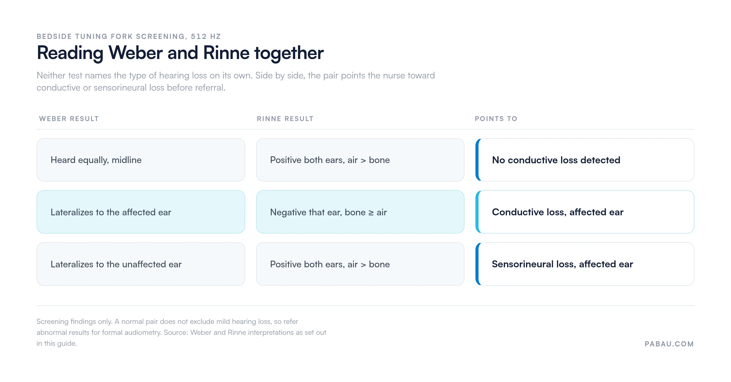
Write down what you heard. “Weber lateralizes left, Rinne negative left” lets the next clinician re-derive the conclusion, while “conductive loss on the left” hides the evidence behind it. Bedside sensitivity is limited, so a normal whisper test does not exclude mild hearing loss.
Telling expected findings from unexpected ones is the clinical core of the examination. The table below covers all three assessment zones.
Ear irrigation is contraindicated in any patient with a known or suspected tympanic membrane perforation. The same applies to a patient with a history of ear surgery, as the British Journal of Nursing confirms. Check the history before any ear cleaning procedure.
Before you document an abnormal tympanic membrane, check that the otoscope light is at full brightness and the speculum is seated correctly. A dim light source makes a healthy membrane look dull and opaque. Check the equipment before you commit the finding to the record.
Pediatric ear assessment differs from adult assessment in three clinically significant ways, each requiring a direct technique adjustment.
Otitis media is the most common pediatric ear finding. In acute otitis media the tympanic membrane appears red, bulging, and opaque, with loss of the normal cone of light. Otitis media with effusion (glue ear) presents differently, with an amber or retracted membrane and a visible fluid level.
Document the child’s cooperation level and any technique modification you used. If the examination stayed incomplete because of distress or movement, that belongs in the record too.
Ear examination documentation has to be systematic, specific, and reproducible. An entry reading “ears checked” carries no clinical value and will not survive an audit or a medico-legal review.
Every ear assessment record should include these components:
Sample documentation phrases for normal findings:
For abnormal findings, document exactly what you observed rather than the diagnosis you suspect. “Tympanic membrane erythematous and bulging with loss of cone of light, right ear” is appropriate nursing documentation. “Patient has otitis media” is a diagnosis and exceeds nursing scope without physician confirmation.
Working from a structured form rather than free text cuts omissions and supports clinical governance requirements. Digital clinical forms that mirror the assessment sequence prompt each component in turn, instead of leaving it to recall after a busy list. Records held that way also meet the retention and access standards HIPAA and GDPR set for patient data.

Certain findings require immediate escalation to a physician or referral to ENT, rather than watchful waiting or nurse-initiated treatment.
Escalation criteria belong in your practice’s clinical protocols and in the way notes are written. Our guide to safer clinical notes covers how to record the reasoning behind a referral, which carries as much weight as the finding itself.
Workflow, more than clinical skill, decides whether an ear assessment record holds up. Nurses fall back on free-text fields, paper charts, or generic note templates that do not follow the examination sequence. The record then arrives incomplete, missing lateralization, with follow-up actions agreed out loud and never written down.
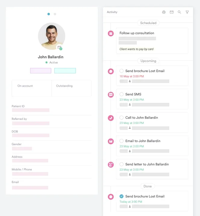
Practice management software like Pabau lets a practice build clinical forms that mirror the examination itself. The form carries external inspection fields, separate right and left otoscopic findings, hearing results with lateralization options, and a follow-up action dropdown.

Because the form sits inside the patient record, findings stay searchable and auditable across locations. A nurse repeating the assessment three months later compares the current membrane appearance against a documented baseline, instead of a verbal handover.
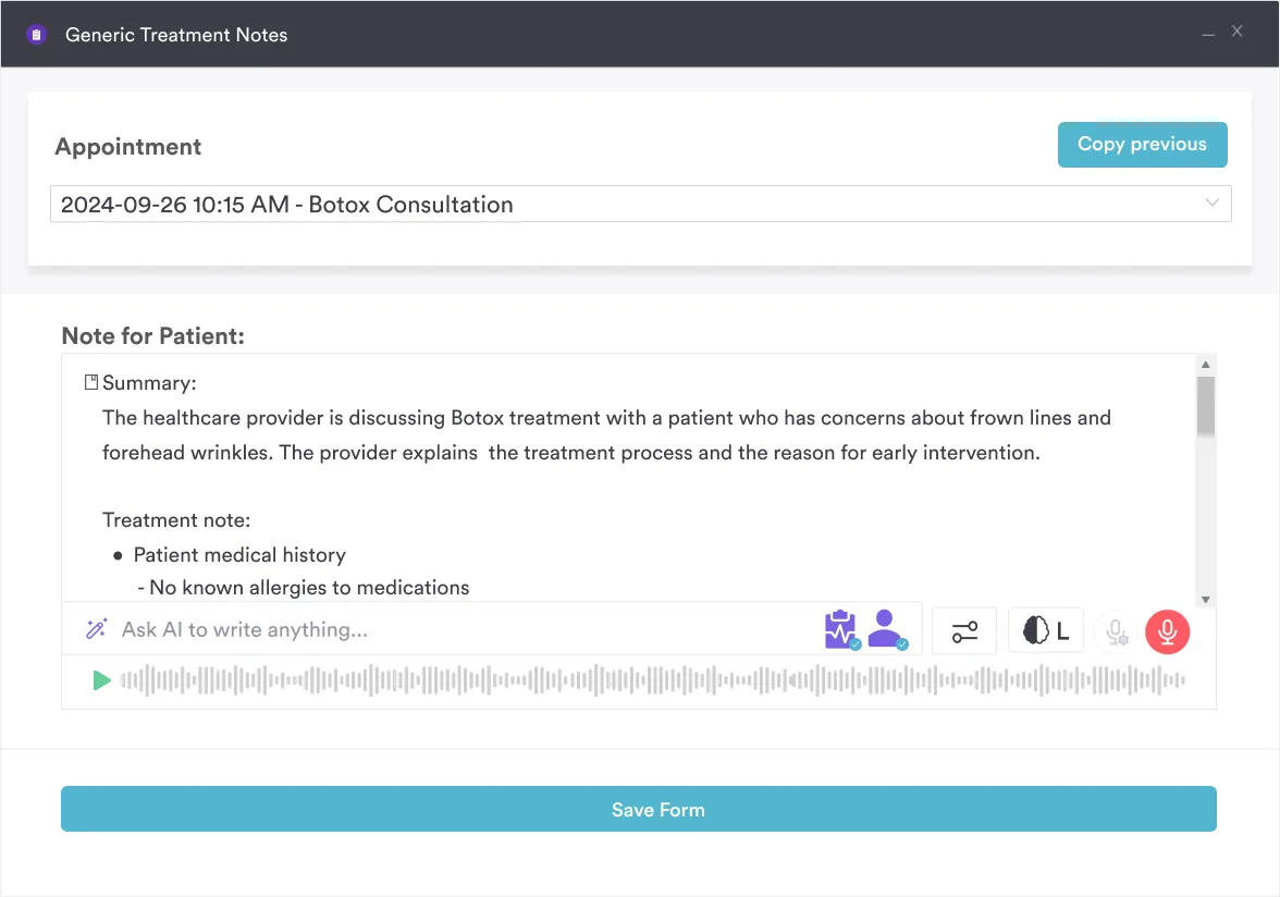
Pabau Scribe, our AI scribe, drafts the note from the consultation itself. That cuts the time spent writing notes after a list and evens out wording between practitioners. Practices running a high volume of ear checks usually standardize the form inside their GP clinic software, so every nurse records the same fields.
If you are still choosing a system, compare how each one handles clinical forms and record keeping. Our roundup of the best primary care EHR options sets the main contenders side by side.

Pabau’s digital forms and patient records help practices capture ear assessment findings consistently, keep records audit-ready, and flag follow-up actions without paper-based workarounds.

A systematic ear assessment proves its worth at the moment someone else reads the note. The four-step sequence is quick to learn. What slips is the record — which ear, which finding, which action, written so the next clinician can act on it.
Standardize the form before you standardize the technique, and the two improve together. Book a demo to see how Pabau keeps every ear assessment finding in the patient record, ready for the next appointment or the next audit.

Want the instrument technique on its own? Otoscope examination walks through speculum choice, canal angle, and what the view should look like.
Need to know how often to screen hearing? Hearing test frequency sets out screening intervals by age and by risk factor.
Examining the ears as part of a wider exam? Head and neck assessment places the ear sequence inside the full regional examination.
Seeing mostly children in your list? Pediatric assessment covers positioning, cooperation, and the findings that differ in young patients.
A nursing ear assessment is a structured clinical examination of the ear. It combines patient history, external inspection and palpation, otoscopic examination of the canal and tympanic membrane, and bedside hearing tests. Nurses use it to identify abnormal findings, record baseline ear health, and decide whether referral is needed.
There are four steps. First, take a targeted history covering pain, discharge, hearing changes, tinnitus, and vertigo. Second, inspect and palpate the external ear structures. Third, examine the canal and tympanic membrane with an otoscope. Fourth, test auditory acuity using the Weber, Rinne, and whisper tests where indicated.
Document each ear separately. Record external inspection findings, otoscopic findings, hearing test results with lateralization, any technique modification, and the clinical action taken. Use specific descriptive language such as “tympanic membrane pearly gray, cone of light visible” rather than “ears normal”. Structured digital forms cut omissions and support audit requirements.
The Weber test checks whether sound lateralizes. A tuning fork on the skull midline that lateralizes to the affected ear suggests conductive loss, while lateralization to the unaffected ear suggests sensorineural loss. The Rinne test compares air conduction with bone conduction in each ear. Air conduction louder than bone conduction is normal, and the reverse indicates conductive loss in that ear.
Escalate immediately for mastoid tenderness with swelling, sudden unilateral hearing loss, or facial nerve palsy alongside ear symptoms. Blood or clear fluid from the canal after head injury and a visible tympanic membrane perforation also need same-day escalation. Recurrent otitis media, or effusion persisting beyond three months in children, warrants ENT referral.
In children under 3, pull the auricle down and back rather than up and back, because the canal is shorter and more horizontal. Children need careful positioning with caregiver support. Their Eustachian tube anatomy makes otitis media far more common, and it presents with a red, bulging, opaque membrane.






