Scapular assistance test: Procedure, interpretation and free template
The scapular assistance test is a manual examination in which the clinician supp...

1
Last Updated: Oct 19, 2026

The external rotation lag sign test (ERLS) detects full-thickness tears of the infraspinatus and supraspinatus. A positive result is an angular drop after the examiner releases a passively rotated arm.
A lag of more than 5 degrees from near-maximal external rotation is a positive result, based on Hertel et al. (1996).
Reported accuracy varies by study. Hertel found 70% sensitivity and 100% specificity, while Castoldi’s 2009 re-examination reported 56% and 98%.
The test has high specificity for full-thickness rotator cuff tears, so it works best as a rule-in test rather than a screening tool.
Practice management software like Pabau helps physical therapy and sports medicine practices document shoulder findings and track lag angles over time.
The external rotation lag sign test (ERLS) is a passive clinical assessment that detects full-thickness tears of the infraspinatus and supraspinatus tendons. Also called the lateral rotation lag sign, it works by holding the shoulder at near-maximal external rotation and then letting go of the wrist. If the arm drops back, the external rotators cannot hold it.
Hertel and colleagues first described the test in 1996, and it is now a standard part of the shoulder examination battery. You will see it used in orthopedics, sports medicine, and physical therapy practices. Its value lies in specificity. When the test is positive, a full-thickness posterior rotator cuff tear is very likely.
The rotator cuff comprises four muscles: supraspinatus, infraspinatus, teres minor, and subscapularis. The ERLS targets the posterior cuff, where infraspinatus does most of the work in external rotation.
The infraspinatus starts at the infraspinous fossa of the scapula and inserts on the middle facet of the greater tuberosity. It is the primary external rotator of the glenohumeral joint and contributes roughly 60% of external rotation force.
A full-thickness tear removes most of that active restraint. The joint then cannot resist gravity once passive support is withdrawn.
The supraspinatus starts at the supraspinous fossa and inserts on the superior facet of the greater tuberosity. It matters more to the ERLS at higher abduction angles, above 20 degrees, where its external rotation torque increases. A dedicated infraspinatus test isolates the posterior cuff further when you need to separate the two.
Teres minor, the smallest external rotator, is assessed by the hornblower’s sign rather than the ERLS. Subscapularis, the only internal rotator, is assessed by the internal rotation lag sign and the lift-off test.
Technique decides whether the result means anything. Small errors in the starting position or the release introduce false positives from pain inhibition or examiner-induced motion. Follow this sequence.
Seat the patient comfortably with the back unsupported. Position the tested shoulder at roughly 20 degrees of abduction and 20 degrees of scapular plane deviation. Keep the elbow flexed to 90 degrees throughout. This reduces humeral head translation and isolates glenohumeral external rotation.
Stand behind or slightly lateral to the patient on the tested side. Support the elbow from below with one hand, holding the 90-degree flexion angle. Grasp the dorsal surface of the wrist with the other hand.
Passively rotate the shoulder to near-maximal external rotation, stopping just short of the end range. Going all the way to end range adds passive tissue tension that can hold the arm up on its own. Near-maximal, not maximum, is the position that matters.
Release wrist support while keeping the elbow supported, and ask the patient to hold the position. An angular drop of more than 5 degrees back toward internal rotation is a positive result. The lag means the infraspinatus and supraspinatus cannot generate enough isometric force to hold the arm against gravity.
A positive ERLS means the posterior rotator cuff cannot hold near-maximal external rotation against gravity. The likely cause is a full-thickness infraspinatus tear, with supraspinatus involvement more probable as the lag angle grows. On its own the test confirms no diagnosis. It is one finding you read alongside imaging, symptom history, and the rest of the examination.
Record the angle you measured, not just a positive or negative. The scale below puts each band of lag on a single axis. That makes the difference between a 6-degree drop and a 40-degree drop obvious.
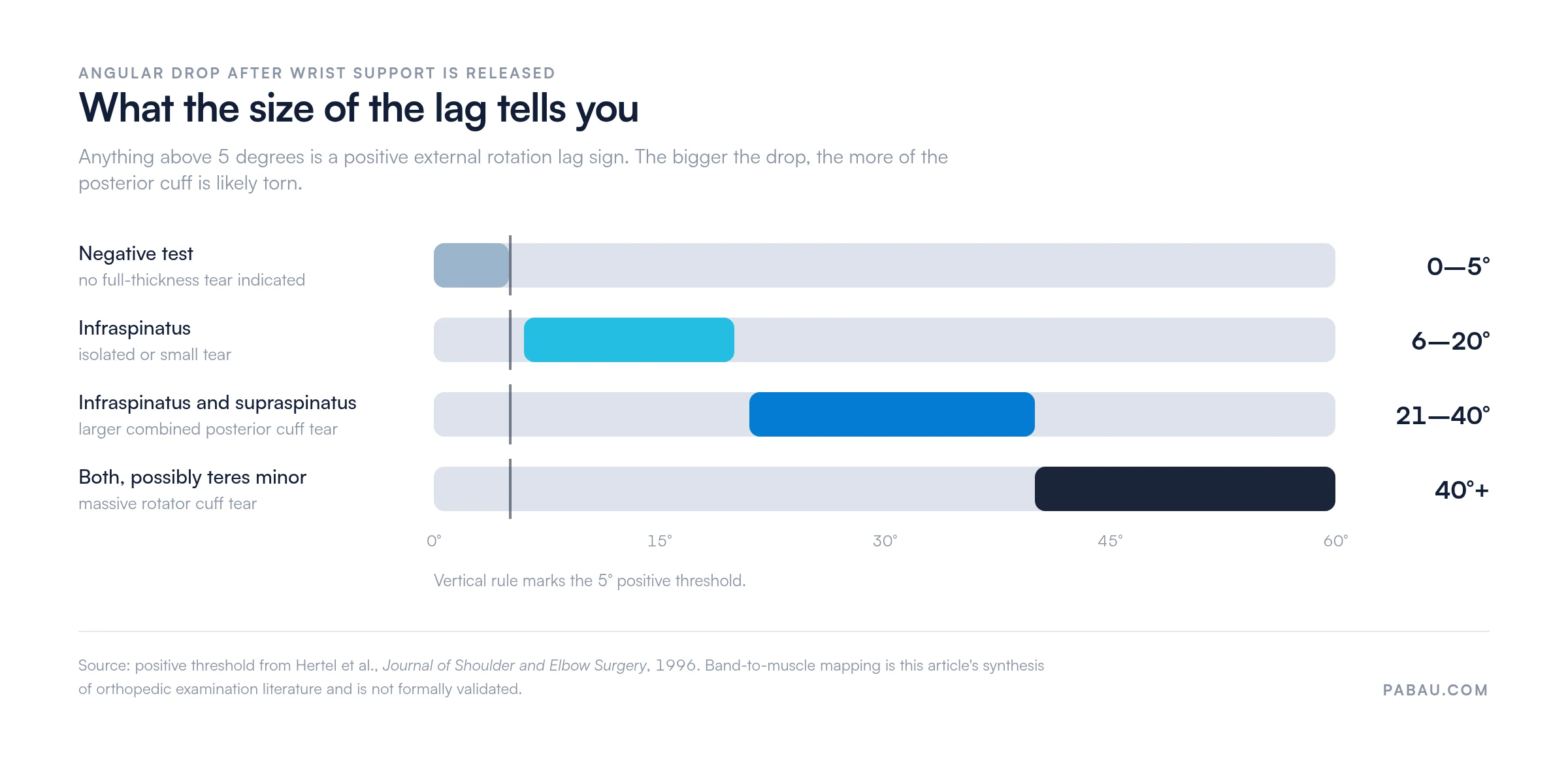
Larger lags correlate with more extensive posterior cuff involvement. The framework below reflects clinical consensus and the grading approach in orthopedic examination texts. Formal study-level evidence mapping each degree range to confirmed tear extent is limited, so treat the bands as a guide rather than a measurement.
Hertel and colleagues first reported the test’s accuracy in the Journal of Shoulder and Elbow Surgery in 1996. Later studies broadly agree that the test rules a tear in well and rules one out poorly. The exact figures move with the study population, the reference standard, and how tears were classified.
Two studies anchor the range. Hertel’s original series reported 70% sensitivity and 100% specificity. Castoldi and colleagues revisited the test in 2009 and reported 56% sensitivity and 98% specificity for full-thickness supraspinatus tears.
The likelihood ratios come from different cohorts and different reference standards, so they do not reconcile arithmetically with the sensitivity and specificity above. Read each figure against its own study rather than combining them.
In practice, treat the ERLS as a rule-in test. A positive result carries strong diagnostic weight. A negative result should not exclude a full-thickness tear in a patient whose history points to one.
Document the exact lag angle (for example, 15 degrees) rather than recording only positive or negative. A graded finding helps track progression, guides imaging decisions, and gives surgeons meaningful pre-operative data.
The ERLS belongs to the lag sign family and is usually run alongside tests for the other cuff muscles. The table below sets out each test’s target muscle, arm position, and positive criterion.
That makes it easier to pick the right combination for a given presentation. The drop arm test is the one most often paired with it in a first-pass shoulder screen.
The hornblower’s sign is the closest relative to the ERLS. Both are lag signs and both test external rotators, but they target different muscles at different arm positions. A positive ERLS with a negative hornblower’s sign points to infraspinatus involvement with teres minor intact, which can shape surgical planning.
The internal rotation lag sign and the lift-off test assess the anterior cuff. Pairing them with the ERLS gives you a view of the whole rotator cuff before anyone orders imaging.
Digital clinical forms let physical therapy and sports medicine teams build a shoulder examination template that captures every test in this battery. Results land on the patient timeline as soon as the clinician saves the form.

Most ERLS errors happen at two points: positioning and interpretation. These are the mistakes that show up when clinicians run the test for the first time or under time pressure.
Timestamped clinical entries keep lag angle measurements from the initial assessment, week 6, and week 12 in one timeline. That saves hunting through separate notes to find the baseline figure.

The ERLS earns its place in a structured shoulder examination when a full-thickness posterior cuff tear is in the differential. It is not a general shoulder screening tool, and using it that way wastes its specificity.
Reach for it when one or more of the following are present:
In a full shoulder examination, the ERLS fits after observation, range of motion, and rotator cuff strength testing. Sequencing it after the drop arm test and before the hornblower’s sign gives you a systematic posterior-to-anterior sweep in one sitting.
Referral decisions need documented clinical reasoning in most jurisdictions. A recorded positive ERLS with a quantified lag angle supplies that objective anchor.
A lag above 20 degrees, external rotation weakness, and a positive drop arm test together justify a direct MRI referral. That combination usually skips the prior ultrasound step in most pathways. Check your local or national guideline, since referral thresholds vary by jurisdiction and health system.
Practices handling a high volume of shoulder referrals can trigger the referral letter and imaging request automatically when a clinician flags a positive special test. That closes the delay between the assessment and the onward referral.
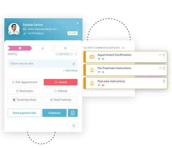
Most practices still record a lag sign as a tick box inside a free-text note. The angle either goes in as a stray number or gets left out, and the next clinician has no baseline to compare against.
Practice management software like Pabau handles this with structured clinical forms. You build one shoulder examination template with a numeric field for the lag angle, and every clinician fills in the same field. The reading saves to the client record, timestamped, so a 12-week comparison takes seconds.
From there, automated workflows can fire the referral letter template when a clinician flags a positive result. If you are weighing up options, our guide to physical therapy practice software covers what to check before you commit.

Pabau helps physical therapy and sports medicine practices build structured examination protocols, capture lag angle findings digitally, and trigger referral workflows automatically. See how it fits your practice.

The external rotation lag sign test gives you a specific, no-equipment way to identify a full-thickness posterior cuff tear at the point of care. Its specificity is what makes a positive result worth acting on. Its moderate sensitivity is why a negative result never closes the question.
The change worth making today is the smallest one. Write down the angle. A number in the record turns a special test into a measure you can track, defend at referral, and hand to a surgeon.
Book a demo to see how Pabau captures lag angles as structured data and moves a positive finding straight into a referral.

Screening for impingement as well as a tear? Neer’s test covers the impingement sign most often run in the same session.
Need to check the anterior cuff? Belly press test isolates subscapularis when the lift-off position is too painful.
Recording the full shoulder examination? Shoulder range of motion template gives you a structured chart for every plane.
Want a palpation check alongside the lag signs? Rent test covers the greater tuberosity technique for spotting a rotator cuff tear.
A positive external rotation lag sign test indicates a full-thickness tear of the infraspinatus, and often the supraspinatus. The arm drops more than 5 degrees once wrist support is released. That means the posterior cuff cannot hold the externally rotated position against gravity.
A lag of more than 5 degrees from the near-maximal external rotation starting position is a positive result, as established by Hertel et al. (1996). Smaller drops within the first 5 degrees fall inside normal variation and do not indicate a positive test.
The ERLS tests infraspinatus, and supraspinatus at higher positions, with the arm in the scapular plane at low abduction. The hornblower’s sign tests teres minor with the shoulder at 90 degrees of abduction. A positive ERLS with a negative hornblower’s sign suggests infraspinatus involvement with teres minor intact.
The lag angle provides a rough guide. Lags of 6 to 20 degrees typically suggest isolated infraspinatus involvement, while lags above 20 degrees increasingly implicate the supraspinatus. Formal tear size mapping still requires MRI or arthroscopic confirmation, so the ERLS alone cannot define the dimensions of a tear.
The ERLS primarily tests infraspinatus, the dominant external rotator of the glenohumeral joint. At higher arm positions, above 20 degrees of abduction, supraspinatus contribution to the test increases. A positive ERLS at those elevated positions implicates both muscles.
The drop arm test assesses supraspinatus by asking the patient to slowly lower a fully abducted arm. Failure to control the descent is a positive result. The ERLS targets infraspinatus through a passive external rotation release. Both tests have high specificity, but the ERLS points more directly at the posterior cuff.