Head to toe assessment cheat sheet: Printable nursing guide
A head to toe assessment cheat sheet gives you one fixed order at the bedside, s...

3
Last Updated: Oct 23, 2026

Circulation assessment checks heart rate, blood pressure, capillary refill, skin perfusion, and peripheral pulses to catch hemodynamic compromise early.
The 5 P’s (pain, pallor, pulselessness, paresthesia, paralysis) screen one limb for compartment syndrome, and they miss early cases.
Normal capillary refill is under 2 seconds in adults. Prolonged refill with tachycardia and hypotension points to shock.
Hypotension is a late sign, so escalate on tachycardia, prolonged refill, and cool peripheries before the blood pressure falls.
Practice management software like Pabau turns each parameter into a required field, so records stay complete and audit-ready.
A circulation assessment is a systematic check of how well the heart and vessels are delivering oxygenated blood to the tissues. It sits in the Resuscitation Council UK’s ABCDE approach as step C, after airway and breathing.
Clinicians measure heart rate, blood pressure, skin perfusion, capillary refill time, and peripheral pulses. Read together, those findings show whether the patient is stable or sliding toward shock.
Speed matters in the acute phase. For every minute defibrillation is delayed in cardiac arrest, survival drops roughly 7-10%. A structured assessment is how deterioration gets caught before it reaches that point.
This guide covers each parameter and its normal range, the mnemonics used in emergency and orthopedic settings, and shock recognition. It also covers the documentation that supports safe escalation. It is written for nurses, paramedics, and clinical staff in emergency, acute, and outpatient settings. GP clinic software keeps consultations, results and recalls against one patient record rather than three systems.
A full circulation assessment covers seven measurable parameters. Each one carries distinct hemodynamic information, and none of them should be read on its own.
Aesthetic and skin practices use the same parameters for pre-treatment screening. They sit alongside the skin assessment tools already used at consultation.
Normal ranges shift with age, clinical context, and the patient’s own baseline. Always read a finding against their history, not just a population reference value.
Press firmly on a fingernail or the central sternum for five seconds, then release. Color should return within 2 seconds.
Refill beyond that suggests reduced peripheral perfusion, usually from shock or peripheral vascular disease. Some guidelines accept up to 3 seconds, so the trend matters as much as one reading.
Central measurement at the sternum beats a fingertip in a cold room, where low ambient temperature falsely prolongs refill. A peer-reviewed review rates refill time as a useful non-invasive marker. It also notes only moderate sensitivity when the sign is used alone.
Skin shows perfusion trouble before the vital signs move. Pallor points to vasoconstriction or anemia, and cyanosis to inadequate oxygenation.
Mottling is a blotchy, livedo-like pattern, most visible over the knees. In critically ill patients it is a reliable sign of poor peripheral perfusion and predicts worse outcomes in sepsis.
The 5 P’s screen limb perfusion in suspected compartment syndrome or acute limb ischemia. It is a focused neurovascular check on one limb, rather than a whole-body circulation assessment.
The 5 P’s have known sensitivity limits. Compartment syndrome can be present without all five features, and pulselessness is often a late finding.
Some references swap paralysis for poikilothermia, the limb’s loss of temperature control. Either version needs backing up with compartment pressure measurement and urgent surgical review.
In an ankle or foot injury, settle the imaging question first with the Ottawa ankle rules calculator. Then repeat the neurovascular check after any manipulation or splinting.
Paresthesia can also have a non-ischemic cause. Brachial neuritis produces similar arm symptoms, and brachial neuritis exercises guide recovery once the diagnosis is clear.
Peripheral circulation assessment looks at blood flow through the limbs and distal vessels. It matters most in vascular, orthopedic, diabetes, and aesthetic settings, where arterial compromise complicates treatment.
Start with pulse palpation. Assess the radial, ulnar, posterior tibial, and dorsalis pedis pulses on both sides, and grade each one from 0 to 3.
Grade 0 is absent, 1 is weak or thready, 2 is normal, and 3 is bounding. Asymmetry between limbs earns further investigation, including Doppler assessment.
In the arm, symptoms that come and go with position point somewhere else. The Morley test is one of the bedside checks for thoracic outlet syndrome.
For suspected peripheral arterial disease, the ankle-brachial index (ABI) gives a ratio of ankle to brachial systolic pressure. A ratio below 0.9 suggests arterial stenosis, per NICE guidance.
Musculoskeletal caseloads generate these checks constantly. Physical therapy EMR software holds the findings in one template, so the next clinician compares like with like instead of guessing at a colleague’s shorthand.
The CMS triad is the orthopedic and trauma nursing standard for monitoring an injured or post-operative limb. It adds neurological screening to the circulation check, so it catches more than pulses alone.
Record CMS findings at set intervals after fracture reduction, casting, splinting, or vascular intervention. Deterioration in any component warrants immediate escalation. Worsening CMS in a limb cast is a cast compression emergency until proven otherwise.
Sports medicine and orthopedic outpatient teams run this cycle weekly. Sports medicine software keeps the observation record beside the treatment plan, and automated escalation workflows chase a due check instead of relying on memory.
Shock is inadequate tissue perfusion, and a circulation assessment is the main way to catch it early. The four main types share findings but differ in the detail.
Hypotension is a late sign. By the time systolic pressure falls below 90 mmHg, a healthy adult has usually lost more than 30% of circulating blood volume.
Tachycardia, prolonged refill, and cool peripheries should trigger escalation well before that. For cardiac presentations, a myocardial infarction care plan shows how those findings turn into planned interventions.
Hemorrhage is the leading preventable cause of death in trauma. The ATLS (Advanced Trauma Life Support) framework puts bleeding control inside the circulation step, ahead of formal monitoring.
In prehospital and emergency settings this assessment feeds major hemorrhage protocol activation and damage control decisions. The same escalation discipline applies outside hospital, and crisis intervention strategies cover how clinical teams prepare for acute deterioration.
The ABCDE approach gives a fixed sequence for any acutely ill or injured patient, and circulation is the C step. Each problem is identified and treated before you move on.
Within C you look for active bleeding, check heart rate and rhythm, measure blood pressure, check capillary refill, and judge skin perfusion. IV access and circulatory support come before Disability.
The framework underpins nursing assessment across emergency, acute medical, and critical care, in line with NICE guideline NG94.
NEWS2 (National Early Warning Score 2) folds heart rate and systolic pressure into a scored escalation system. An aggregate score of 5 triggers urgent clinical review, and 7 or above requires emergency escalation.
Teams working from structured client records can build NEWS2-aligned templates that prompt for each parameter in turn.
Incomplete documentation is a clinical governance risk and a medicolegal one. From a legal standpoint, a finding that was never recorded did not happen.
Good documentation says what was found, when, by whom, and what happened next. A complete entry covers:
SBAR (Situation, Background, Assessment, Recommendation) is the standard format for handing an abnormal finding to a senior clinician. Spoken out loud, it sounds like this.
Most teams record circulation findings in free text, and that is where the detail drains away. One clinician writes “CRT 3s, cool peripheries” while the next writes “peripherally shut down”. Neither entry supports an audit.
Practice management software like Pabau replaces free text with structured digital forms. Each parameter gets its own required field, so heart rate, blood pressure, refill time, skin findings, and escalation status are captured every time.
Because the fields are consistent, you can trend one patient across encounters and pull an audit sample in minutes. Nothing has to be transcribed off a paper observation chart first.
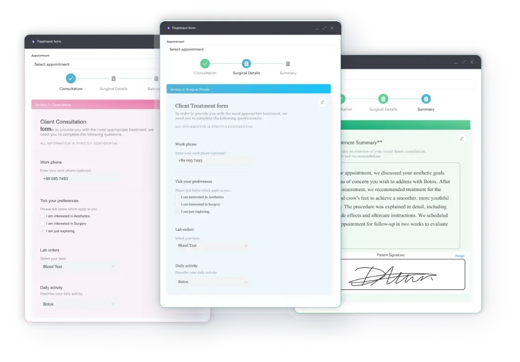
In outpatient and aesthetic practices, pre-treatment screening is part of the consent workflow. A patient booked for IV therapy may need a hemodynamic baseline on file before treatment starts.
Pabau’s compliance management tools build that checklist into the booking, so the record is complete before the patient arrives. Phrasing matters too, and safer clinical notes covers wording that holds up months later.


Pabau’s digital forms and client records let you build structured assessment templates that capture every parameter, prompt escalation, and keep an audit-ready history. See how practice teams standardize their clinical documentation.

A circulation assessment earns its value when the parameters are read together. A pulse of 110 means one thing in a frightened patient and something else in one who is quietly bleeding.
So act on the early signs rather than waiting for blood pressure to confirm what the skin already told you. The clinician who escalates on tachycardia and a 4-second refill will look overcautious some of the time. That is the right trade to make.
Then record what you found as carefully as you assessed it, because the next clinician inherits your notes rather than your memory. Book a demo to see how Pabau keeps circulation findings structured, trended, and ready for audit.

Assessing pain in a patient who cannot speak? Abbey Pain Scale template is a 1-minute observational tool with six categories for non-verbal patients.
Building a diet plan for a GERD patient? acid reflux care plan template covers symptom assessment, trigger identification, and dietary guidance for GERD management.
Suspect peripheral arterial disease from a weak pedal pulse? The ankle-brachial index covers how to perform, interpret, and document the ABI ratio.
Care planning for a patient with reduced physical capacity? activity intolerance nursing care plan gives a structured framework for assessment, diagnosis, and outcome measurement.
Moving from Circulation to Disability in the ABCDE sequence? The neurological vital signs guide covers GCS scoring and escalation triggers.
A circulation assessment is a systematic clinical evaluation of how well the cardiovascular system delivers oxygenated blood to the tissues. It covers heart rate, blood pressure, capillary refill time, skin color and temperature, peripheral pulses, jugular venous pressure, and urine output. Together these findings identify hemodynamic compromise or shock.
During the primary assessment, circulation is evaluated by checking heart rate and rhythm, blood pressure, and capillary refill time. You also assess skin color and temperature, plus the presence and quality of peripheral pulses. Active bleeding is identified and controlled before moving to the next step. This follows the ABCDE approach endorsed by the Resuscitation Council UK.
The 5 P’s are pain, pallor, pulselessness, paresthesia, and paralysis. They form a mnemonic for assessing limb perfusion in suspected compartment syndrome or acute limb ischemia, not for general circulation assessment. Pulselessness is typically a late sign, and pain on passive stretch is often the earliest indicator.
Assess distal circulation by palpating the radial, ulnar, dorsalis pedis, and posterior tibial pulses. Then measure capillary refill time and evaluate skin color and temperature in the extremities. In orthopedic and trauma contexts, apply the CMS triad of circulation, movement, and sensation. The ankle-brachial index is used for suspected peripheral arterial disease.
Signs of poor circulation include tachycardia, hypotension, capillary refill beyond 2 seconds, and cool, pale, or mottled extremities. Weak or absent peripheral pulses and urine output below 0.5 mL/kg/hour point the same way. In trauma, active bleeding alongside these signs indicates hypovolemic shock needing urgent intervention.
Record each parameter with units, such as heart rate in bpm and refill time in seconds. Describe skin findings rather than summarizing them, grade pulses on one consistent scale, and note the time, the clinician, and any interventions. Add the NEWS2 score where it applies, and use SBAR when escalating.