



GLP-1 diet plan
A GLP-1 diet plan is a structured eating framework for patients taking semagluti...

2
Last Updated: Oct 13, 2026

The anterior interosseous nerve test, also called the OK sign or pinch grip test, checks three forearm muscles for motor weakness.
A positive result is a triangular pinch instead of a round circle, with no numbness or tingling anywhere in the hand.
EMG and nerve conduction studies confirm the diagnosis, and they are ordered no earlier than three weeks after symptoms begin.
Conservative care comes first, most patients recover within 3 to 12 months, and surgery is considered only after six months without improvement.
Structured clinical forms and outcome tracking keep findings comparable between visits, so a recovery plateau is easy to spot.
The anterior interosseous nerve test is a bedside exam for motor weakness in the anterior interosseous nerve (AIN).
The AIN is a purely motor branch of the median nerve, so weakness shows up with no numbness at all. You ask the patient to form an OK sign with the thumb and index finger.
A triangular or lateral pinch instead of a round circle is a positive result. The full sequence runs to six steps, needs no equipment, and takes about a minute. A positive test tells you which muscles are weak, not why. Electrodiagnostic studies answer that second question.
The test evaluates motor integrity of the AIN. Failure to form a perfect circle points to weakness in the three muscles the AIN exclusively innervates. Because the AIN carries no sensory fibers, patients present with isolated motor weakness and nothing else.
That absence of sensory involvement is the fastest way to separate AIN palsy from carpal tunnel syndrome and pronator syndrome. Both of those compress the median nerve higher up, and both produce numbness the patient will mention unprompted.
The test goes by several names in practice, including the OK sign test, the pinch grip test, and the circle sign. The syndrome itself is sometimes called Kiloh-Nevin syndrome. Knowing which muscles are at stake makes the deformity easier to read when the findings are subtle.
The AIN arises from the median nerve roughly 5 to 8 cm distal to the lateral epicondyle, in the proximal forearm. From there it runs along the interosseous membrane between the radius and ulna. It travels deep to the flexor digitorum superficialis and alongside the flexor digitorum profundus.
The nerve terminates in the pronator quadratus, which it innervates distally. Three muscles receive their sole motor supply from the AIN. The table below maps each one to the joint it controls and to the finding you expect when that muscle is paralyzed.
The pinch grip test requires no equipment and takes about 60 seconds. Follow these six steps in sequence for a reliable result.
This test targets the third AIN-innervated muscle directly. Flex the elbow fully, to roughly 130 degrees, which mechanically shortens the pronator teres so it cannot contribute to pronation. Then resist the patient’s attempt to pronate from that position.
Compare that result against pronation strength with the elbow closer to extension. Weakness only in the flexed position isolates the pronator quadratus from the pronator teres. It also confirms AIN involvement at the distal forearm level.
A positive anterior interosseous nerve test means the patient cannot form a round circle. Instead they produce a pinch deformity, contacting tip to tip or tip to nail rather than pad to pad. That pattern indicates weakness of the flexor pollicis longus and the FDP to the index finger.
Partial and complete palsy produce different pictures, and the distinction is worth making. In complete palsy all three AIN-innervated muscles are affected. The patient fails the pinch test, cannot flex the index DIP against resistance, and shows pronator quadratus weakness on the isolation test.
In partial palsy only one or two muscles are weak. The most common partial pattern spares the pronator quadratus and affects only the FPL and FDP. That produces a positive pinch test alongside normal pronation strength.
Two limitations apply. Patients with generalized joint hypermobility can appear to form a normal circle through hyperextension rather than through FPL and FDP strength. The test also cannot separate compressive AIN neuropathy, Parsonage-Turner syndrome involving the AIN fascicle, and incomplete median nerve injury.
AIN syndrome presents as painless weakness of pinch grip, usually developing over days to weeks. The classic complaint is difficulty pinching small objects, opening jars, or writing. Some patients report a prodromal ache in the proximal forearm, particularly in cases linked to Parsonage-Turner syndrome, but frank pain is unusual.
There is no numbness, no tingling, and no sensory change in the hand, forearm, or fingers. That is the feature separating AIN syndrome from pronator syndrome and carpal tunnel syndrome. Clinicians in sports medicine practices will recognize it in throwing athletes, gymnasts, and manual workers doing repetitive forearm pronation.
AIN compression occurs at the proximal forearm, where several structures can impinge the nerve. The most frequently implicated are:
Idiopathic AIN syndrome, where no compressing structure is identified, remains the most common presentation in practice.
A positive test narrows the differential considerably, but the picture still has to be weighed against the conditions below. The table compares AIN syndrome with its most common alternatives, using sensory findings and the OK sign as the discriminating criteria.
Electromyography (EMG) and nerve conduction studies (NCS) are the gold standard for confirming AIN syndrome. Both StatPearls and the American Association of Neuromuscular and Electrodiagnostic Medicine treat electrodiagnosis as the confirmatory step. The workup serves three functions.
Key findings include fibrillation potentials and positive sharp waves in the FPL and the FDP to the index finger on needle EMG. Normal sensory nerve action potentials confirm the purely motor distribution. Motor conduction to the pronator quadratus may show reduced amplitude or slowing.
MRI is not first line, but it earns its place when the clinical picture is atypical. Denervation edema on STIR sequences in the FPL, FDP and pronator quadratus correlates with acute or subacute AIN injury. It also helps separate Parsonage-Turner syndrome from compressive neuropathy, since the former shows a patchy, multifocal pattern.
Order EMG and NCS no earlier than three weeks after symptom onset. Denervation changes take time to develop, so a study run too early can look falsely normal and delay the diagnosis.
Management depends on the likely cause, the severity of the weakness, and how long symptoms have been present. Conservative care suits most cases and should be the default first approach.
Most patients are referred on to physical therapy, so the practice taking that referral needs a record it can compare visit to visit. Purpose-built physiotherapy clinic management software keeps those measures in one place rather than scattered across visit notes.
Baseline outcome measures belong in the record before any exercise program starts, so progress is tracked against numbers rather than recall. State licensing rules shape what else has to be documented, and they vary widely. Our guide to physical therapy clinic requirements covers Arizona’s version in full.
Surgical decompression (neurolysis) is considered when there is no clinical or electrodiagnostic improvement after six months of conservative management. It is also considered when imaging identifies a discrete compressing structure. Outcomes are generally good for compressive causes and less predictable for Parsonage-Turner cases, which is why separating the two matters before operating.
Laid out on a single timeline, the decision points sit further apart than they read on the page.
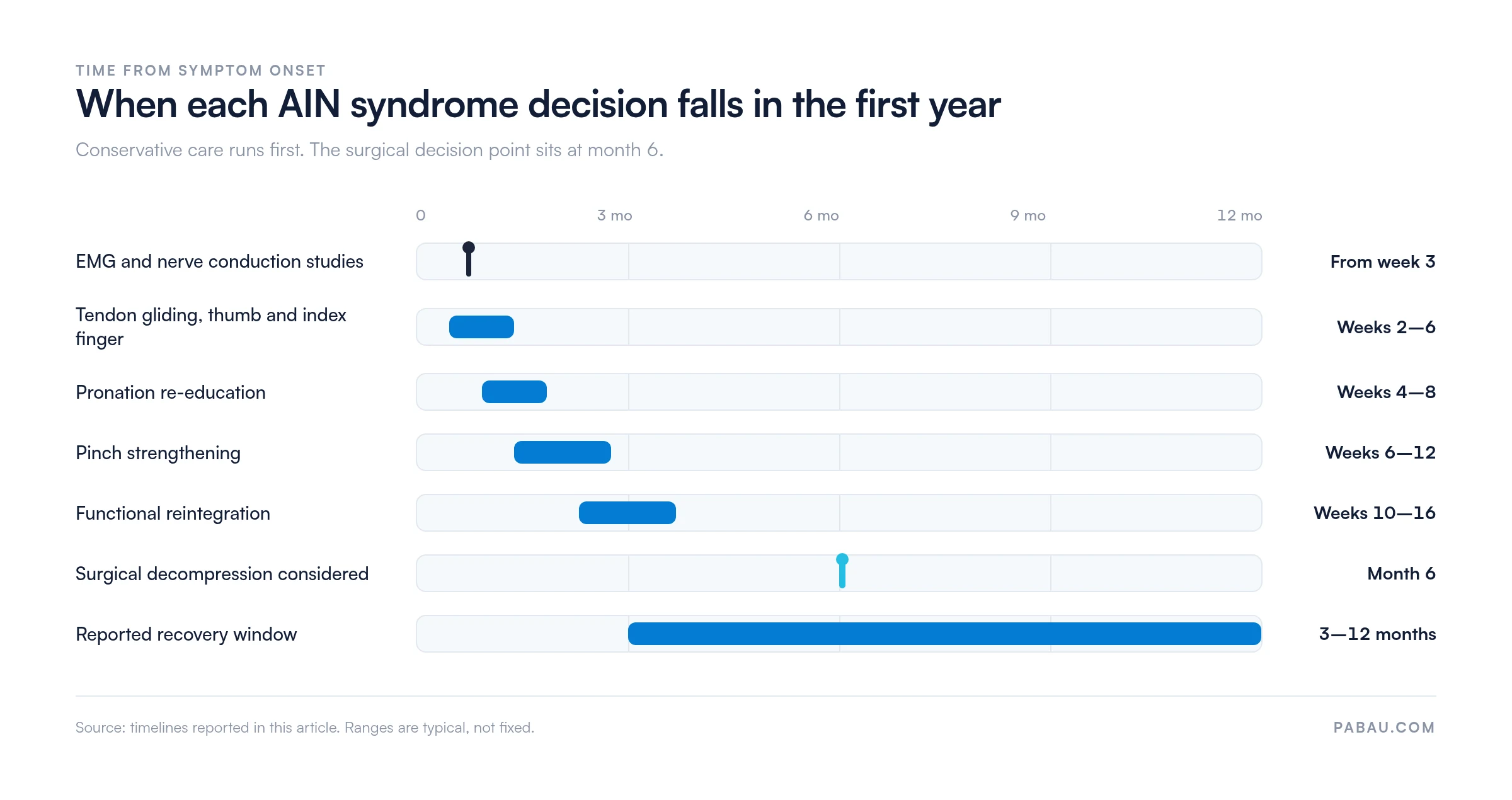
Peripheral motor nerve recovery follows reinnervation rather than the calendar, so the weeks below are a guide and not a schedule. Deliver the program under qualified physical therapist supervision and adapt it to the patient in front of you. A staged return to running protocol applies the same logic to a different injury.
Spontaneous recovery occurs in a meaningful proportion of cases, particularly those attributed to Parsonage-Turner syndrome or idiopathic compression. Recovery timelines run from 3 to 12 months with conservative management. Individual variation is considerable, and some patients take longer than a year.
Published series report outcomes that vary by cause. Compressive cases treated surgically show good to excellent results in the majority. Parsonage-Turner cases recover less predictably, and often incompletely, whichever treatment is chosen.
Several factors shape recovery speed. Younger patients, a shorter delay before treatment, and partial rather than complete palsy all correlate with faster and more complete recovery. Repeating the same measures at set intervals is what shows a plateau early enough to act on.
The maneuver is quick, but it has several interpretive traps that a hurried exam walks straight into. The table below collects the ones worth checking every time.
Recording this exam the same way at every visit is harder than it sounds in a busy physical therapy or sports medicine practice. The deformity pattern, the PQ isolation result, grip strength, and the EMG referral date tend to land in four different places in a free-text note. Six months later, nobody can say whether the pinch has improved.
Practice management software like Pabau replaces that with a structured nerve assessment template. Each field is captured the same way at every appointment, so the second visit is directly comparable with the first. Test results, imaging referrals and treatment notes sit in one longitudinal record, which is what physical therapy EMR software is for.
The practical payoff is timing. When the numbers stop moving, the team can see it in the record and action a surgical referral at month six instead of month nine. Date-stamped outcome records at each visit also stand up to medicolegal scrutiny for patients with peripheral nerve conditions. You can book a demo to see how that workflow fits a nerve assessment practice.


Pabau lets physical therapy and sports medicine practices build structured clinical templates. Every outcome measure and nerve assessment finding stays in one patient record.

AIN syndrome is underdiagnosed because it does not look like median nerve pathology. There is no numbness, no night pain, and no tingling to anchor the diagnosis. What the patient has is a hand that will not pinch, and a test that takes a minute to confirm it.
So run the OK sign on any painless pinch weakness, and add the pronator quadratus isolation test before you call the exam normal. Then commit to the timeline: EMG after three weeks, conservative care first, and a surgical conversation at month six if nothing has moved. The trade-off is patience against nerve recovery, and the record is what tells you which one is winning.
That record only works if every visit is written down the same way. Book a demo to see how Pabau keeps nerve assessment findings comparable from the first visit to discharge.

Building out your upper limb screening? The Wright test covers the neurovascular exam for thoracic outlet compression, a differential worth ruling out alongside AIN palsy.
Testing cranial nerves too? The hypoglossal nerve test walks through another purely motor nerve exam, including what a positive finding rules in and out.
Assessing the shoulder in the same visit? The infraspinatus test explains how to isolate external rotation weakness and grade the result.
Running a sports medicine practice? Sports medicine software compared reviews the systems that handle assessment workflows, recalls, and outcome tracking.
Keeping on top of compliance? Compliance for physical therapy practices sets out the documentation standards that apply to multi-visit musculoskeletal care.
The anterior interosseous nerve test asks the patient to form a circular “OK” shape with the thumb and index finger. It detects weakness of the flexor pollicis longus, the flexor digitorum profundus to the index finger, and the pronator quadratus. A positive test produces a triangular or lateral pinch instead of a round circle, with no sensory loss.
Pronator syndrome involves median nerve compression at the elbow or proximal forearm. It produces both motor and sensory deficits, including numbness of the thumb, index and middle fingers. Anterior interosseous nerve syndrome is a purely motor entrapment distal to that point, with no sensory loss at all. The presence or absence of sensory symptoms is the primary bedside distinction.
Yes. Many cases resolve with conservative management over 3 to 12 months, particularly idiopathic compression and Parsonage-Turner syndrome. Watchful waiting with activity modification, splinting and rehabilitation is the appropriate initial strategy. Surgical decompression is considered only after six months of conservative treatment without improvement, or when imaging identifies a discrete compressing structure.
A positive anterior interosseous nerve test warrants referral for EMG and nerve conduction studies, the gold standard for confirming the diagnosis. Those studies should be timed no earlier than three weeks after symptom onset, so denervation changes have time to develop. MRI is used when the cause is unclear, or when Parsonage-Turner syndrome has to be separated from compressive neuropathy.
Yes. AIN injury is a recognized complication of supracondylar humerus fractures, particularly in children. The nerve can be stretched, contused, or tethered by fracture fragments or post-reduction callus. Reported incidence varies across studies, so check current orthopedic literature for population-specific rates. Spontaneous recovery is common when these injuries are managed conservatively alongside the fracture.
Rehabilitation focuses on FPL tendon gliding, isolated DIP flexion for the index finger, and progressive pronation re-education with the elbow moving from flexion toward extension. Tip-pinch strengthening against increasing resistance follows. A qualified physical therapist should supervise the program and progress it against the patient’s reinnervation, not a fixed schedule. Functional task work begins once pinch grip strength returns.






