Nitrates medication list
A nitrates medication list is a point-of-care reference that sets out the main c...

Last Updated: Oct 6, 2026

Urine chemistry combines physical, chemical, and microscopic examination of a single urine specimen.
Normal dipstick values run pH 4.5 to 8.0 and specific gravity 1.003 to 1.030.
Glucose, protein, ketones, blood, nitrites, bilirubin, and leukocyte esterase should all read negative.
Most abnormal findings fall into the UTI triad, the diabetic screening panel, or kidney disease markers.
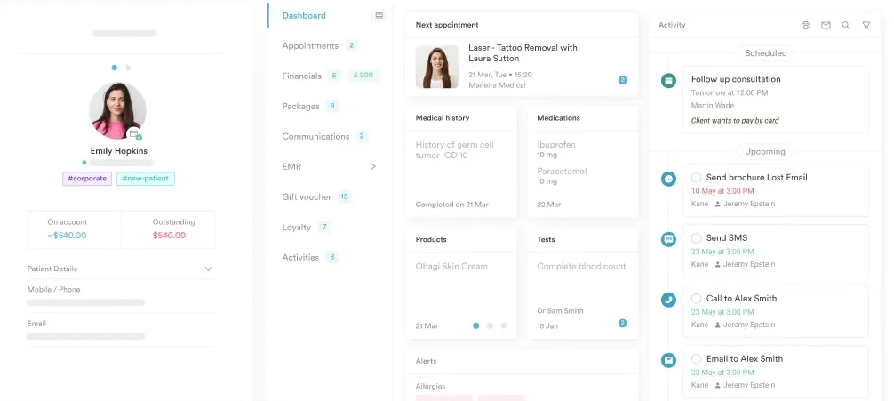
Practice management software like Pabau captures each analyte value in the client record, timestamped for audit.
A structured recording form for the chemical, physical, and microscopic findings from a urine specimen. Print it for point-of-care testing, or file the completed sheet in the client record.
Download templateUrine chemistry testing reads the chemical composition of a urine specimen to flag urinary tract conditions, kidney dysfunction, diabetes, and liver disease. Ten analytes on one reagent strip do most of that work.
This reference covers what each analyte detects and the normal range for it. It also walks through reading an abnormal cluster, collecting the specimen, and documenting the result. The form above gives you somewhere to record all of it.
Urine chemistry is a routine diagnostic test that evaluates the chemical composition of urine to identify abnormalities that point to disease. It runs in three integrated parts:
Together they work as a non-invasive screen for urinary tract infections (UTIs), kidney dysfunction, diabetes mellitus, liver disease, and metabolic disturbance.
The dipstick reagent strip sits at the core of the test. Each color-changing pad is sensitive to one chemical constituent, and ten pads sit on a single strip.
Clinical indications include routine health screening and acute symptom investigation for dysuria or hematuria. They also cover chronic disease monitoring in diabetes and chronic kidney disease, plus preoperative assessment. According to StatPearls NCBI, urinalysis remains one of the most commonly ordered laboratory tests in primary and specialty care.
Normal urine is roughly 91 to 96% water, with organic and inorganic solutes making up the rest. Three organic constituents dominate:
Those waste products reflect renal filtration capacity and systemic metabolism. The electrolyte fraction — sodium, potassium, chloride, phosphate, and sulfate — sits under hormonal control from aldosterone and antidiuretic hormone.
The glomerular filtration barrier normally excludes large molecules such as protein and glucose, while small water-soluble metabolites pass freely. So protein or glucose on a dipstick signals either glomerular dysfunction or metabolic dysregulation. Cleveland Clinic’s diagnostic reference sets out the same baseline.
The dipstick evaluates ten analytes, and each one carries its own clinical implication. Here is what every pad detects, and which conditions turn it abnormal:
Reference ranges guide interpretation and clinical decisions. The table below gives standardized normal values for every analyte, along with what an abnormal result usually means.
Ranges vary slightly by laboratory analyzer and methodology, so always check the intervals printed on your own laboratory reports. These values follow consensus ranges from the Medscape eMedicine urinalysis reference and Mayo Clinic diagnostic standards.
Abnormal dipstick findings need clinical correlation with patient history, physical examination, and further testing before you draw a conclusion. One abnormal analyte is not a diagnosis. A cluster of findings is stronger, such as positive leukocyte esterase and nitrites alongside white blood cells on microscopy.
The MedlinePlus medical encyclopedia makes the same point about specimen handling, timing, and clinical context.
Three patterns cover most abnormal results, and the matrix below shows which pads go positive in each one.
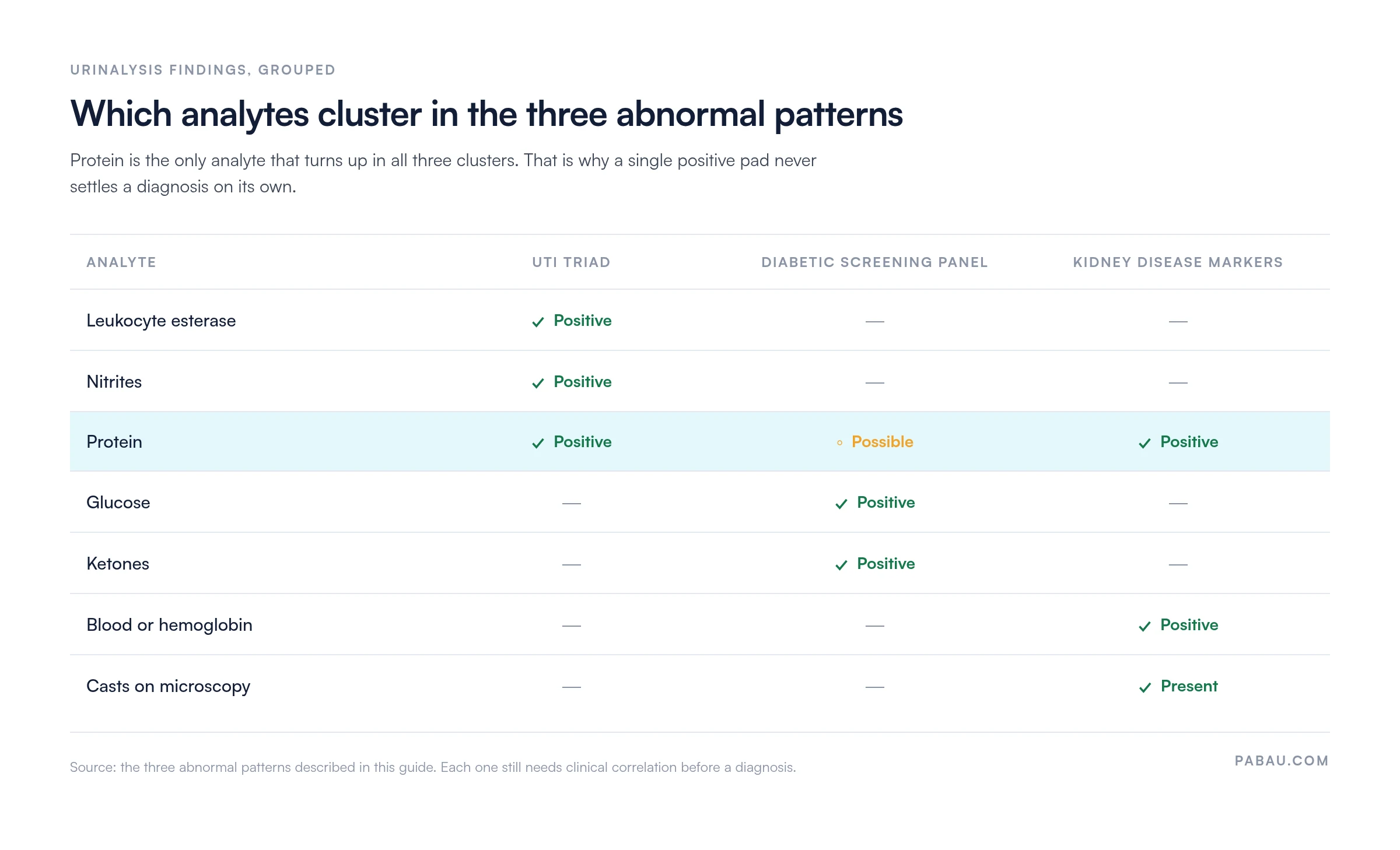
That overlap is why protein rarely points to one condition by itself. Record every abnormal finding and correlate it with the clinical picture before you start treatment.
Practice management software like Pabau captures normal and abnormal values through its patient intake forms. The result and its clinical context then stay in one record.

Proper collection protects analytical accuracy and prevents false positives from contamination. Clean-catch midstream is the standard technique for routine testing, and it is worth talking the patient through it:
Record the collection time and method in the patient’s chart. Delayed processing or poor handling invalidates the findings, so that detail decides whether the result can be read at all.
Pabau’s client record system timestamps every clinical note, which keeps the collection procedure and the result date audit-ready.
Good documentation of a urinalysis supports clinical decisions, regulatory compliance, and continuity of care. Record the collection date and time, the analytical method, every analyte value, and your interpretation of the abnormal findings.
Add the clinical context too. Name the reason for testing, whether that is an acute complaint, chronic disease monitoring, or screening. Note any factor affecting result validity, such as medication effects or recent trauma, and the follow-up you recommend.
Structured EHR documentation lets you spot a trend quickly. Progressive proteinuria in a diabetic patient, for instance, warrants either medication intensification or a nephrology referral.
Pabau Scribe, our AI scribe, transcribes and structures clinical notes from verbal dictation, so writing them up takes less of the appointment. Standard documentation covers specimen quality, every dipstick value, a comparison with prior results, and a clinical impression with the treatment plan.
Practice-performed urinalysis with dipstick reagent strips falls under the Clinical Laboratory Improvement Amendments (CLIA). It needs compliance management software that tracks certification status, quality control testing, and staff competency validation.
Meeting CLIA waiver requirements comes down to four habits. Use only FDA-cleared reagent strips, document quality control results monthly, keep staff training records, and verify analyzer calibration.
Non-waived urinalysis with microscopy, where you count red and white blood cells and casts, needs higher-level CLIA certification and proficiency testing.
Most practices run the dipstick at the point of care and then write the values somewhere else. The strip is read in the treatment room and the numbers go onto a paper form. Someone types them into the record later that day.
Pabau removes that second step. A digital form built for urinalysis holds one field per analyte, so the values land straight in the client record. The appointment, the practitioner, and a timestamp come with them.
Compliance follows the same route. Quality control logs, CLIA certification dates, and staff competency records sit in the same system as the results they cover. That is what an inspector asks to see.
Every Pabau subscription includes every feature, so the forms, the client record, and the compliance tools arrive together rather than as separate line items.

Pabau’s digital forms capture each dipstick value at the point of care, and the client record timestamps it. Your quality control logs and CLIA paperwork live in the same system.

The dipstick is quick, cheap, and easy to run badly. Most of the error sits in collection and documentation rather than in reading the pads.
So standardize the two ends. Talk every patient through clean-catch collection. Then write down the collection time, the method, and the whole analyte set, even when the strip reads normal. A negative result you can trace is worth more later than a positive one you cannot.
Download the reference form above and use it as the recording layer until the values live in your practice’s system. Book a demo to see how Pabau captures urinalysis results and the compliance records that sit behind them.

Need to record vital signs alongside the strip? Vital signs record template gives you one sheet for temperature, pulse, respiration, and blood pressure at the same visit.
Following up a hematuria finding? Kidney stones nursing care plan sets out the assessment, interventions, and patient education for renal calculi.
Building the intake that comes before the test? Medical review of systems template covers the genitourinary questions worth asking before you reach for a dipstick.
Choosing a system to hold the results? Clinical documentation software walks through what to look for when structured results have to reach the client record.
Urine chemistry evaluates the chemical composition of urine in three ways. It uses visual examination, a ten-analyte dipstick, and microscopic analysis of cells and crystals. It screens for UTI, kidney disease, diabetes, and systemic conditions.
A dipstick reads pH, specific gravity, protein, glucose, ketones, blood, nitrites, leukocyte esterase, bilirubin, and urobilinogen. Microscopy adds red and white blood cells, crystals, bacteria, casts, and epithelial cells.
Normal pH runs 4.5 to 8.0, with specific gravity 1.003 to 1.030. Urobilinogen sits at 0.1 to 1.0 EU/dL. Protein, glucose, ketones, blood, nitrites, leukocyte esterase, and bilirubin all read negative. Your facility’s own intervals may differ slightly.
Abnormal findings point to possible disease. Proteinuria suggests kidney dysfunction, glucosuria diabetes, and hematuria infection, stones, or malignancy. Positive leukocyte esterase or nitrites suggest UTI, ketonuria suggests ketosis, and high urobilinogen suggests liver disease. Clinical correlation is essential.
Use clean-catch midstream collection. Cleanse the urethral opening with antiseptic, void the first portion into the toilet, then collect 30 to 50 mL mid-stream. Seal it immediately and send it to the laboratory without delay, refrigerating it if processing is delayed.