

 Maja Popovska

Cytocare consent form
Cytocare is a hyaluronic acid-based mesotherapy skin booster from Revitacare, in...

1
Last Updated: Oct 27, 2026
Free PDF 6 pages, print-ready Clinician-reviewed
The Pirani score is a validated six-component clinical assessment tool used to grade the severity of congenital talipes equinovarus (CTEV) in infants and guide Ponseti treatment decisions at every stage of correction. Developed by Dr. Shafique Pirani, it gives pediatric orthopedic teams an objective, reproducible measure of foot deformity that replaces subjective visual estimation. Each of the six clinical signs is scored 0 (normal), 0.5 (moderate abnormality), or 1 (severe abnormality), producing a total between 0 and 6. This reference guide covers every sign, the interpretation thresholds that drive casting and tenotomy decisions, and how to track scores longitudinally across the full correction course.
A ready-to-use assessment sheet covering all six clinical signs (hindfoot and midfoot subscales), scoring rubric (0, 0.5, 1 per sign), total score calculation, and longitudinal tracking across Ponseti casting cycles.
Start from scratch, or upload this PDF or any form you already use, and Pabau's AI Form rebuilds it with tick boxes, text fields and a signature. Attach it to the treatment once, and every patient gets it on time.
AI Form rebuilds the consent, history and aftercare forms you already use. You review each one, then publish.
Patients complete forms on their phone before they arrive, so the front desk stops retyping paper.
Every signed form is versioned and attached to the treatment record, ready when you need it.
Set how long a consent stays valid. Returning patients skip forms they have already signed.
Supports HIPAA and GDPR compliance AES-256 encryption 4.8 on G2 4.7 on Capterra Trusted by 4,000+ practices worldwide
The Pirani score is a six-sign clinical scale that grades clubfoot severity in infants. Totals run from 0 for a normal foot to 6 for the most severe deformity. Developed by Dr. Shafique Pirani, it scores three hindfoot signs and three midfoot signs at 0, 0.5, or 1 each. Ponseti teams use it at every cast change to judge progress, time the Achilles tenotomy, and spot relapse.
It is a widely used severity measure for congenital talipes equinovarus (CTEV). In an interobserver reliability study, agreement was excellent among trained pediatric orthopedic surgeons and good among other surgeons. What follows covers each sign, how to read the subscales, and a sample score trajectory across a full casting course.

The Pirani score grades six signs across two subscales, hindfoot and midfoot. Each sign scores 0, 0.5, or 1, for a maximum total of 6.
A total score of 0 indicates complete correction. Achilles tenotomy is typically considered once the midfoot subscale reaches 0 and residual hindfoot equinus persists.
The score informs clinical judgment but does not replace it, so tenotomy timing still follows your protocol and the clinical findings.
Reassess both subscales after each Ponseti cast and record them consistently to catch stalled correction or early relapse.
Pabau, the practice management platform we build, lets pediatric teams capture Pirani scores in digital forms and track them across visits without paper records.
Before the Pirani score, severity grading depended on the clinician’s subjective impression. That variability made it hard to compare outcomes across centers, audit treatment decisions, or hand over care between team members. The score reduces a complex three-dimensional deformity to six observable, palpable signs, each with a clearly defined normal and abnormal state.
Pediatric orthopedic surgeons, physical therapists, and orthotists in a Ponseti program all use the score at each clinical contact. Capturing it in digital clinical forms rather than paper sheets cuts transcription errors. It also puts the longitudinal trend in front of the team at a glance.

The six signs are split equally between the hindfoot and midfoot subscales. Each sign scores 0, 0.5, or 1, giving a maximum of 3 per subscale and 6 overall. The table below summarizes what each score value means in practice.
The three hindfoot signs assess what is happening behind the ankle. Posterior crease reflects soft tissue contracture, and rigid equinus reflects how tight the gastrocnemius-soleus complex is. Empty heel indicates calcaneal displacement within the fat pad. All three are assessed with the infant relaxed and the knee extended when judging rigid equinus.
The three midfoot signs capture the forefoot and midfoot deformity components. Curved lateral border reflects the supination and adduction of the forefoot. Medial crease indicates the degree of cavus or adduction. Talar head coverage assesses navicular position relative to the talar head, the key landmark for Ponseti correction sequencing.
Total score interpretation follows a severity gradient, but the subscale breakdown carries equal clinical weight. A foot scoring 3/3 on the hindfoot with only mild midfoot involvement needs a different casting approach from a foot that scores 3/3 on both.
The bands below are an illustrative reading, not part of the validated score. Published studies use different total cutoffs, roughly 3 to 5, when they relate the score to tenotomy.
Tenotomy threshold: Achilles tenotomy is typically considered when the midfoot subscale has reached 0 and residual hindfoot equinus persists. In practice, that means a rigid equinus score of 0.5 or 1. The Ponseti method is the first-line treatment for clubfoot, according to a review of the Ponseti literature. Always verify tenotomy timing against current institutional protocol and individual clinical findings.
The Ponseti method is a serial casting protocol that corrects CTEV in a set order: Cavus first, then adduction and supination, then equinus. A study on Pirani score and Ponseti casting found that a higher initial score correlates with more casts. Individual variation is substantial, though, so one score should not be read as a firm cast count.
The score drives decisions at four points in the workflow.
Teams using measurements tracking tools within their clinical records can plot the subscale trend automatically. Regression then shows between appointments without anyone reviewing a stack of paper sheets.
Few published resources show how a well-correcting foot’s score should move across multiple casts. The downloadable sheet gives each visit its own dated entry, so the trajectory builds as you go. The illustrative table below follows a foot that starts at a total score of 5.5, based on patterns described in the Ponseti literature.
Plotted cast by cast, the pattern is easier to read: the midfoot falls steadily while the hindfoot barely moves until the tenotomy.
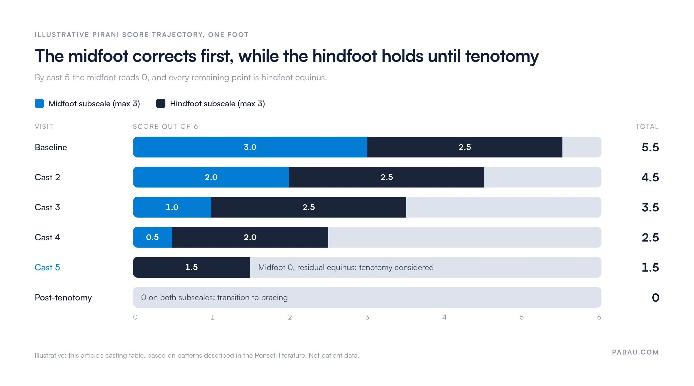
Red-flag patterns: A total score that fails to drop by at least 0.5 points after two consecutive casts suggests inadequate correction. Possible causes include casting technique, skin tolerance limiting abduction, or an underlying stiff deformity that may warrant early orthopedic review. Document the stalled trajectory clearly in the patient record, so escalation decisions rest on longitudinal evidence.
Document the hindfoot and midfoot subscale scores separately at each visit, alongside the total. A foot with a total of 2.0 split as 0 hindfoot / 2.0 midfoot is clinically very different from 1.0 / 1.0. The subscale breakdown is what drives the next casting decision.
Bilateral CTEV accounts for roughly 30 to 50 percent of cases. When both feet are affected, score each one independently. Record them as “right foot” and “left foot” within the same assessment sheet.
Asymmetric bilateral presentations are common. One foot may correct quickly while the other lags, needing extra casts on one side only. A single combined score hides that asymmetry, and casting decisions then fall behind the slower foot.
Pediatric orthopedic teams that keep structured patient records with per-limb fields can compare both feet side by side at follow-up. Nobody has to page back through paper bilateral entries to spot a one-sided change.

The downloadable assessment sheet is built for every Ponseti casting visit. These five steps cover completing and filing it.
The Pirani score is relevant to any clinician involved in CTEV diagnosis and management.
A standardized assessment sheet does more than save time at the appointment. It creates a defensible, auditable record of how clinical decisions were reached.
Consistency across practitioners: When every clinician on the schedule uses the same six-sign rubric, inter-rater variability drops. This matters most in teaching hospitals, where residents and attending surgeons share casting duties.
Outcome benchmarking: Longitudinal score data lets teams compare their correction rates against published benchmarks. It also surfaces outliers that may benefit from technique review and feeds service-level audit cycles.
Parental communication: A completed sheet with a falling score gives parents a progress marker they can see. Parents of infants in serial casting often find that number more reassuring than verbal reassurance alone.
Medicolegal protection: If a relapse occurs or a tenotomy decision is questioned, a complete longitudinal score record shows how each decision was reached. Every step traces back to objective clinical findings.
On paper, every cast change adds another sheet to the file. The trend between visits only appears when someone lines the sheets up and compares them by hand. During bracing, when visits are months apart, a slow rise on one foot is easy to miss.
In Pabau, the assessment sheet becomes a digital form attached to the patient’s record. Each visit’s sign scores, subscale totals and treatment decision are saved against the date, for each foot.
The surgeon, physical therapist and nurse then open the same record and see the same trajectory. A stalled subscale or a one-sided rise shows up at the visit it happens, so you can adjust casting or bring the bracing review forward.

Capture Pirani scores, track casting progress, and store consent forms in one place. Pabau replaces paper assessment sheets with structured digital records your whole team can open.

Score each foot sign by sign at every cast, and record both subscales next to the total. That habit gives your tenotomy timing a documented basis and flags a stalled foot while there is still time to change the casting.
The trade-off is a minute of extra documentation per visit, and it only pays off if every clinician scores the same way. Treat the severity bands as a reading aid and your protocol as the rule.
Book a demo to see how Pabau keeps every Pirani score, cast and brace visit on one patient record your whole team can read.

Checking dorsiflexion as the equinus resolves? Ankle dorsiflexion test explains how to measure and track ankle range of motion between visits.
Assessing foot posture once the child is walking? Foot Posture Index FPI-6 gives you a six-item standing assessment with a free scoring template.
Following development after bracing ends? Pediatric balance scale covers a 14-item balance test with a scoring guide for children.
Building a wider set of outcome scores? Outcome measures template shows how to choose, score and track the measures your team relies on.
Choosing software for a physical therapy team? Physiotherapy practice management software compares the tools musculoskeletal teams use to run records, scheduling and billing.
The Pirani score grades the severity of congenital talipes equinovarus (clubfoot) in infants and guides Ponseti treatment decisions. It gives orthopedic teams an objective, reproducible measure of correction across serial casting visits, in place of a subjective severity estimate.
A Pirani score of 6 indicates maximum deformity, with all six clinical signs scoring 1 (severe). The foot shows deep posterior and medial creases, rigid equinus, an empty heel, a severely curved lateral border and a fully uncovered talar head. These feet typically need more casts before tenotomy is considered.
Tenotomy is typically considered when the midfoot subscale has reached 0 and residual equinus remains on the hindfoot subscale. That usually means a rigid equinus score of 0.5 or 1. The treating surgeon makes the call. The Pirani score informs that judgment but does not set tenotomy timing on its own.
Reassess it at every cast change, typically every five to seven days during the active casting phase in infancy. During bracing, reassess at each follow-up appointment, usually every three months in the first two years. That rhythm catches relapse before it becomes a full recurrence that needs repeat casting.
A useful mnemonic is “PREMCT”: Posterior crease, Rigid equinus, Empty heel (hindfoot), Medial crease, Curved lateral border, Talar head coverage (midfoot). Grouping the three hindfoot signs (PRE) and three midfoot signs (MCT) separately makes the subscale structure easier to recall at the bedside.
The hindfoot subscale (maximum 3) covers the posterior crease, rigid equinus and empty heel, reflecting the calf contracture and calcaneal position. The midfoot subscale (maximum 3) covers the curved lateral border, medial crease and talar head coverage, reflecting supination, adduction and navicular displacement. Document both separately, because they drive different parts of the casting technique.