Chronic kidney disease nursing care plan
A chronic kidney disease nursing care plan is a structured document that guides ...

3
Last Updated: Oct 14, 2026

A fluid and electrolyte imbalance nursing care plan runs one cycle: assessment, NANDA diagnosis, measurable goal, interventions, and evaluation.
One table below covers all six serum electrolytes, with normal ranges, published critical values, the ECG change to expect, and the first nursing action.
Serum sodium should not rise by more than 10 mEq/L in the first 24 hours of correction.
Potassium outside 3.5 to 5.0 mEq/L earns a 12-lead ECG and continuous cardiac monitoring.
The Assess–Document–Reassess loop keeps each shift’s vitals, labs, and NANDA statement on one patient timeline.
A ready-to-use care plan with NANDA diagnoses, assessment prompts, and normal electrolyte reference ranges. Interventions are laid out for hyponatremia, hypernatremia, hypokalemia, hyperkalemia, and fluid volume imbalance.
Download templateA fluid and electrolyte imbalance nursing care plan records five parts: the assessment, the NANDA diagnosis, the goal, the interventions, and the evaluation.
Writing one is rarely the hard part. The hard part is holding it together across three shifts. Sodium drifts overnight, and the 2 AM potassium result lands on a screen the day nurse never opens.
This page carries a complete care plan for electrolyte imbalance in one place. Below you get a six-electrolyte reference table, two worked mini-plans, and a set of if-then escalation rules. The downloadable version above follows the same structure.
A fluid and electrolyte imbalance nursing care plan is a written document that moves a patient from abnormal labs to a measurable goal.
The plan applies the nursing process in order: assess, diagnose, plan, implement, evaluate. Each stage produces something the next stage uses. The assessment produces the defining characteristics. Those characteristics justify the NANDA diagnosis, and the diagnosis sets the goal.
The plan does four jobs at once. The document records the patient’s baseline, names the risk factors, states a measurable target, and gives the next clinician a reason for every intervention. Structured intake and assessment forms make that record consistent between nurses.
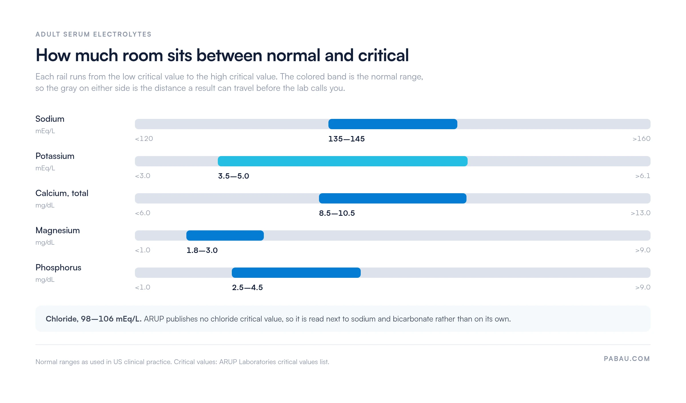
Normal adult serum ranges are sodium 135–145 mEq/L, potassium 3.5–5.0 mEq/L, calcium 8.5–10.5 mg/dL, magnesium 1.5–2.5 mEq/L, phosphorus 2.5–4.5 mg/dL, and chloride 98–106 mEq/L.
The table below adds the three columns most reference lists leave out. You get the critical value that triggers a call from the lab, the ECG change tied to that electrolyte, and the first action.
| Electrolyte | Normal range | Critical value | Signs and ECG change | First nursing action |
|---|---|---|---|---|
| Sodium | 135–145 mEq/L | <120 or >160 mEq/L | Low: headache, confusion, seizures. High: thirst, restlessness. No signature ECG change | Neurologic checks each shift, and escalate any new confusion |
| Potassium | 3.5–5.0 mEq/L | <3.0 or >6.1 mEq/L | Low: weakness, flattened T waves, U waves. High: cramps, peaked T waves | Get a 12-lead ECG and start continuous cardiac monitoring |
| Calcium (total) | 8.5–10.5 mg/dL | <6.0 or >13.0 mg/dL | Low: tetany, positive Chvostek’s or Trousseau’s sign, prolonged QT. High: shortened QT | Test both signs, then set seizure and fall precautions |
| Magnesium | 1.5–2.5 mEq/L (1.8–3.0 mg/dL) | <1.0 or >9.0 mg/dL | Low: tremor, torsades de pointes. High: drowsiness, lost deep tendon reflexes | Check deep tendon reflexes, and recheck potassium alongside |
| Phosphorus | 2.5–4.5 mg/dL | <1.0 or >9.0 mg/dL | Low: muscle weakness, weak respiratory effort. High: tetany when calcium is also low | Watch respiratory effort, and pair the result with calcium |
| Chloride | 98–106 mEq/L | None published by ARUP | Falls with vomiting and gastric suction. Tracks acid-base status | Read it beside sodium and bicarbonate, never on its own |
Reference ranges are not universal. StatPearls, for example, publishes potassium as 3.6 to 5.5 mmol/L. The critical values above come from the published list at ARUP Laboratories. Critical thresholds are set locally, so your facility’s panel governs your care plan.
Read the ranges as distances rather than as numbers. Some electrolytes sit much closer to their low critical value than others, which is what the chart below shows.
Most imbalances come from four sources: fluid lost through the gut or skin, kidneys that cannot regulate, hormones that misfire, and drugs that shift electrolytes.
Renal disease deserves separate attention. A kidney that cannot excrete potassium, phosphate, or free water turns a routine intake into a rising serum level. Chronic kidney disease therefore produces hyperkalemia and hyperphosphatemia far more often than the gut ever does.
Dialysis adds its own pattern. Hemodialysis pulls potassium and phosphate out over a few hours, so the pre-dialysis and post-dialysis panels can differ sharply. Time your electrolyte checks around the treatment, and note in the plan which side of it the sample came from.
A patient with several risk factors stacked together needs the tightest monitoring. An 80-year-old on furosemide who has been vomiting for two days is the standard example. A single record of medications and history is what makes that stacking visible at a glance.
Work the assessment in a fixed order, because the order is what stops findings from going missing at handover.
A fluid deficit shows up as a cluster, not as one finding. Dry mucous membranes on their own mean little in a mouth-breathing patient. Add a heart rate of 110, flat neck veins, urine output under 30 mL per hour, and a 2 kg weight loss. Together they are a fluid deficit.
Weight is the most reliable single number you have. A change of 1 kg in a day is roughly 1 liter of fluid gained or lost. Recording the finding at the point of care keeps that trend intact instead of rebuilding it from memory at the end of the shift.
Both signs test for the neuromuscular irritability of low calcium, and both take under four minutes at the bedside.
For Chvostek’s sign, tap the facial nerve just in front of the earlobe, below the zygomatic arch. Twitching of the upper lip or the side of the face is a positive result.
For Trousseau’s sign, inflate a blood pressure cuff above the patient’s systolic pressure and hold it there for three minutes. Carpopedal spasm, with the wrist and fingers flexing inward, is a positive result.
Trousseau’s sign is the more specific of the two. Chvostek’s sign appears in a share of people whose calcium is entirely normal, so a positive Chvostek’s on its own is not a diagnosis. Document which sign you tested, and record the result either way.
The three electrolyte imbalance nursing diagnosis labels in routine use are Risk for Electrolyte Imbalance, Deficient Fluid Volume, and Excess Fluid Volume.
Older textbooks call these Fluid Volume Deficit and Fluid Volume Excess. The wording is different, the diagnosis is the same, and an examiner will accept either. Pick one form and use it consistently through the plan.
The structure of the statement is where marks and audits are lost. A problem-focused diagnosis takes three parts: the label, a related-to cause, and an as-evidenced-by finding. A risk diagnosis takes two parts only, because the problem has not happened yet.
Hyponatremia nursing care is fluid restriction, regular neurologic checks, seizure precautions, and a correction rate that never exceeds 10 mEq/L in 24 hours.
Hyponatremia is a serum sodium below 135 mEq/L. Below about 125 mEq/L the brain starts to swell. That is why the presenting complaint is usually headache, nausea, or confusion, not a limb sign.
A worked plan for a symptomatic patient looks like this.
Hyponatremia treatment itself belongs to the provider, and the nursing job is to hold the guardrails. Fluid restriction is commonly set between 500 and 800 mL per day. Hypertonic 3% saline is reserved for symptomatic patients in a monitored setting, on a written order.
The rate matters more than the target. Serum sodium should not rise by more than 10 mEq/L in the first 24 hours. European guidance applies that ceiling to acute and chronic hyponatremia alike. A 2024 systematic review of osmotic demyelination reports a tighter limit of 8 mEq/L in 24 hours for higher-risk patients.
Overcorrection is the complication to watch for, not undercorrection. If the sodium climbs faster than the ceiling, stop the infusion and call the provider before the next scheduled check.
A hypokalemia nursing care plan pairs potassium replacement with continuous cardiac monitoring, because the same low potassium that weakens muscles also destabilizes the heart.
Hypokalemia is a serum potassium below 3.5 mEq/L. The early signs are fatigue, muscle weakness, and cramps. On the monitor it shows as flattened T waves and the appearance of U waves.
The safety rules around replacement are the most testable part of the plan. Intravenous potassium is never given as a push. Peripheral infusion is usually capped at 10 mEq per hour, and higher rates need central access with continuous cardiac monitoring.
One nursing intervention for hypokalemia gets missed more than the rest. If potassium will not rise despite replacement, check the magnesium. Low magnesium drives renal potassium loss, so the potassium keeps falling until the magnesium is corrected first.
Oral replacement is gentler on the vein and on the heart, so it is preferred whenever the patient can swallow. Give it with food to reduce nausea, and reinforce dietary sources such as bananas, oranges, potatoes, and spinach.
Goals earn their place only when someone can tell, at a stated time, whether they were met. Write each one with a number and a deadline in it.
Vague outcomes such as “the patient will maintain fluid balance” cannot be evaluated, so they cannot be closed. Every goal above names either a number or an observable behavior.
Electrolyte imbalance nursing interventions fall into three jobs: monitor the trend, replace or restrict what is off, and protect the patient while levels correct.
Monitoring. Check vital signs every one to four hours according to acuity. Record intake and output hourly for unstable patients and each shift for stable ones. Draw the electrolyte panel as ordered, assess neurologic status, and weigh the patient at the same time each morning.
Replacing and restricting. Give IV fluids, oral or IV electrolyte replacement, and diuretics as ordered. Apply the dietary rule that matches the imbalance, whether that is a fluid restriction for hypervolemia or a potassium restriction in renal failure.
Protecting. Set seizure precautions for significant hyponatremia and fall precautions for weakness. Keep the patient on a cardiac monitor for any potassium, magnesium, or calcium result far outside range.
IV fluids for dehydration start with an isotonic crystalloid, usually 0.9% sodium chloride or lactated Ringer’s, at the rate and volume the provider orders.
Isotonic fluid stays in the intravascular space, which is what a hypovolemic patient needs first. Hypotonic fluid such as 0.45% sodium chloride moves into the cells and suits a free-water deficit instead. Hypertonic 3% saline is a symptomatic-hyponatremia drug, not a rehydration fluid.
Once the infusion is running, the nursing job shifts to watching for the opposite problem. Auscultate the lungs, look for new jugular venous distension or peripheral edema, and track urine output. Rising output toward 0.5 mL per kilogram per hour is the sign the deficit is closing.
Care plans usually stop at “monitor and report.” The table below names the specific triggers worth writing into yours, with the action attached to each one.
| What you see | What it means | What you do next |
|---|---|---|
| Potassium above 6.1 mEq/L with peaked T waves | Hyperkalemia with cardiac effects | Call the provider now, keep the monitor on, expect an urgent order |
| Serum sodium has risen more than 10 mEq/L in 24 hours | Overcorrection, with a risk of osmotic demyelination | Stop or slow the infusion and notify the provider |
| New confusion or a seizure with sodium under 125 mEq/L | Symptomatic hyponatremia | Escalate immediately and start seizure precautions |
| Potassium stays low after replacement | Likely untreated hypomagnesemia | Ask for a magnesium level before the next potassium dose |
| Positive Trousseau’s sign | Symptomatic hypocalcemia | Notify the provider, and set seizure and airway precautions |
| Weight changes by 1 kg or more in a day | About 1 liter of fluid gained or lost | Recheck intake and output, then report the trend |
Discharge teaching works when it is tied to the cause of this admission, not to electrolytes in general. A patient who became hypokalemic through diarrhea needs different instructions from one on a loop diuretic.
Evaluation asks three questions each shift: did the labs move toward normal, did the symptoms resolve, and can the patient repeat the plan?
Those questions map onto the nursing responsibilities for monitoring fluid and electrolyte balance. You own the intake and output record, the daily weight, the vital sign trend, and the timing of the repeat panel. You also own the escalation when a value crosses a critical threshold.
A goal that was met gets closed and dated. A goal that was not met gets a reason attached before it is rewritten. The usual reasons are an untreated underlying cause, a missed dose, a fluid restriction nobody explained, or renal function that has not recovered.
Write the evaluation down even when the answer is “no change.” A dated entry in the patient record is what turns a sequence of shifts into an audit trail of clinical reasoning.
Every plan in this article runs the same three-step cycle. We call it the Assess–Document–Reassess loop, and it is the part that breaks first when the record lives in three places.
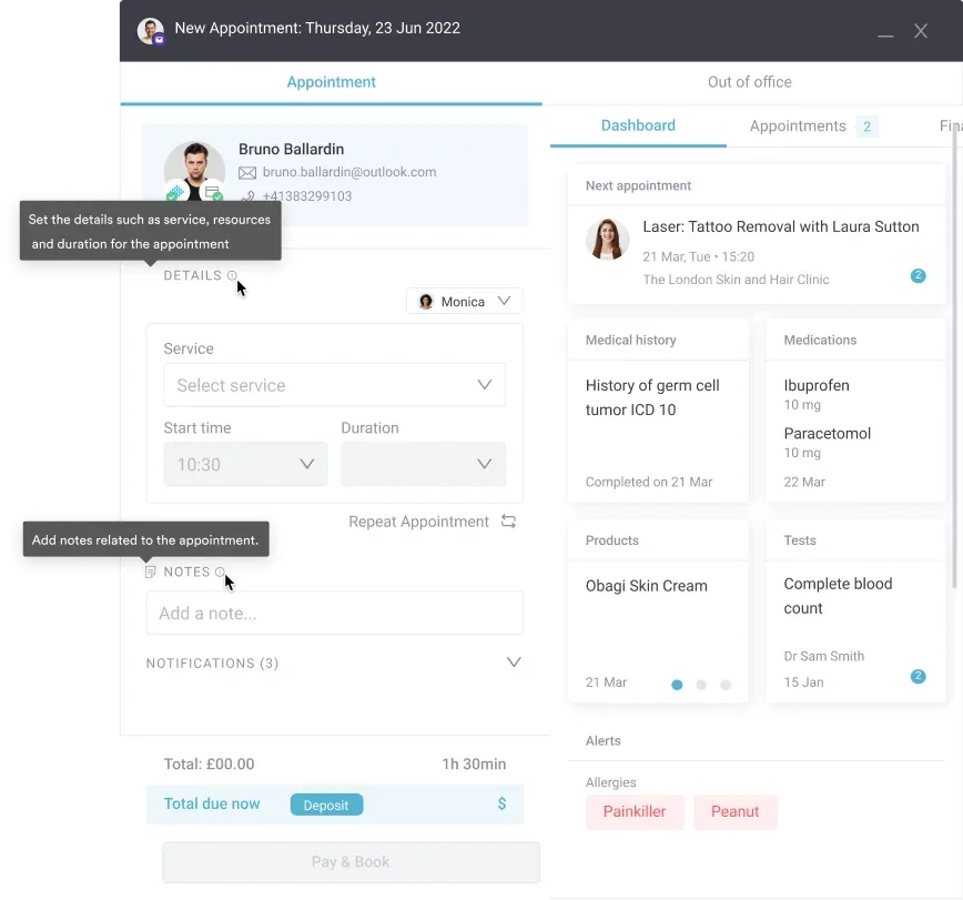
Practice management software like Pabau closes that loop in one system. Digital forms capture the assessment at the bedside, so the finding is typed once. Client records keep the diagnosis, the goal, and the lab values behind them together. The next clinician reads the reasoning instead of reconstructing it.
Automated workflows handle the third step. The recheck, the dietary consult, and the education session each fire as a task at the time the plan sets. The reassessment then happens on schedule, rather than whenever someone remembers.
In the practices we onboard, the step that slips is almost always the reassessment rather than the assessment. Nurses assess well. What gets lost is the follow-up value landing somewhere the next shift never looks. Putting the recheck on the same timeline as the plan is what fixes it.

Pabau’s digital forms, client records, and automated workflows keep each shift’s assessment, diagnosis, and recheck on one patient timeline. Your team stops rebuilding the plan at handover.

A care plan is only as good as its last reassessment. A well-written NANDA statement from 8 AM is worth very little by 8 PM if nobody rechecked the potassium behind it.
So pick the two numbers that will decide this patient’s day, and put a recheck time against each one. The rest of the plan can follow from there. That is the difference between a document that satisfies an audit and one that changes what happens at the bedside.
Download the template above to start from a structure that already carries the diagnoses, the ranges, and the interventions. Book a demo to see how Pabau keeps each assessment, diagnosis, and recheck on one patient timeline.

Need a structured framework for clinical decision-making? Safer clinical notes covers documenting assessments and interventions in a way that holds up under review.
Want your team’s charting to look the same on every shift? Psychiatric evaluation template shows how a structured template stops assessment elements from being skipped.
Still working out how to write the note itself? SOAP notes for social work and clinical practice walks the full ADPIE process with worked examples.
NCP stands for nursing care plan. An NCP for electrolyte imbalance documents the assessment, the NANDA diagnosis, the goal, the interventions, and the evaluation for one patient. An NCP is written when serum levels fall outside the normal range, and reviewed each shift.
The core responsibilities are intake and output, daily weight, vital signs, and the serum electrolyte trend. Weigh the patient at the same time each morning. Report a change of 1 kg or more in a day, because it represents about 1 liter of fluid. Escalate any critical value to the provider straight away.
Interventions split into monitoring, replacement or restriction, and patient safety. Monitor vital signs, intake and output, and the repeat labs. Give fluids, electrolyte replacement, or diuretics as ordered. Then apply the matching precautions, such as seizure precautions for hyponatremia and continuous cardiac monitoring during IV potassium replacement.
Gastrointestinal losses such as vomiting, diarrhea, and nasogastric drainage are the most common cause. Renal causes include chronic kidney disease, acute kidney injury, and dialysis. Other causes are diuretics, corticosteroids, ACE inhibitors, NSAIDs, diabetes insipidus, SIADH, burns, poor oral intake, heart failure, and cirrhosis.
Risk for Electrolyte Imbalance, NANDA code 00195, is defined as being vulnerable to changes in serum electrolyte levels that may compromise health. As a risk diagnosis it has no defining characteristics. Write it with a related-to clause only, never with an as-evidenced-by clause. Related factors include diuretic use, vomiting, diarrhea, renal disease, and endocrine disorders.
Nurses combine three sources. Subjective history covers symptoms, recent illness, medications, and usual fluid intake. Objective findings cover vital signs, skin turgor, mucous membranes, capillary refill, edema, and daily weight. Laboratory data covers the serum electrolyte panel, osmolality, urine specific gravity, and acid-base status.
Normal adult ranges are sodium 135 to 145 mEq/L, potassium 3.5 to 5.0 mEq/L, and calcium 8.5 to 10.5 mg/dL. Magnesium is 1.5 to 2.5 mEq/L and phosphorus 2.5 to 4.5 mg/dL. Chloride is 98 to 106 mEq/L. Reference ranges vary between laboratories, so confirm your facility’s panel.
Hyponatremia is a serum sodium below 135 mEq/L. Nursing care is fluid restriction, regular neurologic checks, and seizure precautions. Hypertonic 3% saline is reserved for symptomatic patients, in a monitored setting, on a provider’s order. Serum sodium should not rise by more than 10 mEq/L in the first 24 hours.
Deficient Fluid Volume is written as a three-part statement: the label, the related-to cause, and the as-evidenced-by findings. An example is Deficient Fluid Volume related to persistent vomiting, as evidenced by urine output of 20 mL per hour and dry mucous membranes. Track daily weight and intake and output to evaluate the plan.