CRM for chiropractors: how to manage patients, bookings and marketing
A CRM for chiropractors is a patient relationship management system built around...

Last Updated: Oct 13, 2026

The duck walk test is a clinical special test used to screen for meniscal tears and assess knee joint stability during weight-bearing squat movement
A positive test is indicated by joint line pain, clicking, or inability to complete the full squat-walk sequence, suggesting posterior horn meniscal pathology
Published sensitivity and specificity range from 66-82%, making it useful as a screening tool but not diagnostic on its own; always follow positive results with imaging or complementary tests
Practice management software like Pabau streamlines documentation of duck walk test findings with digital forms and clinical records, creating searchable assessment archives for ongoing patient care
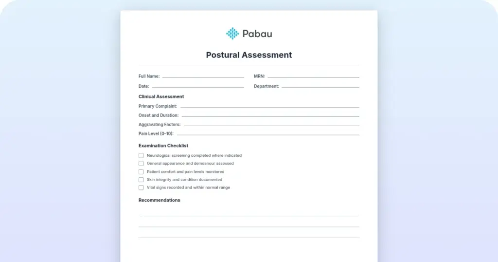
A ready-to-use assessment form documenting patient positioning, test procedure, positive signs (joint line pain, clicking, inability to complete squat), clinical findings, and referral recommendations for meniscal pathology screening.
Download templateThe duck walk test, also called the Childress test or Childress sign, is a clinical assessment tool for screening meniscal pathology during weight-bearing knee movement. Physiotherapists, sports medicine practitioners, and orthopedic clinicians use it as part of a routine knee examination. This guide covers the complete procedure, positive interpretation criteria, and best practices for documenting findings in clinical records.
The duck walk test is a functional movement screening tool that assesses the knee’s response to weight-bearing squat mechanics, specifically targeting the posterior horn of the meniscus. Also known as the Childress test, after the clinician who first described it, the test loads the knee joint through a full squat with internal or external tibial rotation. This creates mechanical stress that provokes pain or clicking when meniscal pathology is present.
Unlike imaging studies like MRI or arthroscopy. It provides immediate clinical feedback and helps guide referral decisions for further diagnostic testing.
The test is widely used in preparticipation physical examinations (PPE), sports physicals, and routine orthopedic screening because it is quick, non-invasive, and requires no special preparation or instrumentation.
Performing the duck walk test correctly requires clear patient instruction and careful observation of movement quality and patient response.
The entire procedure typically takes 30-60 seconds.
A positive duck walk test is indicated by one or more of the following clinical findings:
A positive test suggests meniscal pathology but does not confirm a meniscal tear on its own. The posterior horn of the meniscus is the most commonly affected region when this test is positive, due to the loading mechanics of the full squat position with tibial rotation.
Occupational therapists often score assessments like this directly inside occupational therapy software, keeping results attached to the treatment plan.
Published peer-reviewed studies have evaluated the duck walk test’s ability to detect meniscal tears confirmed by MRI or arthroscopy. The following table summarizes diagnostic accuracy data from clinical research:
These data underscore that the duck walk test is a useful screening tool but should never be relied upon in isolation. A positive test warrants additional clinical assessment and imaging referral.
The duck walk test is valuable for multiple clinical settings and professional groups:
The duck walk test offers several practical advantages in clinical settings. It requires no equipment, can be performed in any clinic environment, and takes less than one minute to complete. Results are immediately available and provide clear clinical direction: if positive, further diagnostic testing is warranted; if negative, the clinician can rule out meniscal pathology with reasonable confidence and pursue alternative diagnoses.
The test also educates patients about their knee mechanics and helps them understand the relationship between movement patterns and symptoms. When documented in clinical records, the duck walk test result becomes part of the patient’s assessment narrative and supports clinical decision-making at future visits. A physical therapy practice tracking range of motion over weeks needs the earlier values beside the current one.
Accurate documentation of the duck walk test is essential for clinical continuity, referral communication, and compliance with documentation standards. Clinicians should record the following elements in the patient’s assessment note:
When imaging confirms a tear, clinicians document it with S83.241.
Using a standardized assessment form (such as the downloadable template above) ensures consistent documentation and makes it easier to write safer clinical notes that hold up for referrals and audits.

The duck walk test is one of several meniscal special tests available to clinicians. Understanding how it compares to McMurray’s test, the Thessaly test, and the Apley Grind test helps guide test selection during a comprehensive knee examination:
Best practice is to use a battery of tests rather than relying on one alone. A structured knee examination typically combines two or more of these tests with comprehensive clinical documentation to build a robust clinical hypothesis before referring for imaging.
Combine the duck walk test with joint line tenderness palpation and the McMurray test for a more complete meniscal screening battery. If two or more tests are positive, the likelihood of meniscal pathology increases significantly, strengthening the case for MRI or orthopedic referral.

Pabau’s digital forms and clinical records help you document special tests like the duck walk test in structured, searchable formats. Build assessment templates once and reuse them for every patient.

The duck walk test is a practical, cost-effective special test for screening meniscal pathology in clinical and sports medicine settings. When performed correctly and interpreted within the context of other examination findings, it helps clinicians build a robust assessment hypothesis and guide appropriate referral for imaging.
Accurate documentation of the duck walk test strengthens clinical communication and supports continuity of care. By using standardized assessment templates, such as the free downloadable form provided above, clinicians create consistent, searchable clinical records that improve patient outcomes and support evidence-based practice. Book a demo with Pabau to see how digital assessment forms and clinical records can streamline your meniscal testing workflows.

Suspecting thoracic outlet syndrome? This guide covers the Halstead maneuver technique, the stop signs, and the test order.
Screening reveals a positive result? A meniscus rehab exercises handout gives patients a clear home program while they await imaging or referral.
Need a place to log symptom location? A body pain chart template helps patients mark exactly where joint line pain occurs.
Missing a hip flexor diagnosis? This guide covers the hip flexor strain test, including the Thomas Test, FADIR, and clinical grading.
Checking for a rotator cuff tear? The infraspinatus test checks for resisted external rotation weakness linked to a cuff tear or nerve neuropathy.
The duck walk test (also called the Childress Test) is a clinical special test in which a patient squats deeply and walks forward in a full squat position. Pain, clicking, or inability to complete the movement may indicate meniscal tears or knee joint pathology. The test takes about 30-60 seconds to perform and requires no equipment.
Published studies report sensitivity of 66-82% and specificity of 70-92%, meaning the test correctly identifies meniscal tears roughly two-thirds to four-fifths of the time and correctly rules them out 70-92% of the time. A positive test warrants further imaging (MRI) or orthopedic referral, but the test cannot diagnose a tear on its own.
A positive duck walk test is indicated by joint line pain, clicking, or inability to complete the full squat-walk movement. These signs suggest meniscal pathology (often posterior horn involvement), but confirmation requires imaging such as MRI or arthroscopy. A positive test should always be followed by additional clinical tests and referral for further evaluation.
The duck walk test is a standard component of preparticipation physical examinations (PPE) and sports physicals because it is quick, requires no equipment, and effectively screens for meniscal injury risk in active patients. A positive result in an athlete prompts follow-up examination and, if indicated, imaging before return to sport clearance.
After a positive duck walk test, perform complementary special tests (McMurray, Thessaly, Apley Grind) and joint line palpation to strengthen your clinical impression. If meniscal pathology remains likely, refer the patient for MRI imaging or orthopedic consultation. Document all findings clearly and communicate your assessment to the referring physician or orthopedic specialist.
Record the test name (Duck Walk Test / Childress Test), patient positioning and instructions followed, specific findings (pain location and severity, clicking, inability to squat), the final result (positive/negative/equivocal), and clinical correlation with other examination findings. Include your next steps, such as imaging referral or follow-up testing. Using a standardized assessment form ensures consistency and defensibility.